
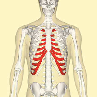
In vertebrate anatomy, ribs are the long curved bones which form the rib cage, part of the axial skeleton. In most tetrapods, ribs surround the chest, enabling the lungs to expand and thus facilitate breathing by expanding the chest cavity. They serve to protect the lungs, heart, and other internal organs of the thorax. In some animals, especially snakes, ribs may provide support and protection for the entire body.

In anatomy, the atlas (C1) is the most superior (first) cervical vertebra of the spine and is located in the neck.
The rib cage is an endoskeletal enclosure in the thorax of most vertebrate animals that comprises the ribs, vertebral column and sternum, which protects vital organs such as the heart, lungs and great vessels. The circumferential enclosure formed by left and right rib cages, together known as the thoracic cage, is a semi-rigid bony and cartilaginous structure which surrounds the thoracic cavity and supports the shoulder girdles to form the core part of the axial skeleton.

The scapula, also known as the shoulder blade, is the bone that connects the humerus with the clavicle. Like their connected bones, the scapulae are paired, with each scapula on either side of the body being roughly a mirror image of the other. The name derives from the Classical Latin word for trowel or small shovel, which it was thought to resemble.

The lumbar vertebrae are, in human anatomy, the five vertebrae between the rib cage and the pelvis. They are the largest segments of the vertebral column and are characterized by the absence of the foramen transversarium within the transverse process and by the absence of facets on the sides of the body. They are designated L1 to L5, starting at the top. The lumbar vertebrae help support the weight of the body, and permit movement.

In anatomy, the axis is the second cervical vertebra (C2) of the spine, immediately inferior to the atlas, upon which the head rests.

In tetrapods, cervical vertebrae are the vertebrae of the neck, immediately below the skull. Truncal vertebrae lie caudal of cervical vertebrae. In sauropsid species, the cervical vertebrae bear cervical ribs. In lizards and saurischian dinosaurs, the cervical ribs are large; in birds, they are small and completely fused to the vertebrae. The vertebral transverse processes of mammals are homologous to the cervical ribs of other amniotes. Most mammals have seven cervical vertebrae, with the only three known exceptions being the manatee with six, the two-toed sloth with five or six, and the three-toed sloth with nine.
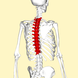
In vertebrates, thoracic vertebrae compose the middle segment of the vertebral column, between the cervical vertebrae and the lumbar vertebrae. In humans, there are twelve thoracic vertebrae and they are intermediate in size between the cervical and lumbar vertebrae; they increase in size going towards the lumbar vertebrae, with the lower ones being much larger than the upper. They are distinguished by the presence of facets on the sides of the bodies for articulation with the heads of the ribs, as well as facets on the transverse processes of all, except the eleventh and twelfth, for articulation with the tubercles of the ribs. By convention, the human thoracic vertebrae are numbered T1–T12, with the first one (T1) located closest to the skull and the others going down the spine toward the lumbar region.

The transverse plane is an anatomical plane that divides the body into superior and inferior sections. It is perpendicular to the coronal and sagittal planes.

The sternal angle is the projecting angle formed between the manubrium and body of a sternum at their junction at the manubriosternal joint.

The internal intercostal muscles are a group of skeletal muscles located between the ribs. They are eleven in number on either side. They commence anteriorly at the sternum, in the intercostal spaces between the cartilages of the true ribs, and at the anterior extremities of the cartilages of the false ribs, and extend backward as far as the angles of the ribs, hence they are continued to the vertebral column by thin aponeuroses, the posterior intercostal membranes. They pull the sternum and ribs upward and inward.
The costal cartilages are bars of hyaline cartilage that serve to prolong the ribs forward and contribute to the elasticity of the walls of the thorax. Costal cartilage is only found at the anterior ends of the ribs, providing medial extension.

The iliolumbar ligament is a strong ligament which attaches medially to the transverse process of the 5th lumbar vertebra, and laterally to back of the inner lip of the iliac crest.
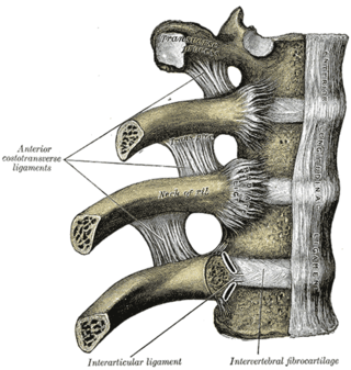
The articulations of the heads of the ribs constitute a series of gliding or arthrodial joints, and are formed by the articulation of the heads of the typical ribs with the costal facets on the contiguous margins of the bodies of the thoracic vertebrae and with the intervertebral discs between them; the first, eleventh and twelfth ribs each articulate with a single vertebra.

The intercostal arteries are a group of arteries passing within an intercostal space. There are 9 anterior and 11 posterior intercostal arteries on each side of the body. The anterior intercostal arteries are branches of the internal thoracic artery and its terminal branch - the musculophrenic artery. The posterior intercostal arteries are branches of the supreme intercostal artery and thoracic aorta.

The superior costal facet is a site where a rib forms a joint with the top of a vertebra.
A costal facet is a site of connection between a rib and a vertebra. The costal facets are located on the vertebrae that the rib articulates with. They are the superior costal facet, the inferior costal facet, and the transverse costal facet. Rib 1 only articulates with a transverse costal facet.

The following outline is provided as an overview of and topical guide to human anatomy:

The sternum or breastbone is a long flat bone located in the central part of the chest. It connects to the ribs via cartilage and forms the front of the rib cage, thus helping to protect the heart, lungs, and major blood vessels from injury. Shaped roughly like a necktie, it is one of the largest and longest flat bones of the body. Its three regions are the manubrium, the body, and the xiphoid process. The word sternum originates from Ancient Greek στέρνον (stérnon) 'chest'.

Each vertebra is an irregular bone with a complex structure composed of bone and some hyaline cartilage, that make up the vertebral column or spine, of vertebrates. The proportions of the vertebrae differ according to their spinal segment and the particular species.

