
In vertebrate anatomy, ribs are the long curved bones which form the rib cage, part of the axial skeleton. In most tetrapods, ribs surround the chest, enabling the lungs to expand and thus facilitate breathing by expanding the chest cavity. They serve to protect the lungs, heart, and other internal organs of the thorax. In some animals, especially snakes, ribs may provide support and protection for the entire body.

In anatomy, the atlas (C1) is the most superior (first) cervical vertebra of the spine and is located in the neck.
The rib cage is an endoskeletal enclosure in the thorax of most vertebrate animals that comprises the ribs, vertebral column and sternum, which protects vital organs such as the heart, lungs and great vessels. The circumferential enclosure formed by left and right rib cages, together known as the thoracic cage, is a semi-rigid bony and cartilaginous structure which surrounds the thoracic cavity and supports the shoulder girdles to form the core part of the axial skeleton.

A joint or articulation is the connection made between bones, ossicles, or other hard structures in the body which link an animal's skeletal system into a functional whole. They are constructed to allow for different degrees and types of movement. Some joints, such as the knee, elbow, and shoulder, are self-lubricating, almost frictionless, and are able to withstand compression and maintain heavy loads while still executing smooth and precise movements. Other joints such as sutures between the bones of the skull permit very little movement in order to protect the brain and the sense organs. The connection between a tooth and the jawbone is also called a joint, and is described as a fibrous joint known as a gomphosis. Joints are classified both structurally and functionally.

In human anatomy, the wrist is variously defined as (1) the carpus or carpal bones, the complex of eight bones forming the proximal skeletal segment of the hand; (2) the wrist joint or radiocarpal joint, the joint between the radius and the carpus and; (3) the anatomical region surrounding the carpus including the distal parts of the bones of the forearm and the proximal parts of the metacarpus or five metacarpal bones and the series of joints between these bones, thus referred to as wrist joints. This region also includes the carpal tunnel, the anatomical snuff box, bracelet lines, the flexor retinaculum, and the extensor retinaculum.

The tibia, also known as the shinbone or shankbone, is the larger, stronger, and anterior (frontal) of the two bones in the leg below the knee in vertebrates ; it connects the knee with the ankle. The tibia is found on the medial side of the leg next to the fibula and closer to the median plane. The tibia is connected to the fibula by the interosseous membrane of leg, forming a type of fibrous joint called a syndesmosis with very little movement. The tibia is named for the flute tibia. It is the second largest bone in the human body, after the femur. The leg bones are the strongest long bones as they support the rest of the body.

The talus, talus bone, astragalus, or ankle bone is one of the group of foot bones known as the tarsus. The tarsus forms the lower part of the ankle joint. It transmits the entire weight of the body from the lower legs to the foot.

The acetabular fossa is the non-articular depressed region at the centre of the floor of the acetabulum. It is surrounded by the articular lunate surface. The floor of the fossa is formed mostly by the ischium; it is rough and thin. The space of the fossa is continuous inferiorly with the acetabular notch.
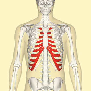
The costal cartilages are bars of hyaline cartilage that serve to prolong the ribs forward and contribute to the elasticity of the walls of the thorax. Costal cartilage is only found at the anterior ends of the ribs, providing medial extension.

The sternoclavicular joint or sternoclavicular articulation is a synovial saddle joint between the manubrium of the sternum, and the clavicle, and the first costal cartilage. The joint possesses a joint capsule, and an articular disc, and is reinforced by multiple ligaments.

The sternocostal joints, also known as sternochondral joints or costosternal articulations, are synovial plane joints of the costal cartilages of the true ribs with the sternum. The only exception is the first rib, which has a synchondrosis joint since the cartilage is directly united with the sternum. The sternocostal joints are important for thoracic wall mobility.

The transverse acetabular ligament bridges the acetabular notch, creating the a foramen. The ligament is one of the sites of attachment of the ligament of head of femur.
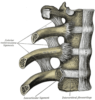
The articulations of the heads of the ribs constitute a series of gliding or arthrodial joints, and are formed by the articulation of the heads of the typical ribs with the costal facets on the contiguous margins of the bodies of the thoracic vertebrae and with the intervertebral discs between them; the first, eleventh and twelfth ribs each articulate with a single vertebra.
The radiate ligament of head of rib is a ligament of the costovertebral joint that typically connects the anterior edge of the head of each rib, and the side of the bodies of two adjacent vertebrae and their intervertebral discs. The ligament is formed as a thickening of the anterior portion of the joint capsule of the costovertebral joint, and thus reinforces it anteriorly.

The first metatarsal bone is the bone in the foot just behind the big toe. The first metatarsal bone is the shortest of the metatarsal bones and by far the thickest and strongest of them.
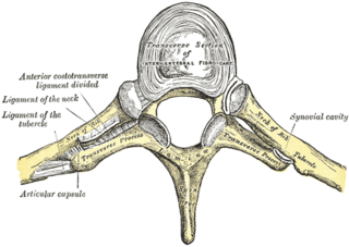
The costotransverse joint is the joint formed between the facet of the tubercle of the rib and the adjacent transverse process of a thoracic vertebra. The costotransverse joint is a plane type of synovial joint which, under physiological conditions, allows only gliding movement.
The lateral costotransverse ligament is a short, thick, though ligament of the costotransverse joint which strengthens the joint posteriorly. It connects the tubercle of a rib, and the transverse process of the corresponding vertebra.

The sternum or breastbone is a long flat bone located in the central part of the chest. It connects to the ribs via cartilage and forms the front of the rib cage, thus helping to protect the heart, lungs, and major blood vessels from injury. Shaped roughly like a necktie, it is one of the largest and longest flat bones of the body. Its three regions are the manubrium, the body, and the xiphoid process. The word sternum originates from Ancient Greek στέρνον (stérnon) 'chest'.
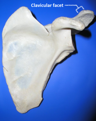
Clavicular facet of scapula is small oval facet on the medial border of the acromion for articulation with the acromial facet on the lateral end of the clavicle. Also called Clavicular articular facet of acromion.

The spinal column, a defining synapomorphy shared by nearly all vertebrates, is a moderately flexible series of vertebrae, each constituting a characteristic irregular bone whose complex structure is composed primarily of bone, and secondarily of hyaline cartilage. They show variation in the proportion contributed by these two tissue types; such variations correlate on one hand with the cerebral/caudal rank, and on the other with phylogenetic differences among the vertebrate taxa.
