Lectins are carbohydrate-binding proteins that are highly specific for sugar groups of other molecules and so cause agglutination of particular cells or precipitation of glycoconjugates and polysaccharides. Lectins have a role in recognition on the cellular and molecular level and play numerous roles in biological recognition phenomena involving cells, carbohydrates, and proteins. Lectins also mediate attachment and binding of bacteria, viruses, and fungi to their intended targets.
A tetrameric protein is a protein with a quaternary structure of four subunits (tetrameric). Homotetramers have four identical subunits, and heterotetramers are complexes of different subunits. A tetramer can be assembled as dimer of dimers with two homodimer subunits, or two heterodimer subunits.

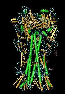
DC-SIGN also known as CD209 is a protein which in humans is encoded by the CD209 gene.

The lectin pathway is a type of cascade reaction in the complement system, similar in structure to the classical complement pathway, in that, after activation, it proceeds through the action of C4 and C2 to produce activated complement proteins further down the cascade. In contrast to the classical complement pathway, the lectin pathway does not recognize an antibody bound to its target. The lectin pathway starts with mannose-binding lectin (MBL) or ficolin binding to certain sugars.

Mannan-binding lectin serine protease 1 also known as mannose-associated serine protease 1 (MASP-1) is an enzyme that in humans is encoded by the MASP1 gene.
Mannose-binding lectin (MBL), also called mannan-binding lectin or mannan-binding protein (MBP), is a lectin that is instrumental in innate immunity as an opsonin and via the lectin pathway.

CD207, langerin is a protein which in humans is encoded by the CD207 gene. Langerin is a type II transmembrane, C-type lectin receptor on Langerhans cells.
The mannose receptor is a C-type lectin primarily present on the surface of macrophages, immature dendritic cells and liver sinusoidal endothelial cells, but is also expressed on the surface of skin cells such as human dermal fibroblasts and keratinocytes. It is the first member of a family of endocytic receptors that includes Endo180 (CD280), M-type PLA2R, and DEC-205 (CD205).
The mannose 6-phosphate receptors (MPRs) are transmembrane glycoproteins that target enzymes to lysosomes in vertebrates.

Peanut agglutinin (PNA) is plant lectin protein derived from the fruits of Arachis hypogaea. Peanut agglutinin may also be referred to as Arachis hypogaea lectin. Lectins recognise and bind particular sugar sequences in carbohydrates; peanut agglutinin binds the carbohydrate sequence Gal-β(1-3)-GalNAc. The name "peanut agglutinin" originates from its ability to stick together (agglutinate) cells, such as neuraminidase-treated erythrocytes, which have glycoproteins or glycolipids on their surface which include the Gal-β(1-3)-GalNAc carbohydrate sequence.

Griffithsin is a protein isolated from the red algae Griffithsia. It has a 121-amino acid sequence which exhibits a Jacalin-like lectin fold. Several structures of this protein have been solved by X-ray crystallography and deposited in the PDB. It has been shown in vitro to be a highly potent HIV entry inhibitor. It is currently being investigated as a potential microbicide for use in the prevention of the transmission of HIV.

Protein ERGIC-53 also known as ER-Golgi intermediate compartment 53 kDa protein or lectin mannose-binding 1 is a protein that in humans is encoded by the LMAN1 gene.

Sialic acid-binding Ig-like lectin 7 is a protein that in humans is encoded by the SIGLEC7 gene. SIGLEC7 has also been designated as CD328.
Jacalin is a plant-based lectin, but not a legume lectin, found in jackfruit. It has been studied for capturing O-glycoproteins such as mucins and IgA1, for potential applications in human immunology.

In the fields of biochemistry and cell biology, the cation-dependent mannose-6-phosphate receptor (CD-MPR) also known as the 46 kDa mannose 6-phosphate receptor is a protein that in humans is encoded by the M6PR gene.
BanLec is a lectin from the jacalin-related lectin family isolated from the fruit of the bananas Musa acuminata and Musa balbisiana. BanLec is one of the predominant proteins in the pulp of ripe bananas and has binding specificity for mannose and mannose-containing oligosaccharides. A 2010 study reported that BanLec was a potent inhibitor of HIV replication.

In molecular biology, the leguminous lectin family is a family of lectin proteins.

In molecular biology the L-like lectin domain is a protein domain found in lectins which are similar to the leguminous plant lectins.
Avadhesha Surolia is a Glycobiologist at Indian Institute of Science (IISc), Bangalore. Presently, he is an Honorary Professor at the Molecular Biophysics Unit (MBU), IISc and holds the Bhatnagar fellowship of the Council of Scientific and Industrial Research (CSIR), India. He is known for his work on lectin structure and interactions, orientation and dynamics of cell surface carbohydrate receptors and protein folding, diabetes, anti-malarials and anti-cancer agents based on curcumin, flavonoids, etc. In addition, neuropathic pain, neurodegenerative disorders and the link between immunity and obsessive compulsive disorder are areas of his current interest
