Related Research Articles

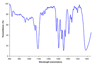
Infrared spectroscopy is the measurement of the interaction of infrared radiation with matter by absorption, emission, or reflection. It is used to study and identify chemical substances or functional groups in solid, liquid, or gaseous forms. It can be used to characterize new materials or identify and verify known and unknown samples. The method or technique of infrared spectroscopy is conducted with an instrument called an infrared spectrometer which produces an infrared spectrum. An IR spectrum can be visualized in a graph of infrared light absorbance on the vertical axis vs. frequency, wavenumber or wavelength on the horizontal axis. Typical units of wavenumber used in IR spectra are reciprocal centimeters, with the symbol cm−1. Units of IR wavelength are commonly given in micrometers, symbol μm, which are related to the wavenumber in a reciprocal way. A common laboratory instrument that uses this technique is a Fourier transform infrared (FTIR) spectrometer. Two-dimensional IR is also possible as discussed below.

Spectroscopy is the field of study that measures and interprets electromagnetic spectra. In narrower contexts, spectroscopy is the precise study of color as generalized from visible light to all bands of the electromagnetic spectrum.
Fourier-transform spectroscopy is a measurement technique whereby spectra are collected based on measurements of the coherence of a radiative source, using time-domain or space-domain measurements of the radiation, electromagnetic or not. It can be applied to a variety of types of spectroscopy including optical spectroscopy, infrared spectroscopy, nuclear magnetic resonance (NMR) and magnetic resonance spectroscopic imaging (MRSI), mass spectrometry and electron spin resonance spectroscopy.

Absorption spectroscopy is spectroscopy that involves techniques that measure the absorption of electromagnetic radiation, as a function of frequency or wavelength, due to its interaction with a sample. The sample absorbs energy, i.e., photons, from the radiating field. The intensity of the absorption varies as a function of frequency, and this variation is the absorption spectrum. Absorption spectroscopy is performed across the electromagnetic spectrum.

Rotational spectroscopy is concerned with the measurement of the energies of transitions between quantized rotational states of molecules in the gas phase. The rotational spectrum of polar molecules can be measured in absorption or emission by microwave spectroscopy or by far infrared spectroscopy. The rotational spectra of non-polar molecules cannot be observed by those methods, but can be observed and measured by Raman spectroscopy. Rotational spectroscopy is sometimes referred to as pure rotational spectroscopy to distinguish it from rotational-vibrational spectroscopy where changes in rotational energy occur together with changes in vibrational energy, and also from ro-vibronic spectroscopy where rotational, vibrational and electronic energy changes occur simultaneously.
Near-infrared spectroscopy (NIRS) is a spectroscopic method that uses the near-infrared region of the electromagnetic spectrum. Typical applications include medical and physiological diagnostics and research including blood sugar, pulse oximetry, functional neuroimaging, sports medicine, elite sports training, ergonomics, rehabilitation, neonatal research, brain computer interface, urology, and neurology. There are also applications in other areas as well such as pharmaceutical, food and agrochemical quality control, atmospheric chemistry, combustion research and knowledge.
Ultrafast laser spectroscopy is a category of spectroscopic techniques using ultrashort pulse lasers for the study of dynamics on extremely short time scales. Different methods are used to examine the dynamics of charge carriers, atoms, and molecules. Many different procedures have been developed spanning different time scales and photon energy ranges; some common methods are listed below.

Attenuated total reflection (ATR) is a sampling technique used in conjunction with infrared spectroscopy which enables samples to be examined directly in the solid or liquid state without further preparation.
Applied spectroscopy is the application of various spectroscopic methods for the detection and identification of different elements or compounds to solve problems in fields like forensics, medicine, the oil industry, atmospheric chemistry, and pharmacology.
Photothermal microspectroscopy (PTMS), alternatively known as photothermal temperature fluctuation (PTTF), is derived from two parent instrumental techniques: infrared spectroscopy and atomic force microscopy (AFM). In one particular type of AFM, known as scanning thermal microscopy (SThM), the imaging probe is a sub-miniature temperature sensor, which may be a thermocouple or a resistance thermometer. This same type of detector is employed in a PTMS instrument, enabling it to provide AFM/SThM images: However, the chief additional use of PTMS is to yield infrared spectra from sample regions below a micrometer, as outlined below.

Two-dimensional infrared spectroscopy is a nonlinear infrared spectroscopy technique that has the ability to correlate vibrational modes in condensed-phase systems. This technique provides information beyond linear infrared spectra, by spreading the vibrational information along multiple axes, yielding a frequency correlation spectrum. A frequency correlation spectrum can offer structural information such as vibrational mode coupling, anharmonicities, along with chemical dynamics such as energy transfer rates and molecular dynamics with femtosecond time resolution. 2DIR experiments have only become possible with the development of ultrafast lasers and the ability to generate femtosecond infrared pulses.

Fourier-transform infrared spectroscopy (FTIR) is a technique used to obtain an infrared spectrum of absorption or emission of a solid, liquid, or gas. An FTIR spectrometer simultaneously collects high-resolution spectral data over a wide spectral range. This confers a significant advantage over a dispersive spectrometer, which measures intensity over a narrow range of wavelengths at a time.
The technique of vibrational analysis with scanning probe microscopy allows probing vibrational properties of materials at the submicrometer scale, and even of individual molecules. This is accomplished by integrating scanning probe microscopy (SPM) and vibrational spectroscopy. This combination allows for much higher spatial resolution than can be achieved with conventional Raman/FTIR instrumentation. The technique is also nondestructive, requires non-extensive sample preparation, and provides more contrast such as intensity contrast, polarization contrast and wavelength contrast, as well as providing specific chemical information and topography images simultaneously.
Stationary-wave integrated Fourier-transform spectrometry (SWIFTS), or standing-wave integrated Fourier-transform spectrometry, is an analytical technique used for measuring the distribution of light across an optical spectrum. SWIFTS technology is based on a near-field Lippmann architecture. An optical signal is injected into a waveguide and ended by a mirror. The input signal interferes with the reflected signal, creating a standing, or stationary, wave.
Biomedical spectroscopy is a multidisciplinary research field involving spectroscopic tools for applications in the field of biomedical science. Vibrational spectroscopy such as Raman or infrared spectroscopy is used to determine the chemical composition of a material based on detection of vibrational modes of constituent molecules. Some spectroscopic methods are routinely used in clinical settings for diagnosis of disease; an example is Magnetic resonance imaging (MRI). Fourier transform infrared (FTIR) spectroscopic imaging is a form of chemical imaging for which the contrast is provided by composition of the material.
An infrared interferometer spectrometer and radiometer (IRIS) is a scientific instrument of the Voyager space probes which enables the measurement of three distinct properties. The instrument itself consists of two separate instruments that together share a single large-aperture telescope system.
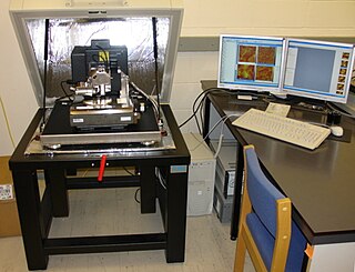
AFM-IR or infrared nanospectroscopy is one of a family of techniques that are derived from a combination of two parent instrumental techniques. AFM-IR combines the chemical analysis power of infrared spectroscopy and the high-spatial resolution of scanning probe microscopy (SPM). The term was first used to denote a method that combined a tuneable free electron laser with an atomic force microscope equipped with a sharp probe that measured the local absorption of infrared light by a sample with nanoscale spatial resolution.

Nano-FTIR is a scanning probe technique that utilizes as a combination of two techniques: Fourier transform infrared spectroscopy (FTIR) and scattering-type scanning near-field optical microscopy (s-SNOM). As s-SNOM, nano-FTIR is based on atomic-force microscopy (AFM), where a sharp tip is illuminated by an external light source and the tip-scattered light is detected as a function of tip position. A typical nano-FTIR setup thus consists of an atomic force microscope, a broadband infrared light source used for tip illumination, and a Michelson interferometer acting as Fourier-transform spectrometer. In nano-FTIR, the sample stage is placed in one of the interferometer arms, which allows for recording both amplitude and phase of the detected light. Scanning the tip allows for performing hyperspectral imaging with nanoscale spatial resolution determined by the tip apex size. The use of broadband infrared sources enables the acquisition of continuous spectra, which is a distinctive feature of nano-FTIR compared to s-SNOM. Nano-FTIR is capable of performing infrared (IR) spectroscopy of materials in ultrasmall quantities and with nanoscale spatial resolution. The detection of a single molecular complex and the sensitivity to a single monolayer has been shown. Recording infrared spectra as a function of position can be used for nanoscale mapping of the sample chemical composition, performing a local ultrafast IR spectroscopy and analyzing the nanoscale intermolecular coupling, among others. A spatial resolution of 10 nm to 20 nm is routinely achieved.

Dirubidium is a molecular substance containing two atoms of rubidium found in rubidium vapour. Dirubidium has two active valence electrons. It is studied both in theory and with experiment. The rubidium trimer has also been observed.
References
- 1 2 "CWP at physics.UCLA.edu // Janine Connes". cwp.library.ucla.edu. Retrieved 2020-05-04.
- ↑ James Lovelock (2009). The Vanishing Face of Gaia: A Final Warning. Penguin books. ISBN 978-0141039251 . Retrieved 27 March 2014.
- ↑ B. Joerges, T. Shinn (2001). Instrumentation Between Science, State and Industry. Springer. p. 127. ISBN 0792367367 . Retrieved 27 March 2014.
- 1 2 McLean, Ian (1997). "Janine Connes". Contributions of 20th Century Women to Physics. University of California . Retrieved 27 March 2014.
- ↑ R. A. Hanel (2003). Exploration of the Solar System by Infrared Remote Sensing. Cambridge University Press. p. 222. ISBN 0521818974 . Retrieved 27 March 2014.
- ↑ National Research Council (U.S.). Panel on Remote Atmospheric Probing (1969). Atmospheric Exploration by Remote Probes: Final Report of the Panel on Remote Atmospheric Probing. Vol. 2. National Academies . Retrieved 27 March 2014.
- ↑ Da-Wen Sun (2009). Infrared Spectroscopy for Food Quality Analysis and Control. Academic Press. p. 151. ISBN 978-0080920870 . Retrieved 27 March 2014.
| International | |
|---|---|
| National | |
| Academics | |
| Other | |
| | This article about a French astronomer is a stub. You can help Wikipedia by expanding it. |