Related Research Articles

Microscopy is the technical field of using microscopes to view objects and areas of objects that cannot be seen with the naked eye. There are three well-known branches of microscopy: optical, electron, and scanning probe microscopy, along with the emerging field of X-ray microscopy.

A microscope is a laboratory instrument used to examine objects that are too small to be seen by the naked eye. Microscopy is the science of investigating small objects and structures using a microscope. Microscopic means being invisible to the eye unless aided by a microscope.

Spectroscopy is the field of study that measures and interprets the electromagnetic spectra that result from the interaction between electromagnetic radiation and matter as a function of the wavelength or frequency of the radiation. Matter waves and acoustic waves can also be considered forms of radiative energy, and recently gravitational waves have been associated with a spectral signature in the context of the Laser Interferometer Gravitational-Wave Observatory (LIGO)

Biophysics is an interdisciplinary science that applies approaches and methods traditionally used in physics to study biological phenomena. Biophysics covers all scales of biological organization, from molecular to organismic and populations. Biophysical research shares significant overlap with biochemistry, molecular biology, physical chemistry, physiology, nanotechnology, bioengineering, computational biology, biomechanics, developmental biology and systems biology.

Förster resonance energy transfer (FRET), resonance energy transfer (RET) or electronic energy transfer (EET) is a mechanism describing energy transfer between two light-sensitive molecules (chromophores). A donor chromophore, initially in its electronic excited state, may transfer energy to an acceptor chromophore through nonradiative dipole–dipole coupling. The efficiency of this energy transfer is inversely proportional to the sixth power of the distance between donor and acceptor, making FRET extremely sensitive to small changes in distance.
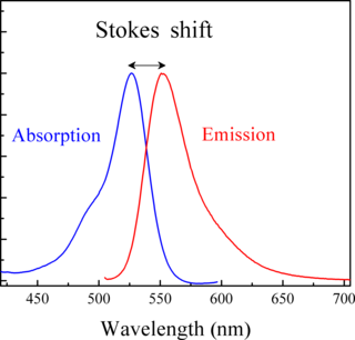
Stokes shift is the difference between positions of the band maxima of the absorption and emission spectra of the same electronic transition. It is named after Irish physicist George Gabriel Stokes. Sometimes Stokes shifts are given in wavelength units, but this is less meaningful than energy, wavenumber or frequency units because it depends on the absorption wavelength. For instance, a 50 nm Stokes shift from absorption at 300 nm is larger in terms of energy than a 50 nm Stokes shift from absorption at 600 nm.
Fluorescence-lifetime imaging microscopy or FLIM is an imaging technique based on the differences in the exponential decay rate of the photon emission of a fluorophore from a sample. It can be used as an imaging technique in confocal microscopy, two-photon excitation microscopy, and multiphoton tomography.

The Max Planck Institute of Biochemistry (MPIB) is a research institute of the Max Planck Society located in Martinsried, a suburb of Munich. The institute was founded in 1973 by the merger of three formerly independent institutes: the Max Planck Institute of Biochemistry, the Max Planck Institute of Protein and Leather Research, and the Max Planck Institute of Cell Chemistry.
Medical optical imaging is the use of light as an investigational imaging technique for medical applications. Examples include optical microscopy, spectroscopy, endoscopy, scanning laser ophthalmoscopy, laser Doppler imaging, and optical coherence tomography. Because light is an electromagnetic wave, similar phenomena occur in X-rays, microwaves, and radio waves.

The Max Planck Institute for Biophysical Chemistry, also known as the Karl-Friedrich Bonhoeffer Institute, was a research institute of the Max Planck Society, located in Göttingen, Germany. On January 1, 2022, the institute merged with the Max Planck Institute for Experimental Medicine in Göttingen to form the Max Planck Institute for Multidisciplinary Sciences.

Stimulated emission depletion (STED) microscopy is one of the techniques that make up super-resolution microscopy. It creates super-resolution images by the selective deactivation of fluorophores, minimizing the area of illumination at the focal point, and thus enhancing the achievable resolution for a given system. It was developed by Stefan W. Hell and Jan Wichmann in 1994, and was first experimentally demonstrated by Hell and Thomas Klar in 1999. Hell was awarded the Nobel Prize in Chemistry in 2014 for its development. In 1986, V.A. Okhonin had patented the STED idea. This patent was unknown to Hell and Wichmann in 1994.
An excitation filter is a high quality optical-glass filter commonly used in fluorescence microscopy and spectroscopic applications for selection of the excitation wavelength of light from a light source. Most excitation filters select light of relatively short wavelengths from an excitation light source, as only those wavelengths would carry enough energy to cause the object the microscope is examining to fluoresce sufficiently. The excitation filters used may come in two main types — short pass filters and band pass filters. Variations of these filters exist in the form of notch filters or deep blocking filters. Other forms of excitation filters include the use of monochromators, wedge prisms coupled with a narrow slit and the use of holographic diffraction gratings, etc. [for beam diffraction of white laser light into the required excitation wavelength ].

Stefan Walter Hell HonFRMS is a Romanian-German physicist and one of the directors of the Max Planck Institute for Biophysical Chemistry in Göttingen, Germany. He received the Nobel Prize in Chemistry in 2014 "for the development of super-resolved fluorescence microscopy", together with Eric Betzig and William Moerner.
Stefan Seeger is a German chemist and professor at the University of Zurich in Switzerland.
Christoph Cremer is a German physicist and emeritus at the Ruprecht-Karls-University Heidelberg, former honorary professor at the University of Mainz and was a former group leader at Institute of Molecular Biology (IMB) at the Johannes Gutenberg University of Mainz, Germany, who has successfully overcome the conventional limit of resolution that applies to light based investigations by a range of different methods. In the meantime, according to his own statement, Christoph Cremer is a member of the Max Planck Institute for Chemistry and the Max Planck Institute for Polymer Research.
Robert Alfano is an Italian-American experimental physicist. He is a Distinguished Professor of Science and Engineering at the City College and Graduate School of New York of the City University of New York, where he is also the founding Director of the Institute for Ultrafast Spectroscopy and Lasers (1982). He is a pioneer in the fields of Biomedical Imaging and Spectroscopy, Ultrafast lasers and optics, tunable lasers, semiconductor materials and devices, optical materials, biophysics, nonlinear optics and photonics; he has also worked extensively in nanotechnology and coherent backscattering. His discovery of the white-light supercontinuum laser is at the root of optical coherence tomography, which is breaking barriers in ophthalmology, cardiology, and oral cancer detection among other applications. He initiated the field known now as Optical Biopsy
Gregorio Weber was an Argentinian scientist who made significant contributions to the fields of fluorescence spectroscopy and protein chemistry. Weber was elected to the National Academy of Sciences in 1975.
Food physical chemistry is considered to be a branch of Food chemistry concerned with the study of both physical and chemical interactions in foods in terms of physical and chemical principles applied to food systems, as well as the applications of physical/chemical techniques and instrumentation for the study of foods. This field encompasses the "physiochemical principles of the reactions and conversions that occur during the manufacture, handling, and storage of foods"
Pierre-Michel Duffieux (1891–1976) was a French physicist, known as the founder of Fourier optics.

Robert Eric Betzig is an American physicist who works as a professor of physics and professor of molecular and cell biology at the University of California, Berkeley. He is also a senior fellow at the Janelia Farm Research Campus in Ashburn, Virginia.
References
- ↑ "Joseph R. Lakowicz Is May Speaker." (PDF). Archived from the original (PDF) on March 4, 2016. Retrieved May 22, 2018. Puget Sound Chemist: Bulletin of the Puget Sound Section of the American Chemical Society, Vol. 36, Nr. 5, May 1975, S. 3.
- 1 2 3 Kathy Kincade: Joseph R. Lakowicz. BioOptics World, 1 May 2008. Retrieved 8 July 2014.
- ↑ Thomas A. Klar, Stefan W. Hell: Subdiffraction resolution in far-field fluorescence microscopy. In: Optics Letters. Vol. 24, Nr. 14, 1999, S. 954–956, doi:10.1364/OL.24.000954.