Related Research Articles

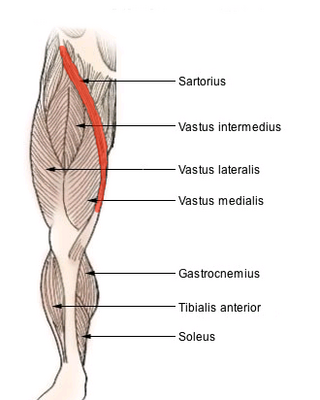
The sartorius muscle is the longest muscle in the human body. It is a long, thin, superficial muscle that runs down the length of the thigh in the anterior compartment.

The foramen spinosum is a small open hole in the greater wing of the sphenoid bone that gives passage to the middle meningeal artery and vein, and the meningeal branch of the mandibular nerve.

In anatomy, the abdominal wall represents the boundaries of the abdominal cavity. The abdominal wall is split into the anterolateral and posterior walls.

The superior gluteal nerve is a mixed nerve of the sacral plexus that originates in the pelvis. It provides motor innervation to the gluteus medius, gluteus minimus, tensor fasciae latae, and piriformis muscles; it also has a cutaneous branch.

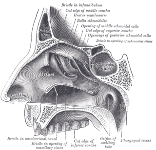
The semilunar hiatus is a crescent-shaped/semicircular/ curved slit/groove upon the lateral wall of the nasal cavity at the middle nasal meatus just inferior to the ethmoidal bulla. It is the location of the openings for the frontal sinus, maxillary sinus, and anterior ethmoidal sinus. It is bounded inferiorly and anteriorly by the sharp concave margin of the uncinate process of the ethmoid bone, superiorly by the ethmoidal bulla, and posteriorly by the ethmoidal process of the inferior nasal concha. It leads into the ethmoidal infundibulum; it marks the medial limit of the ethmoidal infundibulum.

The medial circumflex femoral artery is an artery in the upper thigh that arises from the profunda femoris artery. It supplies arterial blood to several muscles in the region, as well as the femoral head and neck.

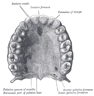
The greater palatine foramen is, along with the lesser palatine foramen, one of two foramina (holes) in each of the left and right palatine bones which form the posterior roof of the human mouth, known as the palate. It is sometimes known as the major palatine foramen.

The lesser palatine foramina are a pair of foramina (openings) on each of the palatine bones, which form the posterior roof of the human mouth. They are sometimes referred to as the minor palatine foramina. The lesser palatine foramina form a passage through the bone for the lesser palatine nerves and the lesser palatine arteries so that they may reach the soft palate from above. They are positioned posterior to the greater palatine foramen, in the pyramidal process of the palatine bone.

The circumflex branch of left coronary artery is a branch of the left coronary artery. It winds around the left side of the heart along the atrioventricular groove. It supplies the posterolateral portion of the left ventricle.

The nerve to obturator internus is a mixed nerve providing motor innervation to the obturator internus muscle and gemellus superior muscle, and sensory innervation to the hip joint. It is a branch of the sacral plexus. It is one of the group of deep gluteal nerves.

The nerve to quadratus femoris is a nerve of the sacral plexus that provides motor innervation to the quadratus femoris muscle and gemellus inferior muscle, and an articular branch to the hip joint. The nerve leaves the pelvis through the greater sciatic foramen.

The transverse acetabular ligament bridges the acetabular notch, creating the a foramen. The ligament is one of the sites of attachment of the ligament of head of femur.

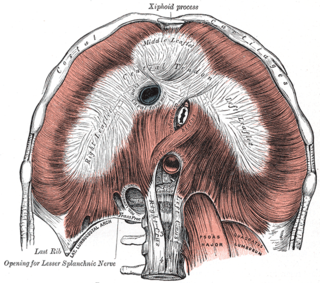
The aortic hiatus is a midline opening in the posterior part of the diaphragm giving passage to the descending aorta as well as the thoracic duct, and variably the azygos and hemiazygos veins. It is the lowest and most posterior of the large apertures.

The supravesical fossa is a depression upon the inner surface of the anterior abdominal wall superior to the bladder formed by a reflection of the peritoneum onto the superior surface of the bladder. It is bounded by the medial umbilical fold and median umbilical fold.

The anterior compartment of the forearm contains the following muscles:

In human anatomy, the adductor hiatus also known as hiatus magnus is a hiatus (gap) between the adductor magnus muscle and the femur that allows the passage of the femoral vessels from the anterior thigh to the posterior thigh and then the popliteal fossa. It is the termination of the adductor canal and lies about 8–13.5 cm (3.1–5.3 in) superior to the adductor tubercle.
A costotransverse ligament is ligament of the costotransverse joint which attaches at the neck of a rib, and at the transverse process of its corresponding vertebra. It extends posteriorly from the rib to the vertebra.

The iliac tubercle is located approximately 5 cm (2 in) posterior to the anterior superior iliac spine on the iliac crest in humans. The transverse plane that includes each of the tubercles is called the transtubercular plane. The origin of the iliotibial tract is the iliac tubercle. The iliac tubercle is also the widest point of the iliac crest, and lies at the level of the L5 spinous process.
A prosection is the dissection of a cadaver or part of a cadaver by an experienced anatomist in order to demonstrate for students anatomic structure. In a dissection, students learn by doing; in a prosection, students learn by either observing a dissection being performed by an experienced anatomist or examining a specimen that has already been dissected by an experienced anatomist.
References
- 1 2 3 "Keith L. Moore: My 60 years as a Clinical Anatomist". American Association of Anatomists. Archived from the original on 19 March 2012. Retrieved 29 June 2011.
- ↑ "DR. KEITH LEON MOORE Obituary (1925 - 2019) Toronto Star". Legacy.com. Retrieved 20 December 2021.
- ↑ "Honored Member Award 1994 Keith L. Moore, MSc, PhD, FIAC, FRSM". American Association of Clinical Anatomists. Retrieved 29 June 2011.
- 1 2 "Keith L. Moore". American Association of Anatomists. Retrieved 29 June 2011.
- ↑ "American Association of Clinical Anatomists – Past Presidents". American Association of Clinical Anatomists. Retrieved 29 June 2011.
- ↑ Moore, Keith L.; Dalley, Arthur F.; Agur, Anne M. R. (2009). Clinically Oriented Anatomy. ISBN 978-1605476520.
- ↑ Moore, Keith L.; Agur, A. M. R.; Dalley, Arthur F. (2011). Essential Clinical Anatomy. ISBN 978-0781799157.
- ↑ "AACA Awards – Honored Member". American Association of Clinical Anatomists. Retrieved 29 June 2009.
- ↑ "Honored Member Award 1994 Keith L. Moore, MSc, PhD, FIAC, FRSM". American Association of Clinical Anatomists. Archived from the original on 19 March 2012. Retrieved 29 June 2009.
- ↑ "Past and Current Award Winners". American Association of Anatomists. Retrieved 29 June 2009.
- ↑ Jeffery, Kim. "Queens Diamond Jubille comes to Barrie". Snap Magazine. Snap Newspaper company. Retrieved 13 February 2013.
- 1 2 Loring M. Danforth (2016). Crossing the Kingdom: Portraits of Saudi Arabia. University of California Press. pp. 129–130. ISBN 9780520290273.
- ↑ Keith L. Moore and Abdul-Majeed A. Zindani, The Developing Human with Islamic Additions, 3rd ed. (Philadelphia: Saunders with Dar al-Qiblah for Islamic Literature, Jeddah, 1983, 1982). page viii insert c.
- ↑ A Scientist's Interpretation of References to Embryology in the Qur'an by Keith L. Moore. Journal of the Islamic Medical Association of North America, Vol. 18, Jan-June 1986.
- ↑ Stenberg, Leif; Wood, Philip, eds. (2023). What Is Islamic Studies?: European and North American Approaches to a Contested Field. Edinburgh University Press. p. 134. ISBN 9781399500012.
- ↑ Human Development as Described in the Qurʼan and Sunnah Archived 20 February 2020 at the Wayback Machine . Editors A.A Zindani, Mustafa A. Ahmed, M.B. Tobin, and T. V. N. Persaud: Islamic Academy for Scientific Research, 1994. Collection of papers that were originally presented in international conferences in Riyadh, Saudi Arabia (1983), Cairo, Egypt (1985), Islamabad, Pakistan (1987), and Dakar, Senegal (1991). The presentations were extended and revised and presented in their current form in Moscow, Russia in September 1993.
- ↑ Moore, Keith L. (1983). The Developing Human: Clinically Oriented Emryology with Islamic Additions. Abul Qasim Publishing House (Saudi Arabia). Archived from the original on 29 January 2020. Retrieved 8 August 2020.
- ↑ Golden, Daniel (23 January 2002). "Western Scholars Play Key Role In Touting 'Science' of the Quran". Wall Street Journal . Retrieved 27 January 2013.
- ↑ Paul Zachary Myers(2010), Islamic apologetics in the International Journal of Cardiology, ScienceBlogs. Archived Retrieved 25 April 2024
- ↑ Taner Edis (2007). An Illusion of Harmony: Science and Religion in Islam. Prometheus Books. p. 96. ISBN 9781591024491.
Bibliography
- Moore, Keith L.; Dalley, Arthur F.; Agur, Anne M. R. (2009). Clinically Oriented Anatomy. ISBN 978-1605476520.
- Moore, Keith L.; Agur, A. M. R.; Dalley, Arthur F. (2011). Essential Clinical Anatomy. ISBN 978-0781799157.
- Moore, Keith L.; Persaud, T. V. N.; Torchia, Mark G. (2018). The Developing Human: Clinically Oriented Embryology. Elsevier Health Sciences. ISBN 9780323611565. Archived from the original on 17 June 2022. Retrieved 8 August 2020.
- Moore, Keith L.; Azzindani, Abdul-Majeed A. (1983). The Developing Human: Clinically Oriented Emryology with Islamic Additions. Abul Qasim Publishing House (Saudi Arabia). Archived from the original on 8 July 2001. Retrieved 8 August 2020.