
The larynx, commonly called the voice box, is an organ in the top of the neck involved in breathing, producing sound and protecting the trachea against food aspiration. The opening of larynx into pharynx known as the laryngeal inlet is about 4–5 centimeters in diameter. The larynx houses the vocal cords, and manipulates pitch and volume, which is essential for phonation. It is situated just below where the tract of the pharynx splits into the trachea and the esophagus. The word ʻlarynxʼ comes from the Ancient Greek word lárunx ʻlarynx, gullet, throat.ʼ

The Adam's apple or laryngeal prominence is the protrusion in the human neck formed by the angle of the thyroid cartilage surrounding the larynx, most visible in men.
The lateral cricoarytenoid muscles extend from the lateral cricoid cartilage to the muscular process of the arytenoid cartilage. By rotating the arytenoid cartilages medially, these muscles adduct the vocal cords and thereby close the rima glottidis, protecting the airway. The lateral cricoarytenoid muscles receive innervation from the recurrent laryngeal branch of the vagus nerve.

The posterior cricoarytenoid muscles are small, paired intrinsic muscles of the larynx that extend between cricoid cartilage to the arytenoid cartilages in the larynx.
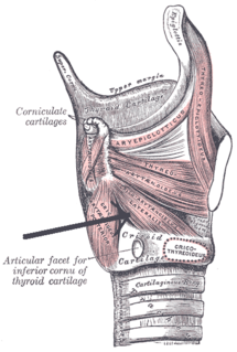
The thyroid cartilage is the largest of the nine cartilages that make up the laryngeal skeleton, the cartilage structure in and around the trachea that contains the larynx. It does not completely encircle the larynx.

The cricothyroid muscle is the only tensor muscle of the larynx aiding with phonation. It is innervated by the superior laryngeal nerve. Its action tilts the thyroid forward to help tense the vocal cords.
Laryngeal paralysis in animals is a condition in which the nerves and muscles that control the movements of one or both arytenoid cartilages of the larynx cease to function, and instead of opening during aspiration and closing during swallowing, the arytenoids remain stationary in a somewhat neutral position. Specifically, the muscle that causes abduction of the arytenoid cartilage, the cricoarytenoideus dorsalis muscle, ceases to function. This leads to inadequate ventilation during exercise and during thermoregulatory panting as well as incomplete protection of the airway during swallowing.

The arytenoid cartilages are a pair of small three-sided pyramids which form part of the larynx. They are the site of attachment of the vocal cords. Each is pyramidal or ladle-shaped and has three surfaces, a base, and an apex. The arytenoid cartilages allow for movement of the vocal cords by articulating with the cricoid cartilage. It may be affected by arthritis, dislocations, or sclerosis.
The laryngeal inlet is the opening that connects the pharynx and the larynx.
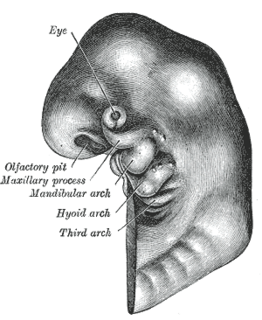
The pharyngeal arches, also known as visceral arches, are structures seen in the embryonic development of vertebrates that are recognisable precursors for many structures. In fish, the arches are known as the branchial arches, or gill arches.
Terminologia Anatomica is the international standard for human anatomical terminology. It is developed by the Federative International Programme on Anatomical Terminology, a program of the International Federation of Associations of Anatomists (IFAA). The second edition was released in 2019 and approved and adopted by the IFAA General Assembly in 2020. Terminologia Anatomica supersedes the previous standard, Nomina Anatomica. It contains terminology for about 7500 human anatomical structures.

The arytenoid muscle is a single muscle of the larynx. It passes from one arytenoid cartilage to the opposite arytenoid cartilage. It has oblique and transverse fibres. It is supplied by the recurrent laryngeal nerve. It approximates the arytenoid cartilages. Continuous electromyography may be used during neck surgeries such as thyroidectomy.
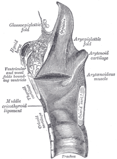
The thyrohyoid membrane is a broad, fibro-elastic sheet of the larynx. It connects the upper border of the thyroid cartilage to the hyoid bone.

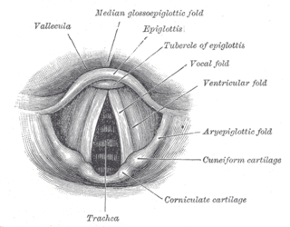
In the human larynx, the cuneiform cartilages are two small, elongated pieces of yellow elastic cartilage, placed one on either side, in the aryepiglottic fold.
The aryepiglottic muscle, or aryepiglotticus muscle is an intrinsic muscle of the larynx.
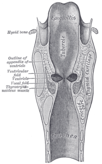
The laryngeal ventricle, is a fusiform fossa, situated between the vestibular and vocal folds on either side, and extending nearly their entire length. There is also a sinus of Morgagni in the pharynx.
A laryngeal cleft or laryngotracheoesophageal cleft is a rare congenital abnormality in the posterior laryngo-tracheal wall. It occurs in approximately 1 in 10,000 to 20,000 births. It means there is a communication between the oesophagus and the trachea, which allows food or fluid to pass into the airway.
Laryngeal cysts are cysts involving the larynx or more frequently supraglottic locations, such as epiglottis and vallecula. Usually they do not extend to the thyroid cartilage. They may be present congenitally or may develop eventually due to degenerative cause. They often interfere with phonation.
Laryngotracheal reconstruction is a surgical procedure that involves expanding or removing parts of the airway to widen a narrowing within it, called laryngotracheal stenosis or subglottic stenosis.
This page is based on this
Wikipedia article Text is available under the
CC BY-SA 4.0 license; additional terms may apply.
Images, videos and audio are available under their respective licenses.
