Lingual papillae

Last updated
Lingual papillae
Illu04 tongue2.jpg
Anatomic landmarks of the tongue. Filiform papillae cover most of the dorsal surface of the anterior 2/3 of the tongue, with fungiform interspaced. Just in front of the sulcus terminalis lies a V-shaped line of circumvallate papillae, and on the posterior aspects of the lateral margins of the tongue lie the foliate papillae.
Gray1018.png
Semidiagrammatic view of a portion of the mucous membrane of the tongue. Two fungiform papillae are shown. On some of the filiform papillae the epithelial prolongations stand erect, in one they are spread out, and in three they are folded in.
Details
Part of Tongue
Identifiers
Latin papillae linguales
NeuroLex ID birnlex_4102
TA98 A05.1.04.013
TA2 2837
TH H3.04.01.0.03006
FMA 54819
Anatomical terminology

Lingual papillae (sg.: papilla) are small structures on the upper surface of the tongue that give it its characteristic rough texture. The four types of papillae on the human tongue have different structures and are accordingly classified as circumvallate (or vallate), fungiform, filiform, and foliate. All except the filiform papillae are associated with taste buds. [1]

Contents

Structure

In living subjects, lingual papillae are more readily seen when the tongue is dry. [2] There are four types of papillae present on the tongue in humans:

Filiform papillae

Filiform papilla, magnified. Gray1016.png
Filiform papilla, magnified.

Filiform papillae are the most numerous of the lingual papillae. [1] They are fine, small, cone-shaped papillae found on the anterior surface of the tongue. [3] They are responsible for giving the tongue its texture and are responsible for the sensation of touch. Unlike the other kinds of papillae, filiform papillae do not contain taste buds. [1] They cover most of the front two-thirds of the tongue's surface. [2]

They appear as very small, conical or cylindrical surface projections, [2] and are arranged in rows which lie parallel to the sulcus terminalis. At the tip of the tongue, these rows become more transverse. [2]

Histologically, they are made up of irregular connective tissue cores with a keratin–containing epithelium which has fine secondary threads. [2] Heavy keratinization of filiform papillae, occurring for instance in cats, gives the tongue a roughness that is characteristic of these animals.

These papillae have a whitish tint, owing to the thickness and density of their epithelium. This epithelium has undergone a peculiar modification as the cells have become cone–like and elongated into dense, overlapping, brush-like threads. They also contain a number of elastic fibers, which render them firmer and more elastic than the other types of papillae. The larger and longer papillae of this group are sometimes termed papillae conicae.

Fungiform papillae

Fungiform papillae, magnified and sectional diagram. Gray1017.png
Fungiform papillae, magnified and sectional diagram.

The fungiform papillae are club shaped projections on the tongue, generally red in color. They are found on the tip of the tongue, scattered amongst the filiform papillae but are mostly present on the tip and sides of the tongue. They have taste buds on their upper surface which can distinguish the five tastes: sweet, sour, bitter, salty, and umami. They have a core of connective tissue. The fungiform papillae are innervated by the seventh cranial nerve, more specifically via the submandibular ganglion, chorda tympani, and geniculate ganglion ascending to the solitary nucleus in the brainstem.

Foliate papillae

Magnified diagram of a vertical section through some foliate papillae in a rabbit. Gray1021.png
Magnified diagram of a vertical section through some foliate papillae in a rabbit.

Foliate papillae are short vertical folds and are present on each side of the tongue. [2] They are located on the sides at the back of the tongue, just in front of the palatoglossal arch of the fauces, [4] [2] There are four or five vertical folds, [2] and their size and shape is variable. [4] The foliate papillae appear as a series of red colored, leaf–like ridges of mucosa. [2] They are covered with epithelium, lack keratin and so are softer, and bear many taste buds. [2] They are usually bilaterally symmetrical. Sometimes they appear small and inconspicuous, and at other times they are prominent. Because their location is a high risk site for oral cancer, and their tendency to occasionally swell, they may be mistaken as tumors or inflammatory disease. Taste buds, the receptors of the gustatory sense, are scattered over the mucous membrane of their surface. Serous glands drain into the folds and clean the taste buds. Lingual tonsils are found immediately behind the foliate papillae and, when hyperplastic, cause a prominence of the papillae.

Circumvallate papillae

Circumvallate papilla in vertical section, showing arrangement of the taste-buds and nerves Gray1015.png
Circumvallate papilla in vertical section, showing arrangement of the taste-buds and nerves

The circumvallate papillae (or vallate papillae) are dome-shaped structures on the human tongue that vary in number from 8 to 12. They are situated on the surface of the tongue immediately in front of the foramen cecum and sulcus terminalis, forming a row on either side; the two rows run backward and medially, and meet in the midline. Each papillae consists of a projection of mucous membrane from 1 to 2 mm. wide, attached to the bottom of a circular depression of the mucous membrane; the margin of the depression is elevated to form a wall (vallum), and between this and the papilla is a circular sulcus termed the fossa. The papilla is shaped like a truncated cone, the smaller end being directed downward and attached to the tongue, the broader part or base projecting a little above the surface of the tongue and being studded with numerous small secondary papillae and covered by stratified squamous epithelium. Ducts of lingual salivary glands, known as Von Ebner's glands empty a serous secretion into the base of the circular depression, which acts like a moat. The function of the secretion is presumed to flush materials from the base of circular depression to ensure that taste buds can respond to changing stimuli rapidly. [5] The circumvallate papillae get special afferent taste innervation from cranial nerve IX, the glossopharyngeal nerve, even though they are anterior to the sulcus terminalis. The rest of the anterior two-thirds of the tongue gets taste innervation from the chorda tympani of cranial nerve VII, distributed with the lingual nerve of cranial nerve V.

Function

Lingual papillae, particularly filiform papillae, are thought to increase the surface area of the tongue and to increase the area of contact and friction between the tongue and food. [2] This may increase the tongue's ability to manipulate a bolus of food, and also to position food between the teeth during mastication (chewing) and swallowing.

Clinical significance

Depapillation

In some diseases, there can be depapillation of the tongue, where the lingual papillae are lost, leaving a smooth, red and possibly sore area. Examples of depapillating oral conditions include geographic tongue, median rhomboid glossitis and other types of glossitis. The term glossitis, particularly atrophic glossitis is often used synonymously with depapillation. Where the entire dorsal surface of the tongue has lost its papillae, this is sometimes termed "bald tongue". [4] Nutritional deficiencies of iron, folic acid, and B vitamins may cause depapillation of the tongue. [4]

Papillitis/hypertrophy

Papillitis refers to inflammation of the papillae, and sometimes the term hypertrophy is used interchangeably.[ citation needed ]

In foliate papillitis the foliate papillae appear swollen. This may occur due to mechanical irritation, or as a reaction to an upper respiratory tract infection. [4] Other sources state that foliate papilitis refers to inflammation of the lingual tonsil, which is lymphoid tissue. [6]

History

Etymology

The term lingual is derived from the Latin word lingua meaning "tongue" or "speech". Papilla is from Latin, meaning "nipple".

Vallate (pronounced /ˈvælt/ VAL-ayt) is from the Latin word vallum (rampart, wall), and means "having a raised edge surrounding a depression". This refers to the circular mucosal elevation which surrounds the circumvallate papillae.

Fungiform (pronounced /ˈfʌnɪfɔːrm/ FUN-jif-orm) is from the Latin words fungus (mushroom) and forma, and means "shaped like a mushroom or fungus".

Foliate (pronounced /ˈfliət/ FOH-lee-ət) is from the Latin word foliatus (leafy), and means "shaped like a leaf".

Filiform (pronounced /ˈfɪlɪfɔːrm/ FIL-if-orm) is from the Latin word filum (thread), and means "shaped like a filament or thread".

Other animals

Seven types of papillae are described in domestic mammals, with their presence and distribution being species-specific [7] : -Mechanical papillae: filiform, conical, lentiform, marginal; -Taste papillae: fungiform, circumvallate, foliate

Foliate papillae are fairly rudimentary structures in humans, [1] representing evolutionary vestiges of similar structures in many other mammals. [2]

Additional images

Related Research Articles

<span class="mw-page-title-main">Tongue</span> Muscular organ in the mouth of most vertebrates

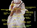
The tongue is a muscular organ in the mouth of a typical tetrapod. It manipulates food for chewing and swallowing as part of the digestive process, and is the primary organ of taste. The tongue's upper surface (dorsum) is covered by taste buds housed in numerous lingual papillae. It is sensitive and kept moist by saliva and is richly supplied with nerves and blood vessels. The tongue also serves as a natural means of cleaning the teeth. A major function of the tongue is the enabling of speech in humans and vocalization in other animals.

<span class="mw-page-title-main">Salivary gland</span> Exocrine glands that produce saliva through a system of ducts

The salivary glands in many vertebrates including mammals are exocrine glands that produce saliva through a system of ducts. Humans have three paired major salivary glands, as well as hundreds of minor salivary glands. Salivary glands can be classified as serous, mucous, or seromucous (mixed).

Articles related to anatomy include:

<span class="mw-page-title-main">Glossopharyngeal nerve</span> Cranial nerve IX, for the tongue and pharynx

The glossopharyngeal nerve, also known as the ninth cranial nerve, cranial nerve IX, or simply CN IX, is a cranial nerve that exits the brainstem from the sides of the upper medulla, just anterior to the vagus nerve. Being a mixed nerve (sensorimotor), it carries afferent sensory and efferent motor information. The motor division of the glossopharyngeal nerve is derived from the basal plate of the embryonic medulla oblongata, whereas the sensory division originates from the cranial neural crest.

<span class="mw-page-title-main">Taste bud</span> Taste receptor cells

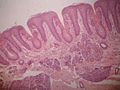
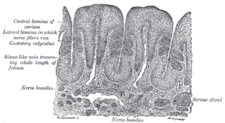
Taste buds are clusters of taste receptor cells, which are also known as gustatory cells. The taste receptors are located around the small structures known as papillae found on the upper surface of the tongue, soft palate, upper esophagus, the cheek, and epiglottis. These structures are involved in detecting the five elements of taste perception: saltiness, sourness, bitterness, sweetness and savoriness (umami). A popular myth assigns these different tastes to different regions of the tongue; in fact, these tastes can be detected by any area of the tongue. Via small openings in the tongue epithelium, called taste pores, parts of the food dissolved in saliva come into contact with the taste receptors. These are located on top of the taste receptor cells that constitute the taste buds. The taste receptor cells send information detected by clusters of various receptors and ion channels to the gustatory areas of the brain via the seventh, ninth and tenth cranial nerves.

<span class="mw-page-title-main">Submandibular gland</span> Human salivary gland

The paired submandibular glands are major salivary glands located beneath the floor of the mouth. In adult humans, they each weigh about 15 grams and contribute some 60–67% of unstimulated saliva secretion; on stimulation their contribution decreases in proportion as parotid gland secretion rises to 50%. The average length of the normal adult human submandibular salivary gland is approximately 27 mm, while the average width is approximately 14.3 mm.

<span class="mw-page-title-main">Sublingual gland</span> Major salivary glands in the mouth

The sublingual gland is a seromucous polystomatic exocrine gland. Located underneath the oral diaphragm, the sublingual gland is the smallest and most diffuse of the three major salivary glands of the oral cavity, with the other two being the submandibular and parotid. The sublingual gland provides approximately 3-5% of the total salivary volume.

<span class="mw-page-title-main">Chorda tympani</span> Nerve carrying taste sensations

Chorda tympani is a branch of the facial nerve that carries gustatory (taste) sensory innervation from the front of the tongue and parasympathetic (secretomotor) innervation to the submandibular and sublingual salivary glands.

<span class="mw-page-title-main">Glossitis</span> Medical condition

Glossitis can mean soreness of the tongue, or more usually inflammation with depapillation of the dorsal surface of the tongue, leaving a smooth and erythematous (reddened) surface,. In a wider sense, glossitis can mean inflammation of the tongue generally. Glossitis is often caused by nutritional deficiencies and may be painless or cause discomfort. Glossitis usually responds well to treatment if the cause is identified and corrected. Tongue soreness caused by glossitis is differentiated from burning mouth syndrome, where there is no identifiable change in the appearance of the tongue, and there are no identifiable causes.

The oral mucosa is the mucous membrane lining the inside of the mouth. It comprises stratified squamous epithelium, termed "oral epithelium", and an underlying connective tissue termed lamina propria. The oral cavity has sometimes been described as a mirror that reflects the health of the individual. Changes indicative of disease are seen as alterations in the oral mucosa lining the mouth, which can reveal systemic conditions, such as diabetes or vitamin deficiency, or the local effects of chronic tobacco or alcohol use. The oral mucosa tends to heal faster and with less scar formation compared to the skin. The underlying mechanism remains unknown, but research suggests that extracellular vesicles might be involved.

<span class="mw-page-title-main">Mucous gland</span>

Mucous gland, also known as muciparous glands, are found in several different parts of the body, and they typically stain lighter than serous glands during standard histological preparation. Most are multicellular, but goblet cells are single-celled glands.

<span class="mw-page-title-main">Intermediate nerve</span> Portion of the facial nerve

The intermediate nerve, nervus intermedius, nerve of Wrisberg or Glossopalatine nerve is the part of the facial nerve located between the motor component of the facial nerve and the vestibulocochlear nerve. It contains the sensory and parasympathetic fibers of the facial nerve. Upon reaching the facial canal, it joins with the motor root of the facial nerve at the geniculate ganglion. Alex Alfieri postulates that the intermediate nerve should be considered as a separate cranial nerve and not a part of the facial nerve.

<span class="mw-page-title-main">Von Ebner's gland</span> Exocrine glands found in the mouth

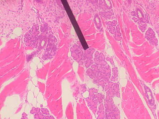
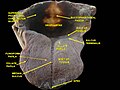
Von Ebner's glands, also called Ebner's glands or gustatory glands, are exocrine glands found in the mouth. More specifically, they are serous salivary glands which reside adjacent to the moats surrounding the circumvallate and foliate papillae just anterior to the posterior third of the tongue, anterior to the terminal sulcus.

<span class="mw-page-title-main">Taste receptor</span> Type of cellular receptor that facilitates taste

A taste receptor or tastant is a type of cellular receptor which facilitates the sensation of taste. When food or other substances enter the mouth, molecules interact with saliva and are bound to taste receptors in the oral cavity and other locations. Molecules which give a sensation of taste are considered "sapid".

Dental pertains to the teeth, including dentistry. Topics related to the dentistry, the human mouth and teeth include:

Solitary chemosensory cells (SCCs) are isolated elements located in epithelia of the apparatuses of endodermic origin. In the aquatic vertebrates, SCCs are also present in the skin. In oral cavity, SCCs precedes the development of taste buds. For long time, SCCs were considered to be typical of aquatic vertebrates. Recently, these elements were also demonstrated in mammals.

<span class="mw-page-title-main">Median rhomboid glossitis</span> Medical condition

Median rhomboid glossitis is a condition characterized by an area of redness and loss of lingual papillae on the central dorsum of the tongue, sometimes including lesions of the tongue and palate. It is seen in patients using inhaled steroids and smokers, and is usually a kind of chronic atrophic oral candidiasis, but hematinic deficiency and diabetes should be excluded.

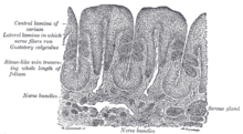
The papilla, in certain kinds of fish, particularly rays, sharks, and catfish, are small lumps of dermal tissue found in the mouth, where they are "distributed uniformly on the tongue, palate, and pharynx". They "project slightly above the surrounding multi-layered epithelium", and the taste buds of the fish are "situated along the crest or at the apex of the papillae".

<span class="mw-page-title-main">Human mouth</span> Part of human anatomy

In human anatomy, the mouth is the first portion of the alimentary canal that receives food and produces saliva. The oral mucosa is the mucous membrane epithelium lining the inside of the mouth.

<span class="mw-page-title-main">Horse tongue</span> Anatomy of the equine tongue

The horse tongue, like that of most mammals, is pink in color and plays an important role in taste perception. With its long, narrow shape typical of herbivorous animals, it enables the horse to grasp its vegetable food, with the help of its lips and teeth. This tongue is sensitive to pressure and temperature, and is involved in licking and chewing behaviors. Although the mare licks its foal for a long time immediately after birth, there is little research into the gustatory sensitivity of horses and the social use these animals make of their tongues.

References

  1. 1 2 3 4 Norton N (2007). Netter's head and neck anatomy for dentistry. illustrations by Netter FH. Philadelphia, Pa.: Saunders Elsevier. p. 402. ISBN   978-1929007882.
  2. 1 2 3 4 5 6 7 8 9 10 11 12 Susan Standring (editor in chief)] (2008). "Chapter 33: NECK AND UPPER AERODIGESTIVE TRACT". Gray's anatomy : the anatomical basis of clinical practice (40th ed.). [Edinburgh]: Churchill Livingstone/Elsevier. ISBN   978-0443066849.{{cite book}}: |last= has generic name (help)
  3. "Tongue | Gastrointestinal Tract". histologyguide.com. Retrieved 2023-02-19.
  4. 1 2 3 4 5 Scully C (2013). Oral and maxillofacial medicine : the basis of diagnosis and treatment (3rd ed.). Edinburgh: Churchill Livingstone. pp. 401, 402. ISBN   9780702049484.
  5. Ross, H R; Pawlina, W (2011). Histology: A text and atlas. Baltimore, MD.: Lippincott, Williams, and Wilkins. ISBN   978-0-7817-7200-6.
  6. Rajendran A; Sundaram S (10 February 2014). Shafer's Textbook of Oral Pathology (7th ed.). Elsevier Health Sciences APAC. p. 34. ISBN   978-81-312-3800-4.
  7. König, Liebich (2020). Veterinary anatomy of domestic animals: textbook and colour atlas (7th, updated and extended ed.). Stuttgart ; New York: Georg Thieme Verlag. ISBN   978-3-13-242933-8.