
Horse teeth refers to the dentition of equine species, including horses and donkeys. Equines are both heterodontous and diphyodontous, which means that they have teeth in more than one shape, and have two successive sets of teeth, the deciduous and permanent sets.

In mammalian oral anatomy, the canine teeth, also called cuspids, dogteeth, eye teeth, vampire teeth, or fangs, are the relatively long, pointed teeth. In the context of the upper jaw, they are also known as fangs. They can appear more flattened, however, causing them to resemble incisors and leading them to be called incisiform. They developed and are used primarily for firmly holding food in order to tear it apart, and occasionally as weapons. They are often the largest teeth in a mammal's mouth. Individuals of most species that develop them normally have four, two in the upper jaw and two in the lower, separated within each jaw by incisors; humans and dogs are examples. In most species, canines are the anterior-most teeth in the maxillary bone. The four canines in humans are the two upper maxillary canines and the two lower mandibular canines. They are specially prominent in dogs (Canidae), hence the name.
Ferugliotherium is a genus of fossil mammals in the family Ferugliotheriidae from the Campanian and/or Maastrichtian period of Argentina. It contains a single species, Ferugliotherium windhauseni, which was first described in 1986. Although originally interpreted on the basis of a single brachydont (low-crowned) molar as a member of Multituberculata, an extinct group of small, rodent-like mammals, it was recognized as related to the hypsodont (high-crowned) Sudamericidae following the discovery of additional material in the early 1990s. After a jaw of the sudamericid Sudamerica was described in 1999, these animals were no longer considered to be multituberculates and a few fossils that were previously considered to be Ferugliotherium were assigned to unspecified multituberculates instead. Since 2005, a relationship between gondwanatheres and multituberculates has again received support. A closely related animal, Trapalcotherium, was described in 2009 on the basis of a single tooth.

Incisors are the front teeth present in most mammals. They are located in the premaxilla above and on the mandible below. Humans have a total of eight. Opossums have 18, whereas armadillos have none.
Hypodontia is defined as the developmental absence of one or more teeth excluding the third molars. It is one of the most common dental anomalies, and can have a negative impact on function, and also appearance. It rarely occurs in primary teeth and the most commonly affected are the adult second premolars and the upper lateral incisors. It usually occurs as part of a syndrome that involves other abnormalities and requires multidisciplinary treatment.

In orthodontics, a malocclusion is a misalignment or incorrect relation between the teeth of the upper and lower dental arches when they approach each other as the jaws close. The English-language term dates from 1864; Edward Angle (1855–1930), the "father of modern orthodontics", popularised it. The word derives from mal- 'incorrect' and occlusion 'the manner in which opposing teeth meet'.

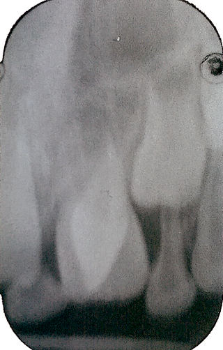
The maxillary central incisor is a human tooth in the front upper jaw, or maxilla, and is usually the most visible of all teeth in the mouth. It is located mesial to the maxillary lateral incisor. As with all incisors, their function is for shearing or cutting food during mastication (chewing). There is typically a single cusp on each tooth, called an incisal ridge or incisal edge. Formation of these teeth begins at 14 weeks in utero for the deciduous (baby) set and 3–4 months of age for the permanent set.

The maxillary lateral incisors are a pair of upper (maxillary) teeth that are located laterally from both maxillary central incisors of the mouth and medially from both maxillary canines. As with all incisors, their function is for shearing or cutting food during mastication, commonly known as chewing. There are generally no cusps on the teeth, but the rare condition known as talon cusps are most prevalent on the maxillary lateral incisors. The surface area of the tooth used in eating is called an incisal ridge or incisal edge. Though relatively the same, there are some minor differences between the deciduous (baby) maxillary lateral incisor and that of the permanent maxillary lateral incisor. The maxillary lateral incisors occlude in opposition to the mandibular lateral incisors.

The mandibular central incisor is the tooth located on the jaw, adjacent to the midline of the face. It is mesial from both mandibular lateral incisors. As with all incisors, its function includes shearing or cutting food during mastication, commonly known as chewing. There are no cusps on the tooth. Instead, the surface area of the tooth used in eating is called an incisal ridge or incisal edge. Though the two are similar, there are some minor differences between the deciduous (baby) mandibular central incisor and that of the permanent mandibular central incisor. The mandibular central incisors are usually the first teeth to appear in the mouth, typically around the age of 6–8 months.
Dilaceration is a developmental disturbance in shape of teeth. It refers to an angulation, or a sharp bend or curve, in the root or crown of a formed tooth. This disturbance is more likely to affect the maxillary incisors and occurs in permanent dentition. Although this may seem more of an aesthetics issue, an impacted maxillary incisor will cause issues related to occlusion, phonetics, mastication, and psychology on young patients.
Dens evaginatus is a rare odontogenic developmental anomaly that is found in teeth where the outer surface appears to form an extra bump or cusp.

Shovel-shaped incisors are incisors whose lingual surfaces are scooped as a consequence of lingual marginal ridges, crown curvature, or basal tubercles, either alone or in combination.
Talon cusp is a rare dental anomaly resulting in an extra cusp or cusp-like projection on an anterior tooth, located on the inside surface of the affected tooth. Sometimes it can also be found on the facial surface of the anterior tooth.

Dental anatomy is a field of anatomy dedicated to the study of human tooth structures. The development, appearance, and classification of teeth fall within its purview. Tooth formation begins before birth, and the teeth's eventual morphology is dictated during this time. Dental anatomy is also a taxonomical science: it is concerned with the naming of teeth and the structures of which they are made, this information serving a practical purpose in dental treatment.
This is a list of definitions of commonly used terms of location and direction in dentistry. This set of terms provides orientation within the oral cavity, much as anatomical terms of location provide orientation throughout the body.
The Dahl effect or Dahl concept is used in dentistry where a localized appliance or localized restoration is used to increase the available interocclusal space for restorations.

A tooth is a hard, calcified structure found in the jaws of many vertebrates and used to break down food. Some animals, particularly carnivores and omnivores, also use teeth to help with capturing or wounding prey, tearing food, for defensive purposes, to intimidate other animals often including their own, or to carry prey or their young. The roots of teeth are covered by gums. Teeth are not made of bone, but rather of multiple tissues of varying density and hardness that originate from the outermost embryonic germ layer, the ectoderm.

Teeth are common to most vertebrates, but mammalian teeth are distinctive in having a variety of shapes and functions. This feature first arose among early therapsids during the Permian, and has continued to the present day. All therapsid groups with the exception of the mammals are now extinct, but each of these groups possessed different tooth patterns, which aids with the classification of fossils.
The infundibulum of a tooth is the funnel-like center that is filled with cementum. The funnel is widest at the top (crown) which is the grinding (occlusal) surface. The infundibulum is also known as the dental cup. Simple tooth infundibula occur most notably in the incisors of horses and other equids, but they also occur in the premolars and molars of ruminants and camelids. The infundibula found in ruminants can get quite complex some with two funneling centers, and with multiple folding in the sides of the cup. These folds produce greater amounts of enamel in vertical curtains that substantially increase the durability of the tooth. The cheek teeth of elephants express this in a slightly different form with the vertical curtains of enamel coming in from the sides and meeting in the middle.

Alemoatherium is an extinct genus of prozostrodontian cynodont which lived in the Late Triassic of Brazil. It contains a single species, A. huebneri, named in 2017 by Agustín Martinelli and colleagues. The genus is based on UFSM 11579b, a left lower jaw (dentary) found in the Alemoa Member of the Santa Maria Formation, preserving the late Carnian-age Hyperodapedon Assemblage Zone. Alemoatherium was among the smallest species of cynodonts found in the rich synapsid fauna of the Santa Maria Formation. Its blade-like four-cusped postcanine teeth show many similarities with those of dromatheriids, an obscure group of early prozostrodontians.
