Mastoiditis

Last updated
Mastoiditis
Gray1193.png
Side view of head, showing surface relations of bones. (Mastoid process labeled near center.)
Specialty Otorhinolaryngology

Mastoiditis is the result of an infection that extends to the air cells of the skull behind the ear. Specifically, it is an inflammation of the mucosal lining of the mastoid antrum and mastoid air cell system inside [1] the mastoid process. The mastoid process is the portion of the temporal bone of the skull that is behind the ear. The mastoid process contains open, air-containing spaces. [2] [3] Mastoiditis is usually caused by untreated acute otitis media (middle ear infection) and used to be a leading cause of child mortality. With the development of antibiotics, however, mastoiditis has become quite rare in developed countries where surgical treatment is now much less frequent and more conservative, unlike former times. [2]

Contents

There is no evidence that the drop in antibiotic prescribing for otitis media has increased the incidence of mastoiditis, raising the possibility that the drop in reported cases is due to a confounding factor such as childhood immunizations against Haemophilus and Streptococcus. Untreated, the infection can spread to surrounding structures, including the brain, causing serious complications. [4] While the use of antibiotics has reduced the incidence of mastoiditis, the risk of masked mastoiditis, a subclinical infection without the typical findings of mastoiditis has increased with the inappropriate use of antibiotics and the emergence of multidrug-resistant bacteria. [5]

Signs and symptoms

Mastoiditis with subperiostal abscess Mastoiditis1.jpg
Mastoiditis with subperiostal abscess

Some common symptoms and signs of mastoiditis include pain, tenderness, and swelling in the mastoid region. There may be ear pain (otalgia), and the ear or mastoid region may be red (erythematous). Fever or headaches may also be present. Infants usually show nonspecific symptoms, including anorexia, diarrhea, or irritability. Drainage from the ear occurs in more serious cases often manifests as brown discharge on the pillowcase upon waking. [4] [6]

Pathophysiology

Mastoid cells of Lenoir Mastoid cells of Lenoir.jpg
Mastoid cells of Lenoir

The pathophysiology of mastoiditis is straightforward: bacteria spread from the middle ear to the mastoid air cells, where the inflammation causes damage to the bony structures. Streptococcus pneumoniae, Streptococcus pyogenes, Staphylococcus aureus, Haemophilus influenzae, and Moraxella catarrhalis are the most common organisms recovered in acute mastoiditis. Organisms that are rarely found are Pseudomonas aeruginosa and other Gram-negative aerobic bacilli, and anaerobic bacteria. [7] P. aeruginosa, Enterobacteriaceae, S. aureus and anaerobic bacteria ( Prevotella , Bacteroides , Fusobacterium , and Peptostreptococcus spp.) are the most common isolates in chronic mastoiditis. [8] Rarely, Mycobacterium species can also cause the infection. Some mastoiditis is caused by cholesteatoma, which is a sac of keratinizing squamous epithelium in the middle ear that usually results from repeated middle-ear infections. If left untreated, the cholesteatoma can erode into the mastoid process, producing mastoiditis, as well as other complications. [4]

Diagnosis

CT scan: Otitis media (simple arrow) and mastoiditis (double arrow) of the right side (left side in image). The external auditory canal is partially occupied by suppuration (triple arrow). 44-year-old woman. OtitisMedia Mastoiditis RightSide 153.svg
CT scan: Otitis media (simple arrow) and mastoiditis (double arrow) of the right side (left side in image). The external auditory canal is partially occupied by suppuration (triple arrow). 44-year-old woman.

The diagnosis of mastoiditis is clinical—based on the medical history and physical examination. Imaging studies provide additional information; The standard method of diagnosis is via MRI scan although a CT scan is a common alternative as it gives a clearer and more useful image to see how close the damage may have gotten to the brain and facial nerves. Planar (2-D) X-rays are not as useful. If there is drainage, it is often sent for culture, although this will often be negative if the patient has begun taking antibiotics. Exploratory surgery is often used as a last resort method of diagnosis to see the mastoid and surrounding areas. [2] [9]

Treatment

Attack triangle in mastoidectomies Attack triangle in mastoidectomies.jpg
Attack triangle in mastoidectomies

If ear infections are treated in a reasonable amount of time, the antibiotics will usually cure the infection and prevent its spread. For this reason, mastoiditis is rare in developed countries. Most ear infections occur in infants as the eustachian tubes are not fully developed and don't drain readily.[ citation needed ]

In all developed countries with up-to-date modern healthcare the primary treatment for mastoiditis is administration of intravenous antibiotics. Initially, broad-spectrum antibiotics are given, such as ceftriaxone. As culture results become available, treatment can be switched to more specific antibiotics directed at the eradication of the recovered aerobic and anaerobic bacteria. [8] Long-term antibiotics may be necessary to completely eradicate the infection. [4] If the condition does not quickly improve with antibiotics, surgical procedures may be performed (while continuing the medication). The most common procedure is a myringotomy, a small incision in the tympanic membrane (eardrum), or the insertion of a tympanostomy tube into the eardrum. [9] These serve to drain the pus from the middle ear, helping to treat the infection. The tube is extruded spontaneously after a few weeks to months, and the incision heals naturally. If there are complications, or the mastoiditis does not respond to the above treatments, it may be necessary to perform a mastoidectomy: a procedure in which a portion of the bone is removed and the infection drained. [4]

Prognosis

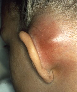
With prompt treatment, it is possible to cure mastoiditis. Seeking medical care early is important. However, it is difficult for antibiotics to penetrate to the interior of the mastoid process and so it may not be easy to cure the infection; it also may recur. Mastoiditis has many possible complications, all connected to the infection spreading to surrounding structures. Hearing loss is likely, or inflammation of the labyrinth of the inner ear (labyrinthitis) may occur, producing vertigo and an ear ringing may develop along with the hearing loss, making it more difficult to communicate. The infection may also spread to the facial nerve (cranial nerve VII), causing facial-nerve palsy, producing weakness or paralysis of some muscles of facial expression, on the same side of the face. Other complications include Bezold's abscess, an abscess (a collection of pus surrounded by inflamed tissue) behind the sternocleidomastoid muscle in the neck, or a subperiosteal abscess, between the periosteum and mastoid bone (resulting in the typical appearance of a protruding ear). Serious complications result if the infection spreads to the brain. These include meningitis (inflammation of the protective membranes surrounding the brain), epidural abscess (abscess between the skull and outer membrane of the brain), dural venous thrombophlebitis (inflammation of the venous structures of the brain), or brain abscess. [2] [4]

Epidemiology

In the United States and other developed countries, the incidence of mastoiditis is quite low, around 0.004%, although it is higher in developing countries. The condition most commonly affects children aged from two to thirteen months, when ear infections most commonly occur. Males and females are equally affected. [3]

See also

Related Research Articles

<span class="mw-page-title-main">Brain abscess</span> Medical condition

Brain abscess is an abscess within the brain tissue caused by inflammation and collection of infected material coming from local or remote infectious sources. The infection may also be introduced through a skull fracture following a head trauma or surgical procedures. Brain abscess is usually associated with congenital heart disease in young children. It may occur at any age but is most frequent in the third decade of life.

<span class="mw-page-title-main">Sinusitis</span> Inflammation of the mucous membrane that lines the sinuses resulting in symptoms

Sinusitis, also known as rhinosinusitis, is inflammation of the mucous membranes that line the sinuses resulting in symptoms that may include thick nasal mucus, a plugged nose, and facial pain. Other signs and symptoms may include fever, headaches, a poor sense of smell, sore throat, a feeling that phlegm is oozing out from the back of the nose to the throat along with a necessity to clear the throat frequently and frequent attacks of cough.

<span class="mw-page-title-main">Cholesteatoma</span> Medical condition

Cholesteatoma is a destructive and expanding growth consisting of keratinizing squamous epithelium in the middle ear and/or mastoid process. Cholesteatomas are not cancerous as the name may suggest, but can cause significant problems because of their erosive and expansile properties. This can result in the destruction of the bones of the middle ear (ossicles), as well as growth through the base of the skull into the brain. They often become infected and can result in chronically draining ears. Treatment almost always consists of surgical removal.

<span class="mw-page-title-main">Lemierre's syndrome</span> Medical condition

Lemierre's syndrome is infectious thrombophlebitis of the internal jugular vein. It most often develops as a complication of a bacterial sore throat infection in young, otherwise healthy adults. The thrombophlebitis is a serious condition and may lead to further systemic complications such as bacteria in the blood or septic emboli.

<span class="mw-page-title-main">Otitis media</span> Inflammation of the middle ear

Otitis media is a group of inflammatory diseases of the middle ear. One of the two main types is acute otitis media (AOM), an infection of rapid onset that usually presents with ear pain. In young children this may result in pulling at the ear, increased crying, and poor sleep. Decreased eating and a fever may also be present. The other main type is otitis media with effusion (OME), typically not associated with symptoms, although occasionally a feeling of fullness is described; it is defined as the presence of non-infectious fluid in the middle ear which may persist for weeks or months often after an episode of acute otitis media. Chronic suppurative otitis media (CSOM) is middle ear inflammation that results in a perforated tympanic membrane with discharge from the ear for more than six weeks. It may be a complication of acute otitis media. Pain is rarely present. All three types of otitis media may be associated with hearing loss. If children with hearing loss due to OME do not learn sign language, it may affect their ability to learn.

<span class="mw-page-title-main">Pharyngitis</span> Inflammation of the back of the throat

Pharyngitis is inflammation of the back of the throat, known as the pharynx. It typically results in a sore throat and fever. Other symptoms may include a runny nose, cough, headache, difficulty swallowing, swollen lymph nodes, and a hoarse voice. Symptoms usually last 3–5 days, but can be longer depending on cause. Complications can include sinusitis and acute otitis media. Pharyngitis is a type of upper respiratory tract infection.

<span class="mw-page-title-main">Tonsillitis</span> Inflammation of the tonsils

Tonsillitis is inflammation of the tonsils in the upper part of the throat. It can be acute or chronic. Acute tonsillitis typically has a rapid onset. Symptoms may include sore throat, fever, enlargement of the tonsils, trouble swallowing, and enlarged lymph nodes around the neck. Complications include peritonsillar abscess (Quinsy).

<span class="mw-page-title-main">Peritonsillar abscess</span> Pus behind the tonsil due to an infection

Peritonsillar abscess (PTA), also known as quinsy, is an accumulation of pus due to an infection behind the tonsil. Symptoms include fever, throat pain, trouble opening the mouth, and a change to the voice. Pain is usually worse on one side. Complications may include blockage of the airway or aspiration pneumonitis.

<span class="mw-page-title-main">Facial nerve paralysis</span> Medical condition

Facial nerve paralysis is a common problem that involves the paralysis of any structures innervated by the facial nerve. The pathway of the facial nerve is long and relatively convoluted, so there are a number of causes that may result in facial nerve paralysis. The most common is Bell's palsy, a disease of unknown cause that may only be diagnosed by exclusion of identifiable serious causes.

<span class="mw-page-title-main">Ear pain</span> Pain in the ear

Ear pain, also known as earache or otalgia, is pain in the ear. Primary ear pain is pain that originates from the ear. Secondary ear pain is a type of referred pain, meaning that the source of the pain differs from the location where the pain is felt.

<span class="mw-page-title-main">Lung abscess</span> Medical condition

Lung abscess is a type of liquefactive necrosis of the lung tissue and formation of cavities containing necrotic debris or fluid caused by microbial infection.

<i>Peptostreptococcus</i> Genus of bacteria

Peptostreptococcus is a genus of anaerobic, Gram-positive, non-spore forming bacteria. The cells are small, spherical, and can occur in short chains, in pairs or individually. They typically move using cilia. Peptostreptococcus are slow-growing bacteria with increasing resistance to antimicrobial drugs. Peptostreptococcus is a normal inhabitant of the healthy lower reproductive tract of women.

<span class="mw-page-title-main">Pericoronitis</span> Inflammation of the soft tissues surrounding the crown of a partially erupted tooth

Pericoronitis is inflammation of the soft tissues surrounding the crown of a partially erupted tooth, including the gingiva (gums) and the dental follicle. The soft tissue covering a partially erupted tooth is known as an operculum, an area which can be difficult to access with normal oral hygiene methods. The hyponym operculitis technically refers to inflammation of the operculum alone.

<span class="mw-page-title-main">Gradenigo's syndrome</span> Medical condition

Gradenigo's syndrome, also called Gradenigo-Lannois syndrome, is a complication of otitis media and mastoiditis involving the apex of the petrous temporal bone. It was first described by Giuseppe Gradenigo in 1904.

<span class="mw-page-title-main">Epitympanic recess</span> Hollow located on the superior/roof aspect of the middle ear

The epitympanic recess is the portion of the tympanic cavity situated superior to the tympanic membrane. The recess lodges the head of malleus, and the body of incus.

Acute infectious thyroiditis (AIT) also known as suppurative thyroiditis, microbial inflammatory thyroiditis, pyrogenic thyroiditis and bacterial thyroiditis.

Anaerobic infections are caused by anaerobic bacteria. Obligately anaerobic bacteria do not grow on solid media in room air ; facultatively anaerobic bacteria can grow in the presence or absence of air. Microaerophilic bacteria do not grow at all aerobically or grow poorly, but grow better under 10% carbon dioxide or anaerobically. Anaerobic bacteria can be divided into strict anaerobes that can not grow in the presence of more than 0.5% oxygen and moderate anaerobic bacteria that are able of growing between 2 and 8% oxygen. Anaerobic bacteria usually do not possess catalase, but some can generate superoxide dismutase which protects them from oxygen.

<span class="mw-page-title-main">Otitis externa</span> Inflammation of the ear canal

Otitis externa, also called swimmer's ear, is inflammation of the ear canal. It often presents with ear pain, swelling of the ear canal, and occasionally decreased hearing. Typically there is pain with movement of the outer ear. A high fever is typically not present except in severe cases.

<span class="mw-page-title-main">Neonatal meningitis</span> Medical condition

Neonatal meningitis is a serious medical condition in infants that is rapidly fatal if untreated. Meningitis is an inflammation of the meninges, the protective membranes of the central nervous system, is more common in the neonatal period than any other time in life, and is an important cause of morbidity and mortality globally. Mortality is roughly half in developing countries and ranges from 8%-12.5% in developed countries.

Osteomyelitis of the jaws is osteomyelitis which occurs in the bones of the jaws. Historically, osteomyelitis of the jaws was a common complication of odontogenic infection. Before the antibiotic era, it was frequently a fatal condition.

References

  1. Diseases of ear nose & throat by PL dhingra & shruti dhingra. published by elsevier
  2. 1 2 3 4 "Mastoiditis". MedlinePlus Medical Encyclopedia. Retrieved July 30, 2003.
  3. 1 2 "Ear Infections – Treatment". webmd.com. Retrieved 24 November 2008.
  4. 1 2 3 4 5 6 Young, Tesfa. "Mastoiditis". eMedicine. Retrieved June 10, 2005.
  5. Omura, T (May 2020). "Meningoencephalitis caused by masked mastoiditis that was diagnosed during a follow-up in an elderly patient with diabetes mellitus: A case report". Geriatrics & Gerontology International. 20 (5): 500–01. doi:10.1111/ggi.13904. PMID   32358876. S2CID   218481126.
  6. "What to Do About Ear infections". webmd.com. Retrieved 24 November 2008.
  7. Nussinovitch M, Yoeli R, Elishkevitz K, Varsano I (2004). "Acute mastoiditis in children: epidemiologic, clinical, microbiologic, and therapeutic aspects over past years". Clin Pediatr (Phila). 43 (3): 261–7. doi:10.1177/000992280404300307. PMID   15094950. S2CID   38653809.
  8. 1 2 Brook I (2005). "The role of anaerobic bacteria in acute and chronic mastoiditis". Anaerobe. 11 (5): 252–7. doi:10.1016/j.anaerobe.2005.03.005. PMID   16701580.
  9. 1 2 Bakhos D, Trijolet JP, Morinière S, Pondaven S, Al Zahrani M, Lescanne E (April 2011). "Conservative management of acute mastoiditis in children". Arch Otolaryngol Head Neck Surg. 137 (4): 346–50. doi: 10.1001/archoto.2011.29 . PMID   21502472.

Further reading