Medium-chain acyl-coenzyme A dehydrogenase deficiency

Last updated
Medium-chain acyl-coenzyme A dehydrogenase deficiency (MCAD)
Other namesCarnitine deficiency secondary to medium-chain acyl-CoA dehydrogenase deficiency, [1] MCAD deficiency or MCADD
Autosomal recessive - en.svg
This condition is inherited in an autosomal recessive manner.
Specialty Endocrinology   OOjs UI icon edit-ltr-progressive.svg

Medium-chain acyl-CoA dehydrogenase deficiency (MCAD deficiency or MCADD) is a disorder of fatty acid oxidation that impairs the body's ability to break down medium-chain fatty acids into acetyl-CoA. The disorder is characterized by hypoglycemia and sudden death without timely intervention, most often brought on by periods of fasting or vomiting.

Contents

Prior to expanded newborn screening, MCADD was an underdiagnosed cause of sudden death in infants. Individuals who have been identified prior to the onset of symptoms have an excellent prognosis.

MCADD is most prevalent in individuals of Northern European Caucasian descent, with an incidence of 1:4000 to 1:17,000 depending on the population. Treatment of MCADD is mainly preventive, by avoiding fasting and other situations where the body relies on fatty acid oxidation to supply energy.

Signs and symptoms

MCAD is one of the enzymes responsible for dehydrogenation of fatty acids as they cycle through the beta-oxidation spiral. BetaOxidationSpiral.tiff
MCAD is one of the enzymes responsible for dehydrogenation of fatty acids as they cycle through the beta-oxidation spiral.

MCADD presents in early childhood with hypoketotic hypoglycemia and liver dysfunction, often preceded by extended periods of fasting or an infection with vomiting. Infants who are exclusively breast-fed may present in this manner shortly after birth, due to poor feeding. In some individuals the first manifestation of MCADD may be sudden death following a minor illness. [2] A number of individuals with MCADD may remain completely asymptomatic, provided they never encounter a situation that sufficiently stresses their metabolism. [2] [3] With the advent of expanded newborn screening, some mothers have been identified with MCADD after their infants had positive newborn screens for low carnitine levels. [4]

The enzyme medium-chain acyl-CoA dehydrogenase (MCAD) is responsible for the dehydrogenation step of fatty acids with chain lengths between 6 and 12 carbons as they undergo beta-oxidation in the mitochondria. Fatty acid beta-oxidation provides energy after the body has used up its stores of glucose and glycogen. This oxidation typically occurs during periods of extended fasting or illness when caloric intake is reduced, and energy needs are increased.[ citation needed ]

Genetics

MCADD is inherited in an autosomal recessive manner, meaning an affected individual must inherit a mutated allele from both of their parents. ACADM is the gene involved, located at 1p31, with 12 exons and coding for a protein of 421 amino acids. [3] There is a common mutation among Northern European Caucasians, replacement of an adenine at position 985 with guanine, which results in a substitution of lysine with glutamic acid at position 304 of the protein. Other mutations have been identified more commonly since newborn screening has expanded the mutation spectrum. [3] The 985A>G common mutation is present in the homozygous state in 80% of Caucasian individuals who presented clinically with MCADD and in 60% of the population identified by screening. [2]

An individual's genotype does not correlate well with their clinical phenotype for MCADD. The clinical presentation of an individual with MCADD depends not only on the presence of the mutations in the ACADM gene, but also on the presence of environmental or physiological stressors that require the body to depend on fatty acid oxidation for energy. Some mutations, identified through newborn screening programs and associated with higher residual enzyme activity have not been seen in individuals with clinical symptoms of MCADD. Despite this, treatment with fasting avoidance remains the norm for all those diagnosed with MCADD. [2]

Diagnosis

Clinically, MCADD or another fatty acid oxidation disorder is suspected in individuals who present with lethargy, seizures, coma and hypoketotic hypoglycemia, particularly if triggered by a minor illness. MCADD can also present with acute liver disease and hepatomegaly, which can lead to a misdiagnosis of Reye syndrome. In some individuals, the only manifestation of MCADD is sudden, unexplained death often preceded by a minor illness that would not usually be fatal. [3]

Acylcarnitine profile of an individual with MCADD, showing characteristic elevation of octanoylcarnitine (C8) MCADD Acylcarnitine Profile.tif
Acylcarnitine profile of an individual with MCADD, showing characteristic elevation of octanoylcarnitine (C8)

In areas with expanded newborn screening using tandem mass spectrometry (MS/MS), MCADD is usually detected shortly after birth, by the analysis of blood spots collected on filter paper. Acylcarnitine profiles with MS/MS will show a very characteristic pattern of elevated hexanoylcarnitine (C6), octanoylcarnitine (C8), decanoylcarnitine (C10) or decenoylcarnitine (C10:1), with C8 being greater than C6 and C10. Secondary carnitine deficiency is sometimes seen with MCADD, and in these cases, acylcarnitine profiles may not be informative. [3] Urine organic acid analysis by gas chromatography-mass spectrometry (GC-MS) will show a pattern of dicarboxylic aciduria with low levels of ketones. Traces of acylglycine species may also be detected. Asymptomatic individuals may have normal biochemical lab results. For these individuals, targeted analysis of acylglycine species by GC-MS, specifically hexanoylglycine and suberylglycine can be diagnostic. [3] [5] After biochemical suspicion of MCADD, molecular genetic analysis of ACADM can be used to confirm the diagnosis. [6] The analysis of MCAD activity in cultured fibroblasts can also be used for diagnosis. [3]

In cases of sudden death where the preceding illness would not usually have been fatal, MCADD is often suspected. The autopsy will often show fatty deposits in the liver. In cases where MCADD is suspected, acylcarnitine analysis of bile and blood can be undertaken postmortem for diagnosis. Where samples are not available, residual blood from newborn screening may be helpful. Biochemical testing of asymptomatic siblings and parents may also be informative. [7] MCADD and other fatty acid oxidation disorders have been recognized in recent years as undiagnosed causes of sudden infant death syndrome. [8] [9]

Treatment

As with most other fatty acid oxidation disorders, individuals with MCADD need to avoid fasting for prolonged periods of time. During illnesses, they require careful management to stave off metabolic decompensation, which can result in death. [2] Supplementation of simple carbohydrates or glucose during illness is key to prevent catabolism. [3] The duration of fasting for individuals with MCADD varies with age, infants typically require frequent feedings or a slow release source of carbohydrates, such as uncooked cornstarch. Illnesses and other stresses can significantly reduce the fasting tolerance of affected individuals. [10]

Individuals with MCADD should have an "emergency letter" that allows medical staff who are unfamiliar with the patient and the condition to administer correct treatment properly in the event of acute decompensation. This letter should outline the steps needed to intervene in a crisis and have contact information for specialists familiar with the individual's care. [3]

Misdiagnosis issues

Prognosis

A 1994 study of the entire population of New South Wales (Australia) found 20 patients. Of these, 5 (25%) had died at or before 30 months of age. Of the survivors, 1 (5%) was severely disabled and the remainder had either suffered mild disability or were making normal progress in school. [11] A 2006 Dutch study followed 155 cases and found that 27 individuals (17%) had died at an early age. Of the survivors, 24 (19%) suffered from some degree of disability, of which most were mild. All the 18 patients diagnosed neonatally were alive at the time of the follow-up. [12]

Incidence

MCADD is most prevalent in individuals of Northern European Caucasian descent. The incidence in Northern Germany is 1:4000, currently the highest in the world. Northern Europe is also the origin of the common mutation in MCADD. For populations without origins in Northern Europe, the incidence is significantly lower, 1:51,000 in Japan and 1:700,000 in Taiwan. The common mutation has not been identified in MCADD cases identified in Asian populations. [3]

Related Research Articles

<span class="mw-page-title-main">Newborn screening</span> Practice of testing infants for diseases

Newborn screening (NBS) is a public health program of screening in infants shortly after birth for conditions that are treatable, but not clinically evident in the newborn period. The goal is to identify infants at risk for these conditions early enough to confirm the diagnosis and provide intervention that will alter the clinical course of the disease and prevent or ameliorate the clinical manifestations. NBS started with the discovery that the amino acid disorder phenylketonuria (PKU) could be treated by dietary adjustment, and that early intervention was required for the best outcome. Infants with PKU appear normal at birth, but are unable to metabolize the essential amino acid phenylalanine, resulting in irreversible intellectual disability. In the 1960s, Robert Guthrie developed a simple method using a bacterial inhibition assay that could detect high levels of phenylalanine in blood shortly after a baby was born. Guthrie also pioneered the collection of blood on filter paper which could be easily transported, recognizing the need for a simple system if the screening was going to be done on a large scale. Newborn screening around the world is still done using similar filter paper. NBS was first introduced as a public health program in the United States in the early 1960s, and has expanded to countries around the world.

<span class="mw-page-title-main">Long-chain 3-hydroxyacyl-coenzyme A dehydrogenase deficiency</span> Medical condition

Long-chain 3-hydroxyacyl-coenzyme A dehydrogenase deficiency is a rare autosomal recessive fatty acid oxidation disorder that prevents the body from converting certain fats into energy. This can become life-threatening, particularly during periods of fasting.

In biochemistry and metabolism, beta oxidation (also β-oxidation) is the catabolic process by which fatty acid molecules are broken down in the cytosol in prokaryotes and in the mitochondria in eukaryotes to generate acetyl-CoA. Acetyl-CoA enters the citric acid cycle, generating NADH and FADH2, which are electron carriers used in the electron transport chain. It is named as such because the beta carbon of the fatty acid chain undergoes oxidation and is converted to a carbonyl group to start the cycle all over again. Beta-oxidation is primarily facilitated by the mitochondrial trifunctional protein, an enzyme complex associated with the inner mitochondrial membrane, although very long chain fatty acids are oxidized in peroxisomes.

<span class="mw-page-title-main">Inborn error of lipid metabolism</span> Medical condition

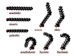
Numerous genetic disorders are caused by errors in fatty acid metabolism. These disorders may be described as fatty oxidation disorders or as a lipid storage disorders, and are any one of several inborn errors of metabolism that result from enzyme defects affecting the ability of the body to oxidize fatty acids in order to produce energy within muscles, liver, and other cell types.

<span class="mw-page-title-main">Carnitine palmitoyltransferase I deficiency</span> Medical condition

Carnitine palmitoyltransferase I deficiency is a rare metabolic disorder that prevents the body from converting certain fats called long-chain fatty acids(LCFA) into energy, particularly during periods without food. It is caused by a mutation in CPT1A on chromosome 11.

<span class="mw-page-title-main">Carnitine palmitoyltransferase II deficiency</span> Medical condition

Carnitine palmitoyltransferase II deficiency, sometimes shortened to CPT-II or CPT2, is an autosomal recessively inherited genetic metabolic disorder characterized by an enzymatic defect that prevents long-chain fatty acids from being transported into the mitochondria for utilization as an energy source. The disorder presents in one of three clinical forms: lethal neonatal, severe infantile hepatocardiomuscular and myopathic.

<span class="mw-page-title-main">Very long-chain acyl-coenzyme A dehydrogenase deficiency</span> Medical condition

Very long-chain acyl-coenzyme A dehydrogenase deficiency is a fatty-acid metabolism disorder which prevents the body from converting certain fats to energy, particularly during periods without food.

<span class="mw-page-title-main">ACADM</span> Mammalian protein found in Homo sapiens

ACADM is a gene that provides instructions for making an enzyme called acyl-coenzyme A dehydrogenase that is important for breaking down (degrading) a certain group of fats called medium-chain fatty acids.

<span class="mw-page-title-main">Short-chain acyl-coenzyme A dehydrogenase deficiency</span> Medical condition

Short-chain acyl-coenzyme A dehydrogenase deficiency (SCADD) is an autosomal recessive fatty acid oxidation disorder which affects enzymes required to break down a certain group of fats called short chain fatty acids.

Acyl-CoA dehydrogenases (ACADs) are a class of enzymes that function to catalyze the initial step in each cycle of fatty acid β-oxidation in the mitochondria of cells. Their action results in the introduction of a trans double-bond between C2 (α) and C3 (β) of the acyl-CoA thioester substrate. Flavin adenine dinucleotide (FAD) is a required co-factor in addition to the presence of an active site glutamate in order for the enzyme to function.

<span class="mw-page-title-main">ACADS</span> Protein-coding gene in humans

Acyl-CoA dehydrogenase, C-2 to C-3 short chain is an enzyme that in humans is encoded by the ACADS gene. This gene encodes a tetrameric mitochondrial flavoprotein, which is a member of the acyl-CoA dehydrogenase family. This enzyme catalyzes the initial step of the mitochondrial fatty acid beta-oxidation pathway. The ACADS gene is associated with short-chain acyl-coenzyme A dehydrogenase deficiency.

<span class="mw-page-title-main">Acyl-CoA</span> Group of coenzymes that metabolize fatty acids

Acyl-CoA is a group of coenzymes that metabolize fatty acids. Acyl-CoA's are susceptible to beta oxidation, forming, ultimately, acetyl-CoA. The acetyl-CoA enters the citric acid cycle, eventually forming several equivalents of ATP. In this way, fats are converted to ATP, the universal biochemical energy carrier.

3-hydroxyacyl-coenzyme A dehydrogenase deficiency is a rare condition that prevents the body from converting certain fats to energy, particularly during fasting. Normally, through a process called fatty acid oxidation, several enzymes work in a step-wise fashion to metabolize fats and convert them to energy. People with 3-hydroxyacyl-coenzyme A dehydrogenase deficiency have inadequate levels of an enzyme required for a step that metabolizes groups of fats called medium chain fatty acids and short chain fatty acids; for this reason this disorder is sometimes called medium- and short-chain 3-hydroxyacyl-coenzyme A dehydrogenase (M/SCHAD) deficiency.

<span class="mw-page-title-main">ACADSB</span> Protein-coding gene in the species Homo sapiens

ACADSB is a human gene that encodes short/branched chain specific acyl-CoA dehydrogenase (SBCAD), an enzyme in the acyl CoA dehydrogenase family.

<span class="mw-page-title-main">ETFA</span> Protein-coding gene in humans

The human ETFA gene encodes the Electron-transfer-flavoprotein, alpha subunit, also known as ETF-α. Together with Electron-transfer-flavoprotein, beta subunit, encoded by the 'ETFB' gene, it forms the heterodimeric electron transfer flavoprotein (ETF). The native ETF protein contains one molecule of FAD and one molecule of AMP, respectively.

<span class="mw-page-title-main">ETFB</span> Protein-coding gene in humans

The human ETFB gene encodes the Electron-transfer-flavoprotein, beta subunit, also known as ETF-β. Together with Electron-transfer-flavoprotein, alpha subunit, encoded by the 'ETFA' gene, it forms the heterodimeric Electron transfer flavoprotein (ETF). The native ETF protein contains one molecule of FAD and one molecule of AMP, respectively.

<span class="mw-page-title-main">ACAD9</span> Protein-coding gene in the species Homo sapiens

Acyl-CoA dehydrogenase family member 9, mitochondrial is an enzyme that in humans is encoded by the ACAD9 gene. Mitochondrial Complex I Deficiency with varying clinical manifestations has been associated with mutations in ACAD9.

<span class="mw-page-title-main">Fatty-acid metabolism disorder</span> Medical condition

A broad classification for genetic disorders that result from an inability of the body to produce or utilize an enzyme or transport protein that is required to oxidize fatty acids. They are an inborn error of lipid metabolism, and when it affects the muscles also a metabolic myopathy.

References

  1. RESERVED, INSERM US14-- ALL RIGHTS. "Orphanet: Medium chain acyl CoA dehydrogenase deficiency". www.orpha.net. Retrieved 14 April 2019.{{cite web}}: CS1 maint: numeric names: authors list (link)
  2. 1 2 3 4 5 Morris, Andrew A.M.; Spiekerkoetter, Ute (2012). "Disorders of Mitochondrial Fatty Acid Oxidation and Related Metabolic Pathways". In Saudubray, Jean-Marie; van den Berghe, Georges; Walter, John H. (eds.). Inborn Metabolic Diseases: Diagnosis and Treatment (5th ed.). New York: Springer. pp.  201–216. ISBN   978-3-642-15719-6.
  3. 1 2 3 4 5 6 7 8 9 10 Matern, D.; Rinaldo, P.; Pagon, R. A.; Bird, T. D.; Dolan, C. R.; Stephens, K.; Adam, M. P. (1993). "Medium-Chain Acyl-Coenzyme a Dehydrogenase Deficiency". PMID   20301597.{{cite journal}}: Cite journal requires |journal= (help)
  4. Leydiker, K. B.; Neidich, J. A.; Lorey, F.; Barr, E. M.; Puckett, R. L.; Lobo, R. M.; Abdenur, J. E. (2011). "Maternal medium-chain acyl-CoA dehydrogenase deficiency identified by newborn screening". Molecular Genetics and Metabolism. 103 (1): 92–95. doi:10.1016/j.ymgme.2011.01.011. PMID   21354840.
  5. Rinaldo, P.; O'Shea, J. J.; Coates, P. M.; Hale, D. E.; Stanley, C. A.; Tanaka, K. (1988). "Medium-Chain Acyl-CoA Dehydrogenase Deficiency". New England Journal of Medicine. 319 (20): 1308–1313. doi:10.1056/NEJM198811173192003. PMID   3054550.
  6. "C8 Elevated + Lesser Elevations of C6 and C10" (PDF). American College of Medical Genetics. Retrieved 2012-06-09.
  7. Rinaldo, P.; Matern, D.; Bennett, M. J. (2002). "Fattyacidoxidationdisorders". Annual Review of Physiology. 64: 477–502. doi:10.1146/annurev.physiol.64.082201.154705. PMID   11826276.
  8. Hegyi, T.; Ostfeld, B.; Gardner, K. (1992). "Medium chain acyl-coenzyme a dehydrogenase deficiency and SIDS". New Jersey Medicine. 89 (5): 385–392. PMID   1635678.
  9. Keppen, L. D.; Randall, B. (1999). "Inborn defects of fatty acid oxidation: A preventable cause of SIDS". South Dakota Journal of Medicine. 52 (6): 187–188, discussion 188–9. PMID   10388343.
  10. Walter, J. H. (2009). "Tolerance to fast: Rational and practical evaluation in children with hypoketonaemia". Journal of Inherited Metabolic Disease. 32 (2): 214–217. doi:10.1007/s10545-009-1087-y. PMID   19255872. S2CID   28507050.
  11. Wilcken, B.; Hammond, J.; Silink, M. (1994-05-01). "Morbidity and mortality in medium chain acyl coenzyme A dehydrogenase deficiency". Archives of Disease in Childhood. 70 (5): 410–412. doi:10.1136/adc.70.5.410. ISSN   1468-2044. PMC   1029830 . PMID   8017963.
  12. Derks, Terry G.J.; Reijngoud, Dirk-Jan; Waterham, Hans R.; Gerver, Willem-Jan M.; Berg, Maarten P. van den; Sauer, Pieter J.J.; Smit, G. Peter A. (2006). "The natural history of medium-chain acyl CoA dehydrogenase deficiency in the Netherlands: Clinical presentation and outcome". The Journal of Pediatrics. 148 (5): 665–670.e3. doi:10.1016/j.jpeds.2005.12.028. PMID   16737882.