Related Research Articles

The pericardium, also called pericardial sac, is a double-walled sac containing the heart and the roots of the great vessels. It has two layers, an outer layer made of strong inelastic connective tissue, and an inner layer made of serous membrane. It encloses the pericardial cavity, which contains pericardial fluid, and defines the middle mediastinum. It separates the heart from interference of other structures, protects it against infection and blunt trauma, and lubricates the heart's movements.

The mesothelium is a membrane composed of simple squamous epithelial cells of mesodermal origin, which forms the lining of several body cavities: the pleura, peritoneum and pericardium.

McBurney's point is the name given to the point over the right side of the abdomen that is one-third of the distance from the anterior superior iliac spine to the umbilicus (navel). This is near the most common location of the appendix.
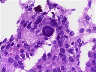
A psammoma body is a round collection of calcium, seen microscopically. The term is derived from the Greek word ψάμμος (psámmos), meaning "sand".

A serous tumour is a neoplasm that typically has papillary to solid formations of tumor cells with crowded nuclei, and which typically arises on the modified Müllerian-derived serous membranes that surround the ovaries in females. Such ovarian tumors are part of the surface epithelial-stromal tumour group of ovarian tumors. They are common neoplasms with a strong tendency to occur bilaterally, and they account for approximately a quarter of all ovarian tumors.

Hyperplasia, or hypergenesis, is an enlargement of an organ or tissue caused by an increase in the amount of organic tissue that results from cell proliferation. It may lead to the gross enlargement of an organ, and the term is sometimes confused with benign neoplasia or benign tumor.

Pseudomyxoma peritonei (PMP) is a clinical condition caused by cancerous cells that produce abundant mucin or gelatinous ascites. The tumors cause fibrosis of tissues and impede digestion or organ function, and if left untreated, the tumors and mucin they produce will fill the abdominal cavity. This will result in compression of organs and will destroy the function of the colon, small intestine, stomach, or other organs. Prognosis with treatment in many cases is optimistic, but the disease is lethal if untreated, with death occurring via cachexia, bowel obstruction, or other types of complications.

The chordae tendineae or tendinous cords, colloquially known as the heart strings, are inelastic cords of fibrous connective tissue that connect the papillary muscles to the tricuspid valve and the mitral valve in the heart.

The cricothyroid ligament is a ligament in the neck. It connects the cricoid cartilage to the thyroid cartilage. It prevents these cartilages from moving too far apart. It is cut during an emergency cricothyrotomy to treat upper airway obstruction.
Epulis fissuratum is a benign hyperplasia of fibrous connective tissue which develops as a reactive lesion to chronic mechanical irritation produced by the flange of a poorly fitting denture. More simply, epulis fissuratum is where excess folds of firm tissue form inside the mouth, as a result of rubbing on the edge of dentures that do not fit well. It is a harmless condition and does not represent oral cancer. Treatment is by simple surgical removal of the lesion, and also by adjustment of the denture or provision of a new denture.
Inflammatory papillary hyperplasia (IPH) is a benign lesion of the oral mucosa which is characterized by the growth of one or more nodular lesions, measuring about 2mm or less. The lesion almost exclusively involves the hard palate, and in rare instances, it also has been seen on the mandible. The lesion is mostly asymptomatic and color of the mucosa may vary from pink to red.

In anatomy, a persistent left superior vena cava is the most common variation of the thoracic venous system. It is present in between 0.3% and 0.5% of the population, and is an embryologic remnant that results from a failure to involute.

The right lymphatic duct is an important lymphatic vessel that drains the right upper quadrant of the body. It forms various combinations with the right subclavian vein and right internal jugular vein.
Pericardiectomy is the surgical removal of part or most of the pericardium. This operation is most commonly used to relieve constrictive pericarditis, or to remove a pericardium that is calcified and fibrous. It may also be used for severe or recurrent cases of pericardial effusion. Post-operative outcomes and mortality are significantly impacted by the disease it is used to treat.
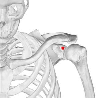
The supraglenoid tubercle is a region of the scapula from which the long head of the biceps brachii muscle originates. It is a small, rough projection superior to the glenoid cavity near the base of the coracoid process. The term supraglenoid is from the Latin supra, meaning above, and glenoid, meaning socket or cavity.

Gingival enlargement is an increase in the size of the gingiva (gums). It is a common feature of gingival disease. Gingival enlargement can be caused by a number of factors, including inflammatory conditions and the side effects of certain medications. The treatment is based on the cause. A closely related term is epulis, denoting a localized tumor on the gingiva.
The tracheoesophageal septum is an embryological structure. It is formed from the tracheoesophageal folds or ridges which fuse in the midline. It divides the oesophagus from the trachea during prenatal development. Developmental abnormalities can lead to a tracheoesophageal fistula.
An exploratory laparotomy is a general surgical operation where the abdomen is opened and the abdominal organs are examined for injury or disease. It is the standard of care in various blunt and penetrating trauma situations in which there may be life-threatening internal injuries. It is also used in certain diagnostic situations, in which the operation is undertaken in search of a unifying cause for multiple signs and symptoms of disease, and in the staging of some cancers.
A pericardial window is a cardiac surgical procedure to create a fistula – or "window" – from the pericardial space to the pleural cavity. The purpose of the window is to allow a pericardial effusion or cardiac tamponade to drain from the space surrounding the heart into the chest cavity.
A peritoneal inclusion cyst is a cyst-like structure that appears in the pelvis due to non neoplastic reactive mesothelial proliferation, often as a consequence of prior episodes of pelvic inflammation, as can occur in pelvic inflammatory disease. It has the potential to mimic ovarian cysts, hydrosalpinx or even malignancy, due to its nonspecific anechoic appearance.
References
- 1 2 3 4 5 Watkins, Jaclyn C.; Nascimento, Alessandra F.; Nucci, Marisa R. (2018). "Disorders of the Peritoneum". Diagnostic Gynecologic and Obstetric Pathology. Elsevier. pp. 800–843. doi:10.1016/b978-0-323-44732-4.00023-6. ISBN 978-0-323-44732-4.