Mesothelin

Last updated
MSLN
MSLN protein.png
Available structures
PDB Ortholog search: PDBe RCSB
Identifiers
Aliases MSLN , MPF, SMRP, mesothelin
External IDs OMIM: 601051 MGI: 1888992 HomoloGene: 4249 GeneCards: MSLN
Orthologs
SpeciesHumanMouse
Entrez
Ensembl
UniProt
RefSeq (mRNA)

NM_001177355
NM_005823
NM_013404

NM_018857
NM_001356286
NM_001374653

RefSeq (protein)

NP_001170826
NP_005814
NP_037536

NP_061345
NP_001343215

Location (UCSC) Chr 16: 0.76 – 0.77 Mb Chr 17: 25.97 – 25.97 Mb
PubMed search [3] [4]
Wikidata
View/Edit Human View/Edit Mouse

Mesothelin, also known as MSLN, is a protein that in humans is encoded by the MSLN gene. [5] [6]

Contents

Function

Mesothelin is a 40 kDa protein that is expressed in mesothelial cells. [7] The protein was first identified by its reactivity with monoclonal antibody K1. [8] Subsequent cloning studies showed that the mesothelin gene encodes a precursor protein that is processed to yield mesothelin which is attached to the cell membrane by a glycophosphatidylinositol linkage and a 31-kDa shed fragment named megakaryocyte-potentiating factor (MPF). Although it has been proposed that mesothelin may be involved in cell adhesion, its biological function is not known. [9] [10] A knockout mouse line that lacks mesothelin reproduces and develops normally. [11]

Mesothelin is over expressed in several human tumors, including mesothelioma, ovarian cancer, pancreatic adenocarcinoma, [7] lung adenocarcinoma, [12] and cholangiocarcinoma. [13] Mesothelin binds MUC16 (also known as CA125), indicating that the interaction of mesothelin and MUC16 may contribute to the implantation and peritoneal spread of tumors by cell adhesion. [14] The region (residues 296-359) consisting of 64 amino acids at the N-terminus of cell surface mesothelin has been identified as the functional binding domain (named IAB) for MUC16/CA125, suggesting the mechanism of mesothelin acting as a MUC16/CA125 functional partner in cancer development. [15]

A protein structure model of human mesothelin and the binding sites of MUC16 (CA125) and antibodies Mesothelin.tif
A protein structure model of human mesothelin and the binding sites of MUC16 (CA125) and antibodies

Medical applications

Mesothelin is a tumor differentiation antigen that is normally present on the mesothelial cells lining the pleura, [17] peritoneum and pericardium. [7] Since mesothelin is overexpressed in several cancers and is immunogenic, the protein could be exploited as tumor marker or as the antigenic target of a therapeutic cancer vaccine. [9] [18] A 2016 review indicates that some immunotherapeutic strategies have shown encouraging results in early-phase clinical trials. [19] Elevations of serum mesothelin specific to ovarian and other cancer patients may be measured using ELISA assays. [20] Soluble mesothelin is identified as the extracellular domain of membrane-bound mesothelin shed from tumor cells according to the mass spectrometry analysis of soluble mesothelin purified from cell culture supernatant. [21]

Assays for blood-borne mesothelin and MPF for tumor diagnosis, especially applied to asbestos-related mesothelioma have been developed. [22] Elevated serum mesothelin was found in most patients with mesothelioma (71%) and ovarian cancer (67%). [23] Blood MPF and mesothelin levels were correlated, with modest accuracy for malignant pleural mesothelioma and lung cancer (sensitivity 74% and 59%, specificity 90% and 86%, respectively for MPF and mesothelin assays). [24] Circulating mesothelin is reported in nearly all pancreatic cancers, [25] however the levels in healthy persons often exceed 80 ng/mL (using 40 kD molecular weight as the conversion factor) and to widely overlap the values in the pancreatic cancer patients. [26] It was noted that the cutoff levels for normal could differ as much as 10-fold among publications, depending on the assay used [26] [24] [23] and thus that normal levels must be determined anew when new assays are introduced. Increase of mesothelin-specific antibodies were also detected in the sera of about 40% of patients with mesothelioma and 42% with ovarian cancer, indicating an antibody response to mesothelin was correlated with high expression of mesothelin on tumor cells. [27]

Human monoclonal antibodies HN1 and SD1 targeting mesothelin have been isolated by phage display. [28] [29] Mitchell Ho and Ira Pastan at the U.S. National Institutes of Health (NIH) generated rabbit monoclonal antibodies targeting rare and poorly immunogenic epitopes of mesothelin, including the C terminus recognized by the YP218 antibody. [16] The rabbit antibodies have been "humanized" by Ho and Zhang using human immunoglobulin germline framework sequences for CDR grafting based on computational structure modeling. [30] The CAR-T cells derived from the humanized YP218 antibody (hYP218) effectively inhibit the growth of human xenograft tumors in mice. [31] [32]

Related Research Articles

<span class="mw-page-title-main">Mesothelioma</span> Cancer associated with asbestos

Mesothelioma is a type of cancer that develops from the thin layer of tissue that covers many of the internal organs. The area most commonly affected is the lining of the lungs and chest wall. Less commonly the lining of the abdomen and rarely the sac surrounding the heart, or the sac surrounding the testis may be affected. Signs and symptoms of mesothelioma may include shortness of breath due to fluid around the lung, a swollen abdomen, chest wall pain, cough, feeling tired, and weight loss. These symptoms typically come on slowly.

<span class="mw-page-title-main">Mucin-16</span> Mammalian protein found in Homo sapiens

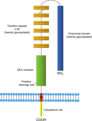
Mucin-16(MUC-16) also known as Ovarian cancer-related tumor marker CA125 is a protein that in humans is encoded by the MUC16 gene. MUC-16 is a member of the mucin family glycoproteins. MUC-16 has found application as a tumor marker or biomarker that may be elevated in the blood of some patients with specific types of cancers, most notably ovarian cancer, or other conditions that are benign.

<span class="mw-page-title-main">Hybridoma technology</span> Method for producing lots of identical antibodies

Hybridoma technology is a method for producing large numbers of identical antibodies. This process starts by injecting a mouse with an antigen that provokes an immune response. A type of white blood cell, the B cell, produces antibodies that bind to the injected antigen. These antibody producing B-cells are then harvested from the mouse and, in turn, fused with immortal myeloma cancer cells, to produce a hybrid cell line called a hybridoma, which has both the antibody-producing ability of the B-cell and the longevity and reproductivity of the myeloma. The hybridomas can be grown in culture, each culture starting with one viable hybridoma cell, producing cultures each of which consists of genetically identical hybridomas which produce one antibody per culture (monoclonal) rather than mixtures of different antibodies (polyclonal). The myeloma cell line that is used in this process is selected for its ability to grow in tissue culture and for an absence of antibody synthesis. In contrast to polyclonal antibodies, which are mixtures of many different antibody molecules, the monoclonal antibodies produced by each hybridoma line are all chemically identical.

<span class="mw-page-title-main">Thymidine kinase</span> Enzyme found in most living cells

Thymidine kinase is an enzyme, a phosphotransferase : 2'-deoxythymidine kinase, ATP-thymidine 5'-phosphotransferase, EC 2.7.1.21. It can be found in most living cells. It is present in two forms in mammalian cells, TK1 and TK2. Certain viruses also have genetic information for expression of viral thymidine kinases. Thymidine kinase catalyzes the reaction:

<span class="mw-page-title-main">Single-domain antibody</span> Antibody fragment

A single-domain antibody (sdAb), also known as a Nanobody, is an antibody fragment consisting of a single monomeric variable antibody domain. Like a whole antibody, it is able to bind selectively to a specific antigen. With a molecular weight of only 12–15 kDa, single-domain antibodies are much smaller than common antibodies which are composed of two heavy protein chains and two light chains, and even smaller than Fab fragments and single-chain variable fragments.

<span class="mw-page-title-main">Glypican</span>

Glypicans constitute one of the two major families of heparan sulfate proteoglycans, with the other major family being syndecans. Six glypicans have been identified in mammals, and are referred to as GPC1 through GPC6. In Drosophila two glypicans have been identified, and these are referred to as dally and dally-like. One glypican has been identified in C. elegans. Glypicans seem to play a vital role in developmental morphogenesis, and have been suggested as regulators for the Wnt and Hedgehog cell signaling pathways. They have additionally been suggested as regulators for fibroblast growth factor and bone morphogenic protein signaling.

<span class="mw-page-title-main">Ira Pastan</span> American scientist

Ira Pastan is an American scientist at the National Cancer Institute. He is a member of the National Academy of Sciences, a Fellow of the AAAS and the American Society of Microbiology. In 2009, he was awarded the prestigious International Antonio Feltrinelli Prize for Medicine. His wife, Linda Pastan, was an American poet.

<span class="mw-page-title-main">CD47</span> Protein-coding gene in humans

CD47 also known as integrin associated protein (IAP) is a transmembrane protein that in humans is encoded by the CD47 gene. CD47 belongs to the immunoglobulin superfamily and partners with membrane integrins and also binds the ligands thrombospondin-1 (TSP-1) and signal-regulatory protein alpha (SIRPα). CD-47 acts as a don't eat me signal to macrophages of the immune system which has made it a potential therapeutic target in some cancers, and more recently, for the treatment of pulmonary fibrosis.

<span class="mw-page-title-main">Glypican 3</span> Protein-coding gene in the species Homo sapiens

Glypican-3 is a protein that, in humans, is encoded by the GPC3 gene. The GPC3 gene is located on human X chromosome (Xq26) where the most common gene encodes a 70-kDa core protein with 580 amino acids. Three variants have been detected that encode alternatively spliced forms termed Isoforms 1 (NP_001158089), Isoform 3 (NP_001158090) and Isoform 4 (NP_001158091).

<span class="mw-page-title-main">CD276</span> Protein-coding gene in the species Homo sapiens

Cluster of Differentiation 276 (CD276) or B7 Homolog 3 (B7-H3) is a human protein encoded by the CD276 gene.

Tumor-associated glycoprotein 72 (TAG-72) is a glycoprotein found on the surface of many cancer cells, including ovary, breast, colon, lung, and pancreatic cancers. It is a mucin-like molecule with a molar mass of over 1000 kDa.

A431 cells are a model human cell line used in biomedical research.

CA 242 is a tumor marker for sialylated Lewis carbohydrates associated with adenocarcinomas and e-selectin-mediated metastatic risk. It is commonly tested along with CEA, CA19-9, and CA242 for detecting pancreatic cancer. The specificity of CA 242 is higher than similar markers. Current research dictates that diagnostic efficiency is highest when various tumor markers are tested for at once.

Folate targeting is a method utilized in biotechnology for drug delivery purposes. This Trojan Horse process, which was created by Drs. Christopher P. Leamon and Philip S. Low, involves the attachment of the vitamin, folate, to a molecule/drug to form a "folate conjugate". Based on the natural high affinity of folate for the folate receptor protein (FR), which is commonly expressed on the surface of many human cancers, folate-drug conjugates also bind tightly to the FR and trigger cellular uptake via endocytosis. Molecules as diverse as small radiodiagnostic imaging agents to large DNA plasmid formulations have successfully been delivered inside FR-positive cells and tissues.

A rabbit hybridoma is a hybrid cell line formed by the fusion of an antibody producing rabbit B cell with a cancerous B-cell (myeloma).

Amatuximab is a chimeric monoclonal antibody designed for the treatment of cancer. It was developed by Morphotek, Inc.

Urelumab is a fully human, non‐ligand binding, CD137 agonist immunoglobulin‐γ 4 (IgG4) monoclonal antibody. It was developed utilizing Medarex's UltiMAb(R) technology by Bristol-Myers Squibb for the treatment of cancer and solid tumors. Urelumab promotes anti-tumor immunity, or an immune response against tumor cells, via CD137 activation. The application of Urelumab has been limited due to the fact that it can cause severe liver toxicity.

<span class="mw-page-title-main">Cancer biomarker</span> Substance or process that is indicative of the presence of cancer in the body

A cancer biomarker refers to a substance or process that is indicative of the presence of cancer in the body. A biomarker may be a molecule secreted by a tumor or a specific response of the body to the presence of cancer. Genetic, epigenetic, proteomic, glycomic, and imaging biomarkers can be used for cancer diagnosis, prognosis, and epidemiology. Ideally, such biomarkers can be assayed in non-invasively collected biofluids like blood or serum.

GL-ONC1 is an investigational therapeutic product consisting of the clinical grade formulation of the laboratory strain GLV-1h68, an oncolytic virus developed by Genelux Corporation. GL-ONC1 is currently under evaluation in Phase I/II human clinical trials in the United States and Europe.

<span class="mw-page-title-main">PD-1 and PD-L1 inhibitors</span> Class of anticancer drugs

PD-1 inhibitors and PD-L1 inhibitors are a group of checkpoint inhibitor anticancer drugs that block the activity of PD-1 and PDL1 immune checkpoint proteins present on the surface of cells. Immune checkpoint inhibitors are emerging as a front-line treatment for several types of cancer.

References

  1. 1 2 3 GRCh38: Ensembl release 89: ENSG00000102854 - Ensembl, May 2017
  2. 1 2 3 GRCm38: Ensembl release 89: ENSMUSG00000063011 - Ensembl, May 2017
  3. "Human PubMed Reference:". National Center for Biotechnology Information, U.S. National Library of Medicine.
  4. "Mouse PubMed Reference:". National Center for Biotechnology Information, U.S. National Library of Medicine.
  5. Kojima T, Oh-eda M, Hattori K, Taniguchi Y, Tamura M, Ochi N, Yamaguchi N (September 1995). "Molecular cloning and expression of megakaryocyte potentiating factor cDNA". The Journal of Biological Chemistry. 270 (37): 21984–21990. doi: 10.1074/jbc.270.37.21984 . PMID   7665620.
  6. Chang K, Pastan I (January 1996). "Molecular cloning of mesothelin, a differentiation antigen present on mesothelium, mesotheliomas, and ovarian cancers". Proceedings of the National Academy of Sciences of the United States of America. 93 (1): 136–140. Bibcode:1996PNAS...93..136C. doi: 10.1073/pnas.93.1.136 . PMC   40193 . PMID   8552591.
  7. 1 2 3 Hassan R, Ho M (January 2008). "Mesothelin targeted cancer immunotherapy". European Journal of Cancer. 44 (1): 46–53. doi:10.1016/j.ejca.2007.08.028. PMC   2265108 . PMID   17945478.
  8. Chang K, Pai LH, Batra JK, Pastan I, Willingham MC (January 1992). "Characterization of the antigen (CAK1) recognized by monoclonal antibody K1 present on ovarian cancers and normal mesothelium". Cancer Research. 52 (1): 181–186. PMID   1727378.
  9. 1 2 Hassan R, Bera T, Pastan I (June 2004). "Mesothelin: a new target for immunotherapy". Clinical Cancer Research. 10 (12 Pt 1): 3937–3942. doi: 10.1158/1078-0432.CCR-03-0801 . PMID   15217923.
  10. Ho M (October 2011). "Advances in liver cancer antibody therapies: a focus on glypican-3 and mesothelin". BioDrugs. 25 (5): 275–284. doi:10.2165/11595360-000000000-00000. PMC   3198870 . PMID   21942912.
  11. Bera TK, Pastan I (April 2000). "Mesothelin is not required for normal mouse development or reproduction". Molecular and Cellular Biology. 20 (8): 2902–2906. doi:10.1128/MCB.20.8.2902-2906.2000. PMC   85523 . PMID   10733593.
  12. Ho M, Bera TK, Willingham MC, Onda M, Hassan R, FitzGerald D, Pastan I (March 2007). "Mesothelin expression in human lung cancer". Clinical Cancer Research. 13 (5): 1571–1575. doi: 10.1158/1078-0432.CCR-06-2161 . PMID   17332303.
  13. Yu L, Feng M, Kim H, Phung Y, Kleiner DE, Gores GJ, et al. (October 2010). "Mesothelin as a potential therapeutic target in human cholangiocarcinoma". Journal of Cancer. 1: 141–149. doi:10.7150/jca.1.141. PMC   2948219 . PMID   20922056.
  14. Rump A, Morikawa Y, Tanaka M, Minami S, Umesaki N, Takeuchi M, Miyajima A (March 2004). "Binding of ovarian cancer antigen CA125/MUC16 to mesothelin mediates cell adhesion". The Journal of Biological Chemistry. 279 (10): 9190–9198. doi: 10.1074/jbc.M312372200 . PMID   14676194.
  15. Kaneko O, Gong L, Zhang J, Hansen JK, Hassan R, Lee B, Ho M (February 2009). "A binding domain on mesothelin for CA125/MUC16". The Journal of Biological Chemistry. 284 (6): 3739–3749. doi: 10.1074/jbc.M806776200 . PMC   2635045 . PMID   19075018.
  16. 1 2 Zhang YF, Phung Y, Gao W, Kawa S, Hassan R, Pastan I, Ho M (May 2015). "New high affinity monoclonal antibodies recognize non-overlapping epitopes on mesothelin for monitoring and treating mesothelioma". Scientific Reports. 5: 9928. Bibcode:2015NatSR...5E9928Z. doi:10.1038/srep09928. PMC   4440525 . PMID   25996440.
  17. Grosso F, Mannucci M, Ugo F, Ferro P, Cassinari M, Vigani A, et al. (October 2021). "Pilot Study to Evaluate Serum Soluble Mesothelin-Related Peptide (SMRP) as Marker for Clinical Monitoring of Pleural Mesothelioma (PM): Correlation with Modified RECIST Score". Diagnostics. 11 (11): 2015. doi: 10.3390/diagnostics11112015 . PMC   8623660 . PMID   34829362.
  18. Hassan R, Ho M (January 2008). "Mesothelin targeted cancer immunotherapy". European Journal of Cancer. 44 (1): 46–53. doi:10.1016/j.ejca.2007.08.028. PMC   2265108 . PMID   17945478.
  19. Morello A, Sadelain M, Adusumilli PS (February 2016). "Mesothelin-Targeted CARs: Driving T Cells to Solid Tumors". Cancer Discovery. 6 (2): 133–146. doi:10.1158/2159-8290.CD-15-0583. PMC   4744527 . PMID   26503962.
  20. Scholler N, Fu N, Yang Y, Ye Z, Goodman GE, Hellström KE, Hellström I (September 1999). "Soluble member(s) of the mesothelin/megakaryocyte potentiating factor family are detectable in sera from patients with ovarian carcinoma". Proceedings of the National Academy of Sciences of the United States of America. 96 (20): 11531–11536. Bibcode:1999PNAS...9611531S. doi: 10.1073/pnas.96.20.11531 . PMC   18068 . PMID   10500211.
  21. Ho M, Onda M, Wang QC, Hassan R, Pastan I, Lively MO (September 2006). "Mesothelin is shed from tumor cells". Cancer Epidemiology, Biomarkers & Prevention. 15 (9): 1751. doi: 10.1158/1055-9965.EPI-06-0479 . PMID   16985043.
  22. Maeda M, Hino O (2006). "Blood tests for asbestos-related mesothelioma". Oncology. 71 (1–2): 26–31. doi:10.1159/000100446. PMID   17344668. S2CID   12953614.
  23. 1 2 Hassan R, Remaley AT, Sampson ML, Zhang J, Cox DD, Pingpank J, et al. (January 2006). "Detection and quantitation of serum mesothelin, a tumor marker for patients with mesothelioma and ovarian cancer". Clinical Cancer Research. 12 (2): 447–453. doi: 10.1158/1078-0432.CCR-05-1477 . PMID   16428485.
  24. 1 2 Iwahori K, Osaki T, Serada S, Fujimoto M, Suzuki H, Kishi Y, et al. (October 2008). "Megakaryocyte potentiating factor as a tumor marker of malignant pleural mesothelioma: evaluation in comparison with mesothelin". Lung Cancer. 62 (1): 45–54. doi:10.1016/j.lungcan.2008.02.012. PMID   18394747.
  25. Johnston FM, Tan MC, Tan BR, Porembka MR, Brunt EM, Linehan DC, et al. (November 2009). "Circulating mesothelin protein and cellular antimesothelin immunity in patients with pancreatic cancer". Clinical Cancer Research. 15 (21): 6511–6518. doi:10.1158/1078-0432.CCR-09-0565. PMC   2782601 . PMID   19843662.
  26. 1 2 Sharon E, Zhang J, Hollevoet K, Steinberg SM, Pastan I, Onda M, et al. (April 2012). "Serum mesothelin and megakaryocyte potentiating factor in pancreatic and biliary cancers". Clinical Chemistry and Laboratory Medicine. 50 (4): 721–725. doi:10.1515/CCLM.2011.816. PMC   6309905 . PMID   22149739.
  27. Ho M, Hassan R, Zhang J, Wang QC, Onda M, Bera T, Pastan I (May 2005). "Humoral immune response to mesothelin in mesothelioma and ovarian cancer patients". Clinical Cancer Research. 11 (10): 3814–3820. doi: 10.1158/1078-0432.CCR-04-2304 . PMID   15897581. S2CID   38327063.
  28. Ho M, Feng M, Fisher RJ, Rader C, Pastan I (May 2011). "A novel high-affinity human monoclonal antibody to mesothelin". International Journal of Cancer. 128 (9): 2020–2030. doi:10.1002/ijc.25557. PMC   2978266 . PMID   20635390.
  29. Tang Z, Feng M, Gao W, Phung Y, Chen W, Chaudhary A, et al. (April 2013). "A human single-domain antibody elicits potent antitumor activity by targeting an epitope in mesothelin close to the cancer cell surface". Molecular Cancer Therapeutics. 12 (4): 416–426. doi:10.1158/1535-7163.MCT-12-0731. PMC   3624043 . PMID   23371858.
  30. Zhang YF, Ho M (April 2017). "Humanization of rabbit monoclonal antibodies via grafting combined Kabat/IMGT/Paratome complementarity-determining regions: Rationale and examples". mAbs. 9 (3): 419–429. doi:10.1080/19420862.2017.1289302. PMC   5384799 . PMID   28165915.
  31. Zhang Z, Jiang D, Yang H, He Z, Liu X, Qin W, et al. (June 2019). "Modified CAR T cells targeting membrane-proximal epitope of mesothelin enhances the antitumor function against large solid tumor". Cell Death & Disease. 10 (7): 476. doi:10.1038/s41419-019-1711-1. PMC   6572851 . PMID   31209210.
  32. Tomar S, Zhang J, Khanal M, Hong J, Venugopalan A, Jiang Q, et al. (July 2022). "Development of Highly Effective Anti-Mesothelin hYP218 Chimeric Antigen Receptor T Cells With Increased Tumor Infiltration and Persistence for Treating Solid Tumors". Molecular Cancer Therapeutics. 21 (7): 1195–1206. doi:10.1158/1535-7163.MCT-22-0073. PMC   9256778 . PMID   35499461.

Further reading