Methanosarcinales S-layer Tile Protein

Last updated
Methanosarcinales S-layer Tile Protein
Identifiers
SymbolS-layer
Pfam PF07752
InterPro IPR006457

The Methanosarcinales S-layer Tile Protein (MSTP) is a protein family found almost exclusively in Methanomicrobia members of the order Methanosarcinales. Typically a tandem repeat of two DUF1608 domains are contained in a single MSTP protein chain and these proteins self-assemble into the protective proteinaceous surface layer (S-layer) structure that encompasses the cell. The S-layer, which is found in most Archaea, and in many bacteria, serves many crucial functions including protection from deleterious extracellular substances. [1]

Contents

Discovery of the Methanosarcinales S-layer

The first S-layers were discovered in bacteria in the 1950s [2] and the presence of S-layers in many Archaea was determined through microscopic (both light and electron) studies of Archaea. The presence of an S-layer in a member of the Methanosarcinales was determined in the 1980s by electron microscope (EM) studies examining the cell morphology of Methanosarcina mazei. [3] This, and other EM studies, confirmed that the cell envelope structure of the Methanosarcinales is composed of a cytoplasmic membrane (CM) with an additional barrier (the S-layer) external to the CM. Under conditions of low osmolarity the S-layer is extensively decorated with a polysaccharide, termed methanochondroitin, and the cells tend to grow in multicellular aggregates. [4] [5] Upon adaptation to high osmolarity conditions the cells disaggregate and grow as single cells that lack the methanochondroitin layer. [5]

Identification of the Methanosarcinales S-layer Protein

The identity of the proteins composing the S-layer of these organisms was subsequently determined by a proteomic approach. [6] The major S-layer proteins of M. acetivorans C2A and M. mazei Gö1 were determined to be MA0829 and MM1976, respectively. Additional proteins with similar characteristics as MA0829 and MM1976 were found to be present in the cell envelopes of these organisms in minor amounts. [6] The genomes of all Methanosarcina species examined thus far have 4-10 paralogous DUF1608 containing proteins. The major and minor S-layer proteins of M. acetivorans C2A and M. mazei Gö1 share many common features including: an N-terminal signal peptide, one or two protein domains of the DUF1608 protein family, [7] a negatively charged tether of ~70 amino acids, and a C-terminal transmembrane helix that likely anchors the S-layer to the CM.

Analysis of protein sequences has determined that members of the DUF1608 protein family contain 250-300 amino acids and are found only in Archaea. [8] With the exception of two halophilic archaea the DUF1608 domain is exclusive to the methanogenic Archaea of the order Methanosarcinales. [9] The DUF1608 has been assigned to the protein family (Pfam), pfam07752. [7]

Structure

The structure of the C-terminal DUF1608 domain of the Methanosarcinales S-layer Tile Protein MA0829. A, the C-terminal DUF1608 domain of MA0829 with domain I colored red, domain II in blue, and the connector subdomain in yellow. B, the crystallographic dimer of the MA0829 CTR. C, a trimer of crystallographic CTR homodimers. Ammonium citrate ligands from the crystallization solution bound at intermolecular interfaces are shown in space filling representation. D, the hexagonal tiles formed from trimeric CTR homodimer assemblies. The primary, asymmetric, and trimer pores are indicated. E, a cutaway side view of the model of the MSTP S-layer (the representation in D is rotated 90deg from the viewer). The position of the primary pore is indicated by arrowheads. Adapted from Methanosarcinales S-layer Tile Protein Figure.jpg
The structure of the C-terminal DUF1608 domain of the Methanosarcinales S-layer Tile Protein MA0829. A, the C-terminal DUF1608 domain of MA0829 with domain I colored red, domain II in blue, and the connector subdomain in yellow. B, the crystallographic dimer of the MA0829 CTR. C, a trimer of crystallographic CTR homodimers. Ammonium citrate ligands from the crystallization solution bound at intermolecular interfaces are shown in space filling representation. D, the hexagonal tiles formed from trimeric CTR homodimer assemblies. The primary, asymmetric, and trimer pores are indicated. E, a cutaway side view of the model of the MSTP S-layer (the representation in D is rotated 90° from the viewer). The position of the primary pore is indicated by arrowheads. Adapted from

The structure of one of the two tandem DUF1608 repeats that comprise the major MSTP of M. acetivorans (MA0829) has been determined at high resolution by X-ray crystallography. [10] The structure of the C-terminal DUF1608 tandem repeat (CTR) of MA0829 revealed that the DUF1608 protein domain is composed of two structurally similar β-sandwich domains connected by a short connector subdomain. The β-sandwich domains are structurally similar not only to each other but also to other proteins associated with envelope structures of disparate species including bacterial, fungal, and viral entities.

While the structure of only one of the two DUF1608 domains of the MA0829 protein was determined the structure of the full-length MA0829 tandem DUF1608 repeat protein (minus the N-terminal signal peptide and C-terminal tether and anchor) could be modeled by virtue of the MA0829 CTR forming the same crystallographic dimer in two different crystal forms. The high degree of primary amino acid sequence identity between the N- and C-terminal DUF1608 domains (79% identical and 87% similar) allowed the homology modeling of the N-terminal DUF1608 amino acid sequence onto one of the DUF1608 domains in the crystallographic DUF1608 CTR dimer thus providing the first high-resolution model of an Archaeal S-layer protein.

A model for the quaternary structure of the M. acetivorans S-layer was proposed based on packing of the MA0829 CTR in a hexagonal lattice in one of the two obtained crystal forms (Protein Data Bank accession number 3U2G). [10] The minimal building block of the S-layer sheet is a trimer of crystallographic MA0829 CTR dimers. Lateral translation of the trimeric unit creates a flat 2-dimensional sheet that has features consistent with the molecular properties of hexagonal archaeal S-layers. [11] [12] [13] The overall appearance of the S-layer resembles a honeycomb structure of hexagonal tiles with center to center spacing between the tiles of ~240 Å and a height of ~45 Å. [10]

Three different types of pores are present in the sheet with "Primary pores" situated on the six-fold symmetry axis and "Trimer pores" on the three-fold symmetry axis. [10] Asymmetric pores are located between the adjacent trimeric building blocks. The size of the pores are sufficiently large to allow the exchange of metabolites between the organism and the external environment. Whereas the protein constituents of lipid-based barriers, such as bacterial outer membranes, can be rapidly modified in response to physiological or environmental stimuli, the large pore sizes of the S-layer composed of MSTP protein subunits are presumably required to allow passage of molecules across a protective barrier whose molecular features are difficult to modify. An interesting feature of the model proposed for the M. acetivorans S-layer is the overwhelmingly negative charge of the surfaces of the S-layer including the pores. The S-layer thus presents a substantial size and charge barrier to the free passage of molecules across the S-layer. [10]

The two structures of the MA0829 CTR have been deposited in the Protein Data Bank: 3U2G is the accession code for the selenomethionine-labeled protein in the P622 space group and 3U2H is the accession code for the unlabeled protein structure in the C2 space group.

Applications

S-layers have many potential biotechnology applications. [14] The use of the high-resolution MA0829 structure to facilitate such studies is complicated by difficulties in reconstituting archaeal S-layers in vitro.

Related Research Articles

Cell nucleus Eukaryotic membrane-bounded organelle containing DNA

In cell biology, the nucleus is a membrane-bound organelle found in eukaryotic cells. Eukaryotes usually have a single nucleus, but a few cell types, such as mammalian red blood cells, have no nuclei, and a few others including osteoclasts have many. The main structures making up the nucleus are the nuclear envelope, a double membrane that encloses the entire organelle and isolates its contents from the cellular cytoplasm; and the nuclear matrix, a network within the nucleus that adds mechanical support, much like the cytoskeleton supports the cell as a whole.

Pyrrolysine

Pyrrolysine is an α-amino acid that is used in the biosynthesis of proteins in some methanogenic archaea and bacteria; it is not present in humans. It contains an α-amino group, a carboxylic acid group. Its pyrroline side-chain is similar to that of lysine in being basic and positively charged at neutral pH.

An S-layer is a part of the cell envelope found in almost all archaea, as well as in many types of bacteria. It consists of a monomolecular layer composed of identical proteins or glycoproteins. This structure is built via self-assembly and encloses the whole cell surface. Thus, the S-layer protein can represent up to 15% of the whole protein content of a cell. S-layer proteins are poorly conserved or not conserved at all, and can differ markedly even between related species. Depending on species, the S-layers have a thickness between 5 and 25 nm and possess identical pores with 2–8 nm in diameter.

Porin (protein)

Porins are beta barrel proteins that cross a cellular membrane and act as a pore, through which molecules can diffuse. Unlike other membrane transport proteins, porins are large enough to allow passive diffusion, i.e., they act as channels that are specific to different types of molecules. They are present in the outer membrane of gram-negative bacteria and some gram-positive mycobacteria, the outer membrane of mitochondria, and the outer chloroplast membrane.

M2 proton channel

The Matrix-2 (M2) protein is a proton-selective viroporin, integral in the viral envelope of the influenza A virus. The channel itself is a homotetramer, where the units are helices stabilized by two disulfide bonds, and is activated by low pH. The M2 protein is encoded on the seventh RNA segment together with the M1 protein. Proton conductance by the M2 protein in influenza A is essential for viral replication.
<i>Methanosarcina</i>

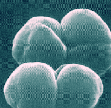
Methanosarcina is a genus of euryarchaeote archaea that produce methane. These single-celled organisms are known as anaerobic methanogens that produce methane using all three metabolic pathways for methanogenesis. They live in diverse environments where they can remain safe from the effects of oxygen, whether on the earth's surface, in groundwater, in deep sea vents, and in animal digestive tracts. Methanosarcina grow in colonies.
Diphtheria toxin

Diphtheria toxin is an exotoxin secreted by Corynebacterium, the pathogenic bacterium that causes diphtheria. The toxin gene is encoded by a prophage. The toxin causes the disease in humans by gaining entry into the cell cytoplasm and inhibiting protein synthesis.

Importin is a type of karyopherin that transports protein molecules from the cell's cytoplasm to the nucleus. It does so by binding to specific recognition sequences, called nuclear localization sequences (NLS).

Epithelial sodium channel

The epithelial sodium channel (short: ENaC, also: amiloride-sensitive sodium channel) is a membrane-bound ion channel that is selectively permeable to the ions of sodium (Na+) and that is assembled as a heterotrimer composed of three homologous subunits α or δ, β, and γ, These subunits are encoded by four genes: SCNN1A, SCNN1B, SCNN1G, and SCNN1D. It is involved primarily in the reabsorption of sodium ions at the collecting ducts of the kidney's nephrons.

DNA clamp

A DNA clamp, also known as a sliding clamp or β-clamp, is a protein complex that serves as a processivity-promoting factor in DNA replication. As a critical component of the DNA polymerase III holoenzyme, the clamp protein binds DNA polymerase and prevents this enzyme from dissociating from the template DNA strand. The clamp-polymerase protein–protein interactions are stronger and more specific than the direct interactions between the polymerase and the template DNA strand; because one of the rate-limiting steps in the DNA synthesis reaction is the association of the polymerase with the DNA template, the presence of the sliding clamp dramatically increases the number of nucleotides that the polymerase can add to the growing strand per association event. The presence of the DNA clamp can increase the rate of DNA synthesis up to 1,000-fold compared with a nonprocessive polymerase.

Colicin

A colicin is a type of bacteriocin produced by and toxic to some strains of Escherichia coli. Colicins are released into the environment to reduce competition from other bacterial strains. Colicins bind to outer membrane receptors, using them to translocate to the cytoplasm or cytoplasmic membrane, where they exert their cytotoxic effect, including depolarisation of the cytoplasmic membrane, DNase activity, RNase activity, or inhibition of murein synthesis.

Protein domain Conserved part of a protein

A protein domain is a conserved part of a given protein sequence and tertiary structure that can evolve, function, and exist independently of the rest of the protein chain. Each domain forms a compact three-dimensional structure and often can be independently stable and folded. Many proteins consist of several structural domains. One domain may appear in a variety of different proteins. Molecular evolution uses domains as building blocks and these may be recombined in different arrangements to create proteins with different functions. In general, domains vary in length from between about 50 amino acids up to 250 amino acids in length. The shortest domains, such as zinc fingers, are stabilized by metal ions or disulfide bridges. Domains often form functional units, such as the calcium-binding EF hand domain of calmodulin. Because they are independently stable, domains can be "swapped" by genetic engineering between one protein and another to make chimeric proteins.
Nuclear envelope

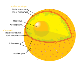
The nuclear envelope, also known as the nuclear membrane, is made up of two lipid bilayer membranes which in eukaryotic cells surrounds the nucleus, which encases the genetic material.

A domain of unknown function (DUF) is a protein domain that has no characterised function. These families have been collected together in the Pfam database using the prefix DUF followed by a number, with examples being DUF2992 and DUF1220. As of 2019, there are almost 4,000 DUF families within the Pfam database representing over 22% of known families. Some DUFs are not named using the nomenclature due to popular usage but are nevertheless DUFs.

CHAP domain

In molecular biology, the CHAP domain is a region between 110 and 140 amino acids that is found in proteins from bacteria, bacteriophages, archaea and eukaryotes of the family Trypanosomidae. The domain is named after the acronym cysteine, histidine-dependent amidohydrolases/peptidases. Many of these proteins are uncharacterised, but it has been proposed that they may function mainly in peptidoglycan hydrolysis. The CHAP domain is found in a wide range of protein architectures; it is commonly associated with bacterial type SH3 domains and with several families of amidase domains. It has been suggested that CHAP domain containing proteins utilise a catalytic cysteine residue in a nucleophilic-attack mechanism.
Tubulin domain

Tubulin/FtsZ family, GTPase domain is an evolutionary conserved protein domain.

An archaellum is a unique whip-like structure on the cell surface of many archaea. The name was proposed in 2012 following studies that showed it to be evolutionarily and structurally different from the bacterial and eukaryotic flagella. The archaellum is functionally the same – it can be rotated and is used to swim in liquid environments. The archaellum was found to be structurally similar to the type IV pilus.

YjeF N terminal protein domain

In molecular biology, the YjeF N terminal is a protein domain found in the N-terminal of the protein, EDC3. The YjeF N-terminal domains occur either as single proteins or fusions with other domains and are commonly associated with enzymes. They help assemble the processing body (P-body) in preparation for mRNAdecay. Structural homology indicated it may have some similarity to the enzyme family, hydrolase.

<i>Methanosarcina barkeri</i>

Methanosarcina barkeri is the most fundamental species of the genus Methanosarcina, and their properties apply generally to the genus Methanosarcina. Methanosarcina barkeri can produce methane anaerobically through different metabolic pathways. M. barkeri can subsume a variety of molecules for ATP production, including methanol, acetate, methylamines, and different forms of hydrogen and carbon dioxide. Although it is a slow developer and is sensitive to change in environmental conditions, M. barkeri is able to grow in a variety of different substrates, adding to its appeal for genetic analysis. Additionally, M. barkeri is the first organism in which the amino acid pyrrolysine was found. Furthermore, two strains of M. barkeri, M. b. Fusaro and M. b. MS have been identified to possess an F-type ATPase along with an A-type ATPase.

Members of the H+, Na+-translocating Pyrophosphatase (M+-PPase) Family (TC# 3.A.10) are found in the vacuolar (tonoplast) membranes of higher plants, algae, and protozoa, and in both bacteria and archaea. They are therefore ancient enzymes.

References

  1. König H (1998). "Archaeobacterial cell envelopes". Can J Microbiol. 34 (4): 395–406. doi:10.1139/m88-071.
  2. Houwink, AL (1953). "A macromolecular mono-layer in the cell wall of Spirillum spec". Biochim Biophys Acta. 10 (3): 360–6. doi:10.1016/0006-3002(53)90266-2. PMID   13058992.
  3. Aldrich HC, Robinson RW, Williams DS (May 1986). "Ultrastructure of Methanosarcina mazei". Systematic and Applied Microbiology. 7 (2–3): 314–9. doi:10.1016/S0723-2020(86)80025-X.
  4. Kreisl P, Kandler O (May 1986). "Chemical structure of the cell wall polymer of methanosarcina". Systematic and Applied Microbiology. 7 (2–3): 293–9. doi:10.1016/S0723-2020(86)80022-4.
  5. 1 2 Sowers KR, Boone JE, Gunsalus RP (1993). "Disaggregation of Methanosarcina spp. and Growth as Single Cells at Elevated Osmolarity". Appl Environ Microbiol. 59 (11): 3832–9. doi:10.1128/AEM.59.11.3832-3839.1993. PMC   182538 . PMID   16349092.
  6. 1 2 Francoleon DR, Boontheung P, Yang Y, Kin U, Ytterberg AJ, Denny PA, Denny PC, Loo JA, Gunsalus RP, Loo RR (April 2009). "S-layer, surface-accessible, and concanavalin A binding proteins of Methanosarcina acetivorans and Methanosarcina mazei". J Proteome Res. 8 (4): 1972–82. doi:10.1021/pr800923e. PMC   2666069 . PMID   19228054.
  7. 1 2 "PF07752". PFAM. Sanger Institute. Archived from the original on 15 October 2013. Retrieved 11 February 2013.
  8. Bateman A, Coin L, Durbin R, Finn RD, Hollich V, Griffiths-Jones S, Khanna A, Marshall M, Moxon S, Sonnhammer EL, Studholme DJ, Yeats C, Eddy SR (2004). "The Pfam protein families database". Nucleic Acids Res. 32 (Database issue): D138–41. doi:10.1093/nar/gkh121. PMC   308855 . PMID   14681378.
  9. Rohlin L, Leon DR, Kim U, Loo JA, Ogorzalek Loo RR, Gunsalus RP (2012). "Identification of the major expressed S-layer and cell surface-layer-related proteins in the model methanogenic archaea: Methanosarcina barkeri Fusaro and Methanosarcina acetivorans C2A". Archaea. 2012: 1–10. doi:10.1155/2012/873589. PMC   3361143 . PMID   22666082. 873589.
  10. 1 2 3 4 5 6 Arbing MA, Chan S, Shin A, Phan T, Ahn CJ, Rohlin L, Gunsalus RP (2012). "Structure of the surface layer of the methanogenic archaean Methanosarcina acetivorans". Proc Natl Acad Sci U S A. 109 (29): 11812–7. Bibcode:2012PNAS..10911812A. doi:10.1073/pnas.1120595109. PMC   3406845 . PMID   22753492.
  11. Sleytr UB, Messner P (1983). "Crystalline surface layers on bacteria". Annu Rev Microbiol. 37: 311–39. doi:10.1146/annurev.mi.37.100183.001523. PMID   6416145.
  12. Cheong GW, Guckenberger R, Fuchs KH, Gross H, Baumeister W (September 1993). "The structure of the surface layer of Methanoplanus limicola obtained by a combined electron microscopy and scanning tunneling microscopy approach". J Struct Biol. 111 (2): 125–34. doi:10.1006/jsbi.1993.1043.
  13. Trachtenberg S, Pinnick B, Kessel M (2000). "The cell surface glycoprotein layer of the extreme halophile Halobacterium salinarum and its relation to Haloferax volcanii: cryo-electron tomography of freeze-substituted cells and projection studies of negatively stained envelopes". J Struct Biol. 130 (1): 10–26. doi:10.1006/jsbi.2000.4215. PMID   10806087.
  14. Sleytr UB, Egelseer EM, Ilk N, Pum D, Schuster B (2007). "S-Layers as a basic building block in a molecular construction kit". FEBS J. 274 (2): 323–34. doi: 10.1111/j.1742-4658.2006.05606.x . PMID   17181542.