Transcription is the process of copying a segment of DNA into RNA. The segments of DNA transcribed into RNA molecules that can encode proteins are said to produce messenger RNA (mRNA). Other segments of DNA are copied into RNA molecules called non-coding RNAs (ncRNAs). mRNA comprises only 1–3% of total RNA samples. Less than 2% of the human genome can be transcribed into mRNA, while at least 80% of mammalian genomic DNA can be actively transcribed, with the majority of this 80% considered to be ncRNA.
A regulatory sequence is a segment of a nucleic acid molecule which is capable of increasing or decreasing the expression of specific genes within an organism. Regulation of gene expression is an essential feature of all living organisms and viruses.

Histone methyltransferases (HMT) are histone-modifying enzymes, that catalyze the transfer of one, two, or three methyl groups to lysine and arginine residues of histone proteins. The attachment of methyl groups occurs predominantly at specific lysine or arginine residues on histones H3 and H4. Two major types of histone methyltranferases exist, lysine-specific and arginine-specific. In both types of histone methyltransferases, S-Adenosyl methionine (SAM) serves as a cofactor and methyl donor group.
The genomic DNA of eukaryotes associates with histones to form chromatin. The level of chromatin compaction depends heavily on histone methylation and other post-translational modifications of histones. Histone methylation is a principal epigenetic modification of chromatin that determines gene expression, genomic stability, stem cell maturation, cell lineage development, genetic imprinting, DNA methylation, and cell mitosis.
In molecular biology and genetics, transcriptional regulation is the means by which a cell regulates the conversion of DNA to RNA (transcription), thereby orchestrating gene activity. A single gene can be regulated in a range of ways, from altering the number of copies of RNA that are transcribed, to the temporal control of when the gene is transcribed. This control allows the cell or organism to respond to a variety of intra- and extracellular signals and thus mount a response. Some examples of this include producing the mRNA that encode enzymes to adapt to a change in a food source, producing the gene products involved in cell cycle specific activities, and producing the gene products responsible for cellular differentiation in multicellular eukaryotes, as studied in evolutionary developmental biology.

DNA methylation is a biological process by which methyl groups are added to the DNA molecule. Methylation can change the activity of a DNA segment without changing the sequence. When located in a gene promoter, DNA methylation typically acts to repress gene transcription. In mammals, DNA methylation is essential for normal development and is associated with a number of key processes including genomic imprinting, X-chromosome inactivation, repression of transposable elements, aging, and carcinogenesis.

A chromomere, also known as an idiomere, is one of the serially aligned beads or granules of a eukaryotic chromosome, resulting from local coiling of a continuous DNA thread. Chromomeres are regions of chromatin that have been compacted through localized contraction. In areas of chromatin with the absence of transcription, condensing of DNA and protein complexes will result in the formation of chromomeres. It is visible on a chromosome during the prophase of meiosis and mitosis. Giant banded (Polytene) chromosomes resulting from the replication of the chromosomes and the synapsis of homologs without cell division is a process called endomitosis. These chromosomes consist of more than 1000 copies of the same chromatid that are aligned and produce alternating dark and light bands when stained. The dark bands are the chromomere.

MECP2 is a gene that encodes the protein MECP2. MECP2 appears to be essential for the normal function of nerve cells. The protein seems to be particularly important for mature nerve cells, where it is present in high levels. The MECP2 protein is likely to be involved in turning off several other genes. This prevents the genes from making proteins when they are not needed. Recent work has shown that MECP2 can also activate other genes. The MECP2 gene is located on the long (q) arm of the X chromosome in band 28 ("Xq28"), from base pair 152,808,110 to base pair 152,878,611.
Histone methylation is a process by which methyl groups are transferred to amino acids of histone proteins that make up nucleosomes, which the DNA double helix wraps around to form chromosomes. Methylation of histones can either increase or decrease transcription of genes, depending on which amino acids in the histones are methylated, and how many methyl groups are attached. Methylation events that weaken chemical attractions between histone tails and DNA increase transcription because they enable the DNA to uncoil from nucleosomes so that transcription factor proteins and RNA polymerase can access the DNA. This process is critical for the regulation of gene expression that allows different cells to express different genes.

Methyltransferases are a large group of enzymes that all methylate their substrates but can be split into several subclasses based on their structural features. The most common class of methyltransferases is class I, all of which contain a Rossmann fold for binding S-Adenosyl methionine (SAM). Class II methyltransferases contain a SET domain, which are exemplified by SET domain histone methyltransferases, and class III methyltransferases, which are membrane associated. Methyltransferases can also be grouped as different types utilizing different substrates in methyl transfer reactions. These types include protein methyltransferases, DNA/RNA methyltransferases, natural product methyltransferases, and non-SAM dependent methyltransferases. SAM is the classical methyl donor for methyltransferases, however, examples of other methyl donors are seen in nature. The general mechanism for methyl transfer is a SN2-like nucleophilic attack where the methionine sulfur serves as the leaving group and the methyl group attached to it acts as the electrophile that transfers the methyl group to the enzyme substrate. SAM is converted to S-Adenosyl homocysteine (SAH) during this process. The breaking of the SAM-methyl bond and the formation of the substrate-methyl bond happen nearly simultaneously. These enzymatic reactions are found in many pathways and are implicated in genetic diseases, cancer, and metabolic diseases. Another type of methyl transfer is the radical S-Adenosyl methionine (SAM) which is the methylation of unactivated carbon atoms in primary metabolites, proteins, lipids, and RNA.

Methyl-CpG-binding domain protein 1 is a protein that in humans is encoded by the MBD1 gene. The protein encoded by MBD1 binds to methylated sequences in DNA, and thereby influences transcription. It binds to a variety of methylated sequences, and appears to mediate repression of gene expression. It has been shown to play a role in chromatin modification through interaction with the histone H3K9 methyltransferase SETDB1. H3K9me3 is a repressive modification.
The family of heterochromatin protein 1 (HP1) consists of highly conserved proteins, which have important functions in the cell nucleus. These functions include gene repression by heterochromatin formation, transcriptional activation, regulation of binding of cohesion complexes to centromeres, sequestration of genes to the nuclear periphery, transcriptional arrest, maintenance of heterochromatin integrity, gene repression at the single nucleosome level, gene repression by heterochromatization of euchromatin, and DNA repair. HP1 proteins are fundamental units of heterochromatin packaging that are enriched at the centromeres and telomeres of nearly all eukaryotic chromosomes with the notable exception of budding yeast, in which a yeast-specific silencing complex of SIR proteins serve a similar function. Members of the HP1 family are characterized by an N-terminal chromodomain and a C-terminal chromoshadow domain, separated by a hinge region. HP1 is also found at some euchromatic sites, where its binding can correlate with either gene repression or gene activation. HP1 was originally discovered by Tharappel C James and Sarah Elgin in 1986 as a factor in the phenomenon known as position effect variegation in Drosophila melanogaster.

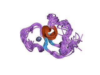
The PHD finger was discovered in 1993 as a Cys4-His-Cys3 motif in the plant homeodomain proteins HAT3.1 in Arabidopsis and maize ZmHox1a. The PHD zinc finger motif resembles the metal binding RING domain (Cys3-His-Cys4) and FYVE domain. It occurs as a single finger, but often in clusters of two or three, and it also occurs together with other domains, such as the chromodomain and the bromodomain.

Methyl-CpG-binding domain protein 2 is a protein that in humans is encoded by the MBD2 gene.

Methyl-CpG-binding domain protein 3 is a protein that in humans is encoded by the MBD3 gene.

Methyl-CpG-binding domain protein 4 is a protein that in humans is encoded by the MBD4 gene.

Transcriptional regulator Kaiso is a protein that in humans is encoded by the ZBTB33 gene. This gene encodes a transcriptional regulator with bimodal DNA-binding specificity, which binds to methylated CGCG and also to the non-methylated consensus KAISO-binding site TCCTGCNA. The protein contains an N-terminal POZ/BTB domain and 3 C-terminal zinc finger motifs. It recruits the N-CoR repressor complex to promote histone deacetylation and the formation of repressive chromatin structures in target gene promoters. It may contribute to the repression of target genes of the Wnt signaling pathway, and may also activate transcription of a subset of target genes by the recruitment of catenin delta-2 (CTNND2). Its interaction with catenin delta-1 (CTNND1) inhibits binding to both methylated and non-methylated DNA. It also interacts directly with the nuclear import receptor Importin-α2, which may mediate nuclear import of this protein. Alternatively spliced transcript variants encoding the same protein have been identified.

Chromobox protein homolog 5 is a protein that in humans is encoded by the CBX5 gene. It is a highly conserved, non-histone protein part of the heterochromatin family. The protein itself is more commonly called HP1α. Heterochromatin protein-1 (HP1) has an N-terminal domain that acts on methylated lysines residues leading to epigenetic repression. The C-terminal of this protein has a chromo shadow-domain (CSD) that is responsible for homodimerizing, as well as interacting with a variety of chromatin-associated, non-histone proteins.

In molecular biology, a Tudor domain is a conserved protein structural domain originally identified in the Tudor protein encoded in Drosophila. The Tudor gene was found in a Drosophila screen for maternal factors that regulate embryonic development or fertility. Mutations here are lethal for offspring, inspiring the name Tudor, as a reference to the Tudor King Henry VIII and the several miscarriages experienced by his wives.

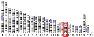
Euchromatic histone-lysine N-methyltransferase 1, also known as G9a-like protein (GLP), is a protein that in humans is encoded by the EHMT1 gene.

Rob Klose is a Canadian researcher and Professor of Genetics at the Department of Biochemistry, University of Oxford. His research investigates how chromatin-based and epigenetic mechanisms contribute to the ways in which gene expression is regulated.


