
A cell wall is a structural layer that surrounds some cell types, found immediately outside the cell membrane. It can be tough, flexible, and sometimes rigid. Primarily, it provides the cell with structural support, shape, protection, and functions as a selective barrier. Another vital role of the cell wall is to help the cell withstand osmotic pressure and mechanical stress. While absent in many eukaryotes, including animals, cell walls are prevalent in other organisms such as fungi, algae and plants, and are commonly found in most prokaryotes, with the exception of mollicute bacteria.
Developmental biology is the study of the process by which animals and plants grow and develop. Developmental biology also encompasses the biology of regeneration, asexual reproduction, metamorphosis, and the growth and differentiation of stem cells in the adult organism.

Plant cells are the cells present in green plants, photosynthetic eukaryotes of the kingdom Plantae. Their distinctive features include primary cell walls containing cellulose, hemicelluloses and pectin, the presence of plastids with the capability to perform photosynthesis and store starch, a large vacuole that regulates turgor pressure, the absence of flagella or centrioles, except in the gametes, and a unique method of cell division involving the formation of a cell plate or phragmoplast that separates the new daughter cells.
Sex is the biological trait that determines whether a sexually reproducing organism produces male or female gametes. During sexual reproduction, a male and a female gamete fuse to form a zygote, which develops into an offspring that inherits traits from each parent. By convention, organisms that produce smaller, more mobile gametes are called male, while organisms that produce larger, non-mobile gametes are called female. An organism that produces both types of gamete is hermaphrodite.

Xylem is one of the two types of transport tissue in vascular plants, the other being phloem; both of these are part of the vascular bundle. The basic function of the xylem is to transport water upward from the roots to parts of the plants such as stems and leaves, but it also transports nutrients. The word xylem is derived from the Ancient Greek word ξύλον (xylon), meaning "wood"; the best-known xylem tissue is wood, though it is found throughout a plant. The term was introduced by Carl Nägeli in 1858.
Phloem is the living tissue in vascular plants that transports the soluble organic compounds made during photosynthesis and known as photosynthates, in particular the sugar sucrose, to the rest of the plant. This transport process is called translocation. In trees, the phloem is the innermost layer of the bark, hence the name, derived from the Ancient Greek word φλοιός (phloiós), meaning "bark". The term was introduced by Carl Nägeli in 1858. Different types of phloem can be distinguished. The early phloem formed in the growth apices is called protophloem. Protophloem eventually becomes obliterated once it connects to the durable phloem in mature organs, the metaphloem. Further, secondary phloem is formed during the thickening of stem structures.

Vascular plants, also called tracheophytes or collectively tracheophyta, are plants that have lignified tissues for conducting water and minerals throughout the plant. They also have a specialized non-lignified tissue to conduct products of photosynthesis. The group includes most land plants other than mosses.

Bark is the outermost layer of stems and roots of woody plants. Plants with bark include trees, woody vines, and shrubs. Bark refers to all the tissues outside the vascular cambium and is a nontechnical term. It overlays the wood and consists of the inner bark and the outer bark. The inner bark, which in older stems is living tissue, includes the innermost layer of the periderm. The outer bark on older stems includes the dead tissue on the surface of the stems, along with parts of the outermost periderm and all the tissues on the outer side of the periderm. The outer bark on trees which lies external to the living periderm is also called the rhytidome.

Cytokinesis is the part of the cell division process and part of mitosis during which the cytoplasm of a single eukaryotic cell divides into two daughter cells. Cytoplasmic division begins during or after the late stages of nuclear division in mitosis and meiosis. During cytokinesis the spindle apparatus partitions and transports duplicated chromatids into the cytoplasm of the separating daughter cells. It thereby ensures that chromosome number and complement are maintained from one generation to the next and that, except in special cases, the daughter cells will be functional copies of the parent cell. After the completion of the telophase and cytokinesis, each daughter cell enters the interphase of the cell cycle.
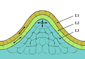
In cell biology, the meristem is a type of tissue found in plants. It consists of undifferentiated cells capable of cell division. Cells in the meristem can develop into all the other tissues and organs that occur in plants. These cells continue to divide until they become differentiated and lose the ability to divide.

The ectoderm is one of the three primary germ layers formed in early embryonic development. It is the outermost layer, and is superficial to the mesoderm and endoderm. It emerges and originates from the outer layer of germ cells. The word ectoderm comes from the Greek ektos meaning "outside", and derma meaning "skin".

The Casparian strip is a band-like thickening in the center of the root endodermis of vascular plants. The composition of the region is mainly suberin, lignin and some structural proteins, which are capable of reducing the diffusive apoplastic flow of water and solutes into the stele and its width varies between species. The Casparian strip is impervious to water so can control the transportation of water and inorganic salts between the cortex and the vascular bundle, preventing water and inorganic salts from being transported to the stele through the apoplast, so that it must enter the cell membrane and move to the stele through the symplastic pathway, blocking the internal and external objects of the cell. The function of mass transportation are similar to that of animal tissues.. The development of the Casparian strip is regulated by transcription factors such as SHORT-ROOT (SHR), SCARECROW (SCR) and MYB36, as well as polypeptide hormone synthesised by midcolumn cells.
A lamella is a small plate or flake, from the Latin, and may also be used to refer to collections of fine sheets of material held adjacent to one another, in a gill-shaped structure, often with fluid in between though sometimes simply a set of 'welded' plates. The term is used in biological contexts to describe thin membranes of plates of tissue. In context of materials science, the microscopic structures in bone and nacre are called lamellae. Moreover, the term lamella is often used as a way to describe crystal structure of some materials.

Plasmodesmata are microscopic channels which traverse the cell walls of plant cells and some algal cells, enabling transport and communication between them. Plasmodesmata evolved independently in several lineages, and species that have these structures include members of the Charophyceae, Charales, Coleochaetales and Phaeophyceae, as well as all embryophytes, better known as land plants. Unlike animal cells, almost every plant cell is surrounded by a polysaccharide cell wall. Neighbouring plant cells are therefore separated by a pair of cell walls and the intervening middle lamella, forming an extracellular domain known as the apoplast. Although cell walls are permeable to small soluble proteins and other solutes, plasmodesmata enable direct, regulated, symplastic transport of substances between cells. There are two forms of plasmodesmata: primary plasmodesmata, which are formed during cell division, and secondary plasmodesmata, which can form between mature cells.

The ground tissue of plants includes all tissues that are neither dermal nor vascular. It can be divided into three types based on the nature of the cell walls. This tissue system is present between the dermal tissue and forms the main bulk of the plant body.
- Parenchyma cells have thin primary walls and usually remain alive after they become mature. Parenchyma forms the "filler" tissue in the soft parts of plants, and is usually present in cortex, pericycle, pith, and medullary rays in primary stem and root.
- Collenchyma cells have thin primary walls with some areas of secondary thickening. Collenchyma provides extra mechanical and structural support, particularly in regions of new growth.
- Sclerenchyma cells have thick lignified secondary walls and often die when mature. Sclerenchyma provides the main structural support to the plant.
- Aerenchyma cells are found in aquatic plants. They are also known to be parenchyma cells with large air cavities surrounded by irregular cells which form columns called trabeculae
A lamella in biology refers to a thin layer, membrane or plate of tissue. This is a very broad definition, and can refer to many different structures. Any thin layer of organic tissue can be called a lamella and there is a wide array of functions an individual layer can serve. For example, an intercellular lipid lamella is formed when lamellar disks fuse to form a lamellar sheet. It is believed that these disks are formed from vesicles, giving the lamellar sheet a lipid bilayer that plays a role in water diffusion.

Mesenchyme is a type of loosely organized animal embryonic connective tissue of undifferentiated cells that give rise to most tissues, such as skin, blood or bone. The interactions between mesenchyme and epithelium help to form nearly every organ in the developing embryo.
The secondary cell wall is a structure found in many plant cells, located between the primary cell wall and the plasma membrane. The cell starts producing the secondary cell wall after the primary cell wall is complete and the cell has stopped expanding. It is most prevalent in the Ground tissue found in vascular plants, with Collenchyma having little to no lignin, and Sclerenchyma having lignified secondary cells walls.

Zygnematophyceae is a class of green algae in the paraphylum streptophyte algae, also referred to as Charophyta, consisting of more than 4000 described species. The Zygnematophyceae are the sister clade of the Embryophyta.

Pits are relatively thinner portions of the cell wall that adjacent cells can communicate or exchange fluid through. Pits are characteristic of cell walls with secondary layers. Generally each pit has a complementary pit opposite of it in the neighboring cell. These complementary pits are called "pit pairs".

