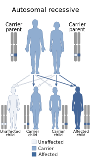
Muscular dystrophies (MD) are a genetically and clinically heterogeneous group of rare neuromuscular diseases that cause progressive weakness and breakdown of skeletal muscles over time. The disorders differ as to which muscles are primarily affected, the degree of weakness, how fast they worsen, and when symptoms begin. Some types are also associated with problems in other organs.
This is a partial list of human eye diseases and disorders.
The Muscular Dystrophy Association (MDA) is an American 501(c)(3) umbrella organization that works to support people with neuromuscular diseases. Founded in 1950 by Paul Cohen, who lived with muscular dystrophy, it works to combat neuromuscular disorders by funding research, providing medical and community services and educating health professionals and the general public and contributed more than $1 billion toward researching therapies and cures, helping to fund the identification of the dystrophin gene responsible for Duchenne muscular dystrophy as well as prospective treatments.

A cone dystrophy is an inherited ocular disorder characterized by the loss of cone cells, the photoreceptors responsible for both central and color vision.

Walker–Warburg syndrome (WWS), also called Warburg syndrome, Chemke syndrome, HARD syndrome, Pagon syndrome, cerebroocular dysgenesis (COD) or cerebroocular dysplasia-muscular dystrophy syndrome (COD-MD), is a rare form of autosomal recessive congenital muscular dystrophy. It is associated with brain and eye abnormalities. This condition has a worldwide distribution. The overall incidence is unknown but a survey in North-eastern Italy has reported an incidence rate of 1.2 per 100,000 live births. It is the most severe form of congenital muscular dystrophy with most children dying before the age of three years.

Corneal dystrophy is a group of rare hereditary disorders characterised by bilateral abnormal deposition of substances in the transparent front part of the eye called the cornea.

Vitelliform macular dystrophy is an irregular autosomal dominant eye disorder which can cause progressive vision loss. This disorder affects the retina, specifically cells in a small area near the center of the retina called the macula. The macula is responsible for sharp central vision, which is needed for detailed tasks such as reading, driving, and recognizing faces. The condition is characterized by yellow, slightly elevated, round structures similar to the yolk of an egg.

Oguchi disease is an autosomal recessive form of congenital stationary night blindness associated with fundus discoloration and abnormally slow dark adaptation.

Bestrophin-1 (Best1) is a protein that, in humans, is encoded by the BEST1 gene.

EEM syndrome is an autosomal recessive congenital malformation disorder affecting tissues associated with the ectoderm, and also the hands, feet and eyes.

A vision disorder is an impairment of the sense of vision.

Macular telangiectasia is a condition of the retina, the light-sensing tissue at the back of the eye that causes gradual deterioration of central vision, interfering with tasks such as reading and driving.
The Macular Society is a UK charity for anyone affected by central vision loss.
Macular dystrophy may refer to any of these eye diseases:

Hypotrichosis with juvenile macular dystrophy is an extremely rare congenital disease characterized by sparse hair growth (hypotrichosis) from birth and progressive macular corneal dystrophy.

Spastic ataxia-corneal dystrophy syndrome is an autosomally resessive disease. It has been found in an inbred Bedouin family. It was first described in 1986. A member of the family who was first diagnosed with this disease also had Bartter syndrome. It was concluded by its first descriptors Mousa-Al et al. that the disease is different from a disease known as corneal-cerebellar syndrome that had been found in 1985.
Occult macular dystrophy (OMD) is a rare inherited degradation of the retina, characterized by progressive loss of function in the most sensitive part of the central retina (macula), the location of the highest concentration of light-sensitive cells (photoreceptors) but presenting no visible abnormality. "Occult" refers to the degradation in the fundus being difficult to discern. The disorder is called "dystrophy" instead of "degradation" to distinguish its genetic origin from other causes, such as age. OMD was first reported by Y. Miyake et al. in 1989.
Macular scarring is formation of the fibrous tissue in place of the normal retinal tissue on the macular area of the retina which provides the sharpest vision in the eyes. It is usually a result of an inflammatory or infectious process.. Some other examples of the etiology include macular pucker, macular hole, and age-related macular degeneration. Macular dystrophies and telangiectasia are among the less common causes.

The human cornea is a transparent membrane which allows light to pass through it. The word corneal opacification literally means loss of normal transparency of cornea. The term corneal opacity is used particularly for the loss of transparency of cornea due to scarring. Transparency of the cornea is dependent on the uniform diameter and the regular spacing and arrangement of the collagen fibrils within the stroma. Alterations in the spacing of collagen fibrils in a variety of conditions including corneal edema, scars, and macular corneal dystrophy is clinically manifested as corneal opacity. The term "corneal blindness" is commonly used to describe blindness due to corneal opacity.


