Related Research Articles

Ophthalmology is a clinical and surgical specialty within medicine that deals with the diagnosis and treatment of eye disorders. A former term is oculism.

Keratoconus (KC) is a disorder of the eye that results in progressive thinning of the cornea. This may result in blurry vision, double vision, nearsightedness, irregular astigmatism, and light sensitivity leading to poor quality-of-life. Usually both eyes are affected. In more severe cases a scarring or a circle may be seen within the cornea.

Laser-Assisted in Situ Keratomileusis (LASIK), commonly referred to as laser eye surgery or laser vision correction, is a type of refractive surgery for the correction of myopia, hyperopia, and an actual cure for astigmatism, since it is in the cornea. LASIK surgery is performed by an ophthalmologist who uses a laser or microkeratome to reshape the eye's cornea in order to improve visual acuity.

Eye surgery, also known as ophthalmic surgery or ocular surgery, is surgery performed on the eye or its adnexa. Eye surgery is part of ophthalmology and is performed by an ophthalmologist or eye surgeon. The eye is a fragile organ, and requires due care before, during, and after a surgical procedure to minimize or prevent further damage. An eye surgeon is responsible for selecting the appropriate surgical procedure for the patient, and for taking the necessary safety precautions. Mentions of eye surgery can be found in several ancient texts dating back as early as 1800 BC, with cataract treatment starting in the fifth century BC. It continues to be a widely practiced class of surgery, with various techniques having been developed for treating eye problems.

Radial keratotomy (RK) is a refractive surgical procedure to correct myopia (nearsightedness). It was developed in 1974 by Svyatoslav Fyodorov, a Russian ophthalmologist. It has been largely supplanted by newer, more accurate operations, such as photorefractive keratectomy, LASIK, Epi-LASIK and the phakic intraocular lens.

Intraocular pressure (IOP) is the fluid pressure inside the eye. Tonometry is the method eye care professionals use to determine this. IOP is an important aspect in the evaluation of patients at risk of glaucoma. Most tonometers are calibrated to measure pressure in millimeters of mercury (mmHg).

An eye examination is a series of tests performed to assess vision and ability to focus on and discern objects. It also includes other tests and examinations pertaining to the eyes. Eye examinations are primarily performed by an optometrist, ophthalmologist, or an orthoptist. Health care professionals often recommend that all people should have periodic and thorough eye examinations as part of routine primary care, especially since many eye diseases are asymptomatic.

Cataract surgery, also called lens replacement surgery, is the removal of the natural lens of the eye that has developed a cataract, an opaque or cloudy area. The eye's natural lens is usually replaced with an artificial intraocular lens (IOL) implant.

Corneal transplantation, also known as corneal grafting, is a surgical procedure where a damaged or diseased cornea is replaced by donated corneal tissue. When the entire cornea is replaced it is known as penetrating keratoplasty and when only part of the cornea is replaced it is known as lamellar keratoplasty. Keratoplasty simply means surgery to the cornea. The graft is taken from a recently deceased individual with no known diseases or other factors that may affect the chance of survival of the donated tissue or the health of the recipient.
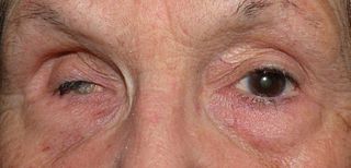
Phthisis bulbi is a shrunken, non-functional eye. It may result from severe eye disease, inflammation or injury, or it may represent a complication of eye surgery. Treatment options include insertion of a prosthesis, which may be preceded by enucleation of the eye.
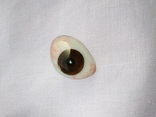
An ocular prosthesis, artificial eye or glass eye is a type of craniofacial prosthesis that replaces an absent natural eye following an enucleation, evisceration, or orbital exenteration. The prosthesis fits over an orbital implant and under the eyelids. Though often referred to as a glass eye, the ocular prosthesis roughly takes the shape of a convex shell and is made of medical grade plastic acrylic. A few ocular prostheses today are made of cryolite glass. A variant of the ocular prosthesis is a very thin hard shell known as a scleral shell which can be worn over a damaged or eviscerated eye. Makers of ocular prosthetics are known as ocularists. An ocular prosthesis does not provide vision; this would be a visual prosthesis. Someone with an ocular prosthesis is altogether blind on the affected side and has monocular vision.
ReLExSmall incision lenticule extraction (SMILE), second generation of ReLEx Femtosecond lenticule extraction (FLEx), is a form of laser based refractive eye surgery developed by Carl Zeiss Meditec used to correct myopia, and cure astigmatism. Although similar to LASIK laser surgery, the intrastromal procedure uses a single femtosecond laser referenced to the corneal surface to cleave a thin lenticule from the corneal stroma for manual extraction.

Bromfenac is a nonsteroidal anti-inflammatory drug (NSAID) marketed in the US as an ophthalmic solution by ISTA Pharmaceuticals for short-term, local use. Prolensa and Bromday are the once-daily formulation of bromfenac, while Xibrom was approved for twice-daily administration. In the European Union, the brand name is Yellox. Bromfenac is indicated for the treatment of ocular inflammation and pain after cataract surgery.

Keratoprosthesis is a surgical procedure where a diseased cornea is replaced with an artificial cornea. Traditionally, keratoprosthesis is recommended after a person has had a failure of one or more donor corneal transplants. More recently, a less invasive, non-penetrating artificial cornea has been developed which can be used in more routine cases of corneal blindness. While conventional cornea transplant uses donor tissue for transplant, an artificial cornea is used in the keratoprosthesis procedure. The surgery is performed to restore vision in patients with severely damaged cornea due to congenital birth defects, infections, injuries and burns.

Gholam A. Peyman is an Iranian American ophthalmologist, retina surgeon, and inventor. He is best known for his invention of LASIK eye surgery, a vision correction procedure designed to allow people to see clearly without glasses. He was awarded the first US patent for the procedure in 1989.
Blast-related ocular trauma comprises a specialized subgroup blast injuries which cause penetrating and blunt force injuries to the eye and its structure. The incidence of ocular trauma due to blast forces has increased dramatically with the introduction of new explosives technology into modern warfare. The availability of these volatile materials, coupled with the tactics of contemporary terrorism, has caused a rise in the number of homemade bombs capable of extreme physical harm.
Bascom Palmer Eye Institute is the University of Miami School of Medicine's ophthalmic care, research, and education center. The institute is based in the Health District of Miami, Florida, and has been ranked consistently as the best eye hospital and vision research center in the nation.
The Intra-Ocular Implant Club was founded in 1966 by the English ophthalmic surgeons Sir Harold Ridley and Peter Choyce, to promote research in the field of intraocular lens (IOL) implantation. At that time there was widespread opposition in the ophthalmic surgery profession to the use of IOLs. The founders saw the club as a forum to allow free and unhindered exchange of ideas about IOLs and implantation surgical techniques. From the outset it was international in its membership and it set itself a parental and advisory role for the then nascent national societies to develop in each country for intraocular implant surgeons. However, this global role was only acknowledged in the name change in July 1975, when the Intra-Ocular Implant Club became The International Intra-Ocular Implant Club (IIIC).

Corneal opacification is a term used when the human cornea loses its transparency. The term corneal opacity is used particularly for the loss of transparency of cornea due to scarring. Transparency of the cornea is dependent on the uniform diameter and the regular spacing and arrangement of the collagen fibrils within the stroma. Alterations in the spacing of collagen fibrils in a variety of conditions including corneal edema, scars, and macular corneal dystrophy is clinically manifested as corneal opacity. The term corneal blindness is commonly used to describe blindness due to corneal opacity.
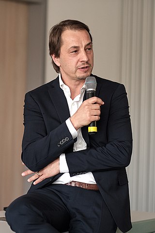
Peter Szurman is a German ophthalmologist, scientist, and professor of ophthalmology in Sulzbach/Saar.
References
- ↑ "TOOTH-IN-EYE (OOKP) SURGERY HELP 8 REGAIN SIGHT". www.ndc.com.sg. Archived from the original on 2008-03-04.
- ↑ Guidera, Anita (28 February 2008). "Son's tooth helps save dad's sight". Irish Independent.
- 1 2 National Dental Centre - 25 May 2005 TOOTH-IN-EYE (OOKP) SURGERY HELP 8 REGAIN SIGHT Archived 4 March 2008 at the Wayback Machine
- ↑ Viitala R, Franklin V, Green D, Liu C, Lloyd A, Tighe B (January 2009). "Towards a synthetic osteo-odonto-keratoprosthesis". Acta Biomater . 5 (1): 438–52. doi:10.1016/j.actbio.2008.07.008. PMID 18706878.
- ↑ Liu C, Okera S, Tandon R, Herold J, Hull C, Thorp S (September 2008). "Visual rehabilitation in end-stage inflammatory ocular surface disease with the osteo-odonto-keratoprosthesis: results from the UK". Br J Ophthalmol . 92 (9): 1211–7. doi:10.1136/bjo.2007.130567. PMID 18511541. S2CID 26716899.
- ↑ Michael R, Charoenrook V, de la Paz MF, Hitzl W, Temprano J, Barraquer RI (August 2008). "Long-term functional and anatomical results of osteo- and osteoodonto-keratoprosthesis". Graefes Arch. Clin. Exp. Ophthalmol. 246 (8): 1133–7. doi:10.1007/s00417-008-0850-3. PMID 18491123. S2CID 13019199.
- ↑ Falcinelli G, Falsini B, Taloni M, Colliardo P, Falcinelli G (October 2005). "Modified osteo-odonto-keratoprosthesis for treatment of corneal blindness: long-term anatomical and functional outcomes in 181 cases". Arch. Ophthalmol. 123 (10): 1319–29. doi:10.1001/archopht.123.10.1319. PMID 16219722.
- ↑ Iannetti L, Liberali M, Armentano M, Alisi L, Visioli G, Mastromarino D, Edoardo B, Iannetti G (May 2022). "Osteo-odonto-keratoprosthesis according to Strampelli original technique: A retrospective study with up to 30 years of follow-up". Am J Ophthalmol . 242: 56–68. doi:10.1016/j.ajo.2022.05.015. hdl: 11573/1635748 . PMID 35618023. S2CID 249035286.
- ↑ Ricci R, Pecorella I, Ciardi A, Della Rocca C, Di Tondo U, Marchi V (April 1992). "Strampelli's osteo-odonto-keratoprosthesis. Clinical and histological long-term features of three prostheses". Br J Ophthalmol . 76 (4): 232llll–4. doi:10.1136/bjo.76.4.232. PMC 504235 . PMID 1390492.