Ovarian squamous cell carcinoma

Last updated
Ovarian squamous cell carcinoma
Squamous Ovarian Cell Carcinoma Squamous Ovarian Cell Carcinoma.png
Squamous Ovarian Cell Carcinoma
Symptoms Vaginal pain and discomfort, pain during sex, abdominal pain, post-menopausal bleeding,
Usual onsetFemales older than 45 years old
CausesNon-pre-existing ovarian lesions (primary) or pre-existing ovarian lesions (Mature cystic teratoma, endometriosis, Brenner tumor)
Diagnostic method CT-scans, Ultrasound, biopsy
Prevention PAP test, Preventive healthcare visits
TreatmentSurgery, Ionizing Radiation, Chemoradiotherapy, Immunotherapy, Platinum-based chemotherapy
Frequencyaccount 1% of ovarian cancer

Ovarian squamous cell carcinoma (oSCC) or squamous ovarian carcinoma (SOC) is a rare tumor that accounts for 1% of ovarian cancers. [1] Included in the World Health Organization's classification of ovarian cancer, [2] it mainly affects women above 45 years of age. Survival depends on how advanced the disease is and how different or similar the individual cancer cells are. [3]

Contents

Squamous ovarian carcinoma is a recognized but uncommon diagnosis, often originating from a transformation of mature cystic teratoma (MCT). Unlike other squamous cell carcinomas, factors like UV exposure and tobacco use play a less significant role. Chronic inflammation in MCT and human papillomavirus (HPV) infection are linked to its development. [3] [4] The tumor emerges through metaplasia of the ovarian surface epithelium. While MCT is the primary source in most cases, others are associated with endometriosis or Brenner tumor, and rare metastasis from other organs can also lead to squamous ovarian carcinoma. [5]

Treatment for oSCC involves surgery, chemotherapy, and radiotherapy, but efficacy of these treatments is unclear. While there is no well-studied chemotherapy regimen for ovarian SCCs, platinum-based chemotherapy is often used. [6]

The earliest reported instance of pure primary SCC was by Ben-Baruchet in 1988; [7] oSSC emerging from pre-existing lesions have been recorded since the early 1950s.

Pathology

Mature cystic teratoma

Ovarian Squamous Cell Carcinoma development from MCTO has been in linked to failure during meiosis I and II. Meiosis Overview new.svg
Ovarian Squamous Cell Carcinoma development from MCTO has been in linked to failure during meiosis I and II.

Mature cystic teratoma (MCT) is a benign (non-cancerous) tumor that develops from at least two of the embryonic germ cell layers. They are characterized by a lining of epithelium and can contain many different tissue types. [8] [3] [9] [10]

Although the majority of MCT cases are non-malignant, approximately 0.17-2% may undergo malignant transformation, with squamous cell carcinoma being the most prevalent, constituting 80% of such transformations. [11] The clinical presentation of oSCC development within MCT lacks specificity, often resulting in the inadvertent discovery of early-stage tumors during routine examinations or postoperative assessments. In advanced cases, patients may exhibit palpable masses, abdominal swelling, and pain, potentially leading to acute abdominal complications due to tumor involvement. [10] [12] [4]

Brenner tumor

Benign mixed Brenner tumor and mucinous cystadenoma of the right ovary. (a) Transvaginal ultrasound images show mixed cystic and solid components, with calcifications in the solid portion (arrow); (b) Macroscopic evaluation revealed an ovary partially replaced by a solid and cystic tumor BT ultrasound.jpg
Benign mixed Brenner tumor and mucinous cystadenoma of the right ovary. (a) Transvaginal ultrasound images show mixed cystic and solid components, with calcifications in the solid portion (arrow); (b) Macroscopic evaluation revealed an ovary partially replaced by a solid and cystic tumor

Brenner tumors (BT) are infrequent ovarian growths composed of specialized ovarian cells surrounded by dense fibrous tissue. Typically small (less than 2 cm), these tumors are often discovered incidentally in women without apparent symptoms. [2] However, when a BT is borderline or malignant, indicating a potential for cancer, it may lead to symptoms such as an abdominal lump, abdominal pain, and post-menopausal bleeding. Less common signs include nausea, vomiting, back pain, bowel problems, reduced appetite, and weight loss.

Differentiating between benign (non-cancerous) and malignant (potentially cancerous) BTs poses a challenge as they appear similarly in medical images. [13] The definitive diagnosis involves examining tissue under a microscope (histopathology). Benign BTs exhibit a consistent fibrous structure, a distinctive characteristic. They may also manifest as a cystic mass with multiple compartments, particularly associated with other ovarian growths. [14]

Endometriosis

oSCC tumors from endometriosis are rare. There are criteria to determine if oSCC comes from endometriosis. Individuals with this type of cancer are usually under 50, have a more severe form of the disease, and often feel abdominal pain. [3]

Conditions for verifying whether the SCC originated as endometriosis or not [6] [15]
Development from the same ovaryGrowth inside endometriosis tissuePresence of endometriosis indicatorsMicroscopic observation of benign epithelium
  • Both endometriosis and squamous epithelial cells must develop in the same ovary.
  • The correlation between tissues from benign endometriosis and malignant SCC should show a pattern similar to a malignant tumor that originates from the endometrium.
  • The malignant tumor has to grow within the tissue of endometriosis.
  • It should not be transferred from the outside of endometriosis to the inside.
  • Tissue characteristics indicative of endometriosis, such as endometrial stroma, evidence of old hemorrhage, hemosiderin pigmentation, etc., need to be present.
  • Microscopic examination should reveal a part changing to benign epithelium.

Screening and diagnosis

Similar to other ovarian cancers patients can undergo a series of tests to identify characteristics and/or markers. This test includes a pelvic exam, imaging (CT, MRI, ultrasound), blood test, and in some cases biopsy with histopathology. Diagnosing this condition remains challenging as preoperative imaging and laboratory tests lack specific indicators.[ medical citation needed ]

Computed tomography

Abdominal CT. Cystic solid mass was detected on the left adnexal and the solid components were enhanced Ovarian Squamous Carcinoma Tumor.jpg
Abdominal CT. Cystic solid mass was detected on the left adnexal and the solid components were enhanced

Computed tomography (CT) scans is a diagnostic x-ray procedure that generates detailed cross-sectional images of the body, facilitating the detection of potential ovarian cancer spread to other organs. [16] While CT scans may not effectively visualize small ovarian tumors, they have the capability to detect larger tumors and evaluate their potential invasion into nearby structures. Additionally, CT scans can identify enlarged lymph nodes, signs of cancer spreading to the liver or other organs, and indications of an ovarian tumor impacting the kidneys or bladder. Although CT scans are not commonly used for biopsying ovarian tumors, they can be utilized in the biopsy of suspected metastases through a technique known as CT-guided needle biopsy. [16]

The difficulty in screening for SCC revolves around the creation of effective strategies with a positive predictive value. The timing aspect is crucial due to the lack of a specific timeframe for the onset of invasive disease or the interval stage between stage I and stage III carcinomas. [17] Ultrasound screening provides a detailed view of the ovaries, identifying morphologic changes as potential signs of malignancy. Key diagnostic factors include the presence of abnormalities, ovarian size, blood flow, and abdominal/pelvic fluid. The persistence of abnormalities after four to six weeks may decrease the occurrence of false positives. Screening protocols, often based on morphologic indices, utilize transvaginal ultrasound findings like cyst wall structure, septation, papillary projections, echogenicity, and ovarian volume to effectively detect malignancy. [17]

Magnetic resonance imaging

Pelvic MRI images. A: Axial T2-weighted image shows multicystic neoplastic masses (white arrows) with solid components in both ovaries. B: A peritoneal implant (blue arrow) with similar characteristics to the ovarian lesions is seen in the peritoneal cavity. C and D: T2-weighted images of the uterine cervix in the axial and sagittal plane reveal the bulky cervical tumor. The cervical stromal ring is well preserved (arrowhead) SCC MRI.png
Pelvic MRI images. A: Axial T2-weighted image shows multicystic neoplastic masses (white arrows) with solid components in both ovaries. B: A peritoneal implant (blue arrow) with similar characteristics to the ovarian lesions is seen in the peritoneal cavity. C and D: T2-weighted images of the uterine cervix in the axial and sagittal plane reveal the bulky cervical tumor. The cervical stromal ring is well preserved (arrowhead)

Although magnetic resonance (MRI) scans are not commonly employed for ovarian cancer detection, the majority of ultrasound-detected ovarian masses are usually benign. The combined approach of ultrasound with Doppler, along with the selective use of contrast-enhanced MRI, proves to be an efficient diagnostic method for identifying benign adnexal masses with distinctive features, including functional masses, dermoid, endometrioma, fibroma, pedunculated fibroid, hydrosalpinx, and peritoneal inclusion cysts. [19] This integrated diagnostic strategy is effective in preventing unnecessary surgical intervention. Additionally, the use of MRI contributes to a decrease in false-positive diagnoses, ultimately reducing the overall number of unnecessary surgical procedures related to ovarian masses.

Advantages and limitations of using MRI to diagnose SCC: [20]
AdvantagesLimitations
1. Higher soft-tissue contrast compared to computed tomography.

2. No radiation exposure, making it suitable for individuals with iodinated contrast agent contraindications, pregnant women, and those of childbearing age.

3. Characterization of tissues through DCE-MRI and DWI allows a move from morphologic assessment to tumor vascularity and cellularity.

4. Improved inter-observer agreement with the addition of DWI to the MRI protocol.

5. DWI and DCE-MRI quantitative parameters can potentially serve as biomarkers of prognosis and response.

1. Long examination time.

2. Technical difficulties covering a large field of view with adequate resolution.

3. Susceptibility to patient and bowel motion.

4. High cost and limited availability.

5. Expertise in interpreting abdominal MRI is usually available only in specialist centers.

6. Limitations based on the use of contrast agents or the technique itself, such as potential false-positive and false-negative results.

7. Need for patient preparation, including fasting and the use of antiperistaltic agents, to improve examination quality.

8. Gadolinium-based contrast agents should be used cautiously in women with renal insufficiency.

9. Limited sensitivity for lesions smaller than 1 cm, especially in areas with similar signal intensity.

10. Limited sensitivity and specificity for assessing lymph node metastasis based on size criteria alone.

Histopathology

(a-c) Seromucinous cystadenoma. A thin-walled paucilocular cyst filled with thin mucinous clear fluid. The cyst wall is lined by endocervical-type columnar mucinous cells admixed with scattered ciliated cells. (d-f) Seromucinous adenofibroma. Mostly solid mass with scattered cystic spaces. Squamous Ovarian Cell Carcinoma.png
(a–c) Seromucinous cystadenoma. A thin-walled paucilocular cyst filled with thin mucinous clear fluid. The cyst wall is lined by endocervical-type columnar mucinous cells admixed with scattered ciliated cells. (d–f) Seromucinous adenofibroma. Mostly solid mass with scattered cystic spaces.

Pathologic examination of biopsies helps distinguish between benign and malignant lesions, aiding in the identification of different cancer types. Larger excisional specimens, known as resections, provide comprehensive information about the cancer, its sub classification, histologic grading, and the extent of its spread, contributing to the pathologic staging. In cases where visible growth [22] or malignant transformation is not apparent, [23] a precise grossing and thorough histopathological examination are essential for an accurate diagnosis. Additionally, conducting situ hybridization for HPV DNA is crucial to rule out the potential involvement of papillomavirus in this condition. To distinguish between primary ovarian serous carcinoma (OSC) and metastatic squamous cell carcinoma (SCC), various immunohistochemical markers such as PAX8, p16, WT1, ER, p53, and CK5/6 are utilized. [23] The p53/p16 index was recognized as a valuable indicator for high-grade OSC, demonstrating diffuse p53 expression or complete absence (null type) along with diffuse p16 expression. [23]

Additional treatment options suggest the use of targeted therapies for squamous cancers, particularly those with elevated epidermal growth factor receptor (EGFR) levels, which remains somewhat limited. [8] [9] Elevated EGFR expression is identified in squamous cell carcinomas of the head and neck as well as the cervix, indicating potential benefits from EGFR inhibition regardless of HPV tumor status. [3] [8]

Management

oSCC exhibits locally invasive characteristics, with tumors demonstrating invasiveness or adherence to the adjacent uterus, pelvic structures, and colonic peritoneum, making surgery the crucial treatment. The standard approach for treating the malignant transformation of mature cystic teratoma of the ovary (MCTO) involves extensive surgery and subsequent chemotherapy, especially for advanced cases. [23] [8] [24] Recommended for patients not seeking fertility, the surgical procedure includes bilateral salpingo-oophorectomy, hysterectomy, comprehensive surgical staging, and lymphadenectomy. [5] [8] [3] Following surgery, advanced cases receive adjuvant chemotherapy with paclitaxel and carboplatin, although the overall prognosis is generally less favorable than that of common epithelial ovarian cancer.

Surgery

Treatment for ovarian cancer often involves surgical procedures to diagnose and remove affected tissues. The choice of surgery depends on the cancer's stage, type, and grade, offering options such as unilateral or bilateral oophorectomy, along with the removal of the fallopian tubes, uterus, and omentum. In cases of low-grade cancers, fertility preservation may be possible, but the presence of microscopic metastases introduces the need for additional surgery. Unilateral surgery is typically employed for germ cell tumors. Achieving complete cytoreduction is essential for managing advanced epithelial ovarian cancer, while debulking surgery aims to remove as much tumor as possible, enhancing the effectiveness of chemotherapy.

An alternative approach involves interval debulking surgery, which combines neoadjuvant chemotherapy with post-debulking chemotherapy. While laparoscopic surgery is suitable for early-stage cancer, advanced cases require more extensive procedures like lymphadenectomy. Secondary surgery may be an option for recurrent cases, effectiveness depending on factors such as surgical technique and the extent of the disease. Oophorectomy's primary side effect, early menopause, can be alleviated with hormone replacement therapy. Hospitals with higher surgical volumes tend to have better surgery outcomes, and the choice between laparoscopy and laparotomy for stage I ovarian cancer remains uncertain. Aggressive surgeries are associated with improved outcomes in advanced stages, despite the presence of perioperative

Lymphadenectomy

Lymphadenectomy, or the surgical removal of lymph nodes, is commonly undertaken in cancer management, with regional and radical dissections differing in the extent of node removal. A meta-analysis research of published randomized controlled trials comparing lymphadenectomy versus no lymphadenectomy in epithelial cancers of the ovary, suggest that overall survival did not demonstrate significance. [25] However, the meta-analysis is unique for focusing on randomized trials, using hazard ratios, and treating ovarian carcinoma as a unified entity. [25] In some cases, complications of lymphadenectomy include lymphedema, and the effectiveness of wound drainage in reducing complications after groin lymph node dissection remains uncertain.

Ionization, radiation and chemotherapy

Typically, a combination of treatments, including surgery, external beam radiotherapy, brachytherapy, and chemotherapy, is employed for these cancers. [26] This understanding could potentially enhance control over the local region, improve quality of life, and increase survival rates. Nevertheless, there is some indication, supported by certain case series, that radiation therapy, particularly when paired with concurrent chemotherapy following aggressive cytoreduction, may be beneficial for certain cases of oSCC originating from teratomas, given the radiosensitive nature of this tumor. [5]

There is no widely accepted chemotherapy regimen used for oSCC, both because of its heterogeneous histology and rarity, but treatment often includes a platinum-based chemotherapy drug. Reported regimens include the combination of paclitaxel and carboplatin (TC) and the combination of bleomycin, etoposide, and cisplatin (BEP). [5] [8] [9]

Immunotherapy

Immunotherapy's adoption in platinum-resistant ovarian cancer remains unsupported by studies, prompting a need for further investigation. [27] Nonetheless, evidence indicates that combining chemotherapy with the humanized anti-VEGF monoclonal antibody, bevacizumab, in the treatment of recurrent, persistent, or metastatic cervical cancer results in a 3.7-month enhancement in overall survival. [3]

Prognosis

Overall survival to different treatments of patients with oSCC a. International Federation of Gynecology and Obstetrics (FIGO) stage and overall survival. b. Hysterectomy and overall survival. c. Chemotherapy and overall survival for stage II, III, IV. d. chemotherapy with platinum and overall survival for stage II, III, IV. Overall survival of treatments for Ovarian Squamous Cell Carcinoma.png
Overall survival to different treatments of patients with oSCC a. International Federation of Gynecology and Obstetrics (FIGO) stage and overall survival. b. Hysterectomy and overall survival. c. Chemotherapy and overall survival for stage II, III, IV. d. chemotherapy with platinum and overall survival for stage II, III, IV.

Ovarian squamous cell carcinoma typically presents with a mean tumor size of 14.8 cm, and overall survival does not vary based on tumor size. [8] Clinical manifestations include abdominal pain and a palpable abdominal mass. The cancer stage is correlated with the time of diagnosis, revealing that higher stages and delayed diagnoses are associated with a worse prognosis. [8] [6] [2] Hysterectomy reduces the risk of death, while lymphadenectomy does not demonstrate improved survival. Fertility-sparing surgery shows no difference in mortality compared to radical surgery in patients under 45. [12] Adjuvant chemotherapy enhances survival in advanced stages, with platinum-based chemotherapy [24] linked to a better prognosis. Radiotherapy and chemoradiotherapy do not improve survival. [3] [6]

Poor prognostic factors include age over 45 or 55, advanced staging, larger tumors, high cancer grade, absence of hysterectomy, presence of residual tumor lesions, lack of adjuvant chemotherapy, and the use of non-platinum-based chemotherapy. In cases of recurrence, secondary cytoreductive surgery may be considered, revealing efficacy in selected cases with recurrent malignant transformation of MCTO. [19] [6] [3]

Related Research Articles

Liver tumors are abnormal growth of liver cells on or in the liver. Several distinct types of tumors can develop in the liver because the liver is made up of various cell types. Liver tumors can be classified as benign (non-cancerous) or malignant (cancerous) growths. They may be discovered on medical imaging, and the diagnosis is often confirmed with liver biopsy. Signs and symptoms of liver masses vary from being asymptomatic to patients presenting with an abdominal mass, hepatomegaly, abdominal pain, jaundice, or some other liver dysfunction. Treatment varies and is highly specific to the type of liver tumor.

<span class="mw-page-title-main">Teratoma</span> Type of germ cell tumor

A teratoma is a tumor made up of several different types of tissue, such as hair, muscle, teeth, or bone. Teratomata typically form in the tailbone, ovary, or testicle.

<span class="mw-page-title-main">Ovarian cyst</span> Fluid-filled sac in the ovary

An ovarian cyst is a fluid-filled sac within the ovary. Often they cause no symptoms. Occasionally they may produce bloating, lower abdominal pain, or lower back pain. The majority of cysts are harmless. If the cyst either breaks open or causes twisting of the ovary, it may cause severe pain. This may result in vomiting or feeling faint, and even cause head aches.

<span class="mw-page-title-main">Ovarian cancer</span> Cancer originating in or on the ovary

Ovarian cancer is a cancerous tumor of an ovary. It may originate from the ovary itself or more commonly from communicating nearby structures such as fallopian tubes or the inner lining of the abdomen. The ovary is made up of three different cell types including epithelial cells, germ cells, and stromal cells. When these cells become abnormal, they have the ability to divide and form tumors. These cells can also invade or spread to other parts of the body. When this process begins, there may be no or only vague symptoms. Symptoms become more noticeable as the cancer progresses. These symptoms may include bloating, vaginal bleeding, pelvic pain, abdominal swelling, constipation, and loss of appetite, among others. Common areas to which the cancer may spread include the lining of the abdomen, lymph nodes, lungs, and liver.

<span class="mw-page-title-main">Dermoid cyst</span> Medical condition

A dermoid cyst is a teratoma of a cystic nature that contains an array of developmentally mature, solid tissues. It frequently consists of skin, hair follicles, and sweat glands, while other commonly found components include clumps of long hair, pockets of sebum, blood, fat, bone, nail, teeth, eyes, cartilage, and thyroid tissue.

<span class="mw-page-title-main">Serous tumour</span> Medical condition

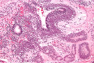
A serous tumour is a neoplasm that typically has papillary to solid formations of tumor cells with crowded nuclei, and which typically arises on the modified Müllerian-derived serous membranes that surround the ovaries in females. Such ovarian tumors are part of the surface epithelial-stromal tumour group of ovarian tumors. They are common neoplasms with a strong tendency to occur bilaterally, and they account for approximately a quarter of all ovarian tumors.

<span class="mw-page-title-main">Surface epithelial-stromal tumor</span> Medical condition

Surface epithelial-stromal tumors are a class of ovarian neoplasms that may be benign or malignant. Neoplasms in this group are thought to be derived from the ovarian surface epithelium or from ectopic endometrial or fallopian tube (tubal) tissue. Tumors of this type are also called ovarian adenocarcinoma. This group of tumors accounts for 90% to 95% of all cases of ovarian cancer; however is mainly only found in postmenopausal women with the exception of the United States where 7% of cases occur in women under the age of 40. Serum CA-125 is often elevated but is only 50% accurate so it is not a useful tumor marker to assess the progress of treatment. 75% of women with epithelial ovarian cancer are found within the advanced-stages; however younger patients are more likely to have better prognoses than older patients.

<span class="mw-page-title-main">Sex cord–gonadal stromal tumour</span> Medical condition

Sex cord–gonadal stromal tumour is a group of tumours derived from the stromal component of the ovary and testis, which comprises the granulosa, thecal cells and fibrocytes. In contrast, the epithelial cells originate from the outer epithelial lining surrounding the gonad while the germ cell tumors arise from the precursor cells of the gametes, hence the name germ cell. In humans, this group accounts for 8% of ovarian cancers and under 5% of testicular cancers. Their diagnosis is histological: only a biopsy of the tumour can make an exact diagnosis. They are often suspected of being malignant prior to operation, being solid ovarian tumours that tend to occur most commonly in post menopausal women.

<span class="mw-page-title-main">Germ cell tumor</span> Medical condition

Germ cell tumor (GCT) is a neoplasm derived from the primordial germ cells. Germ-cell tumors can be cancerous or benign. Germ cells normally occur inside the gonads. GCTs that originate outside the gonads may be birth defects resulting from errors during development of the embryo.

<span class="mw-page-title-main">Ovarian tumor</span> Benign, borderline, or malignant neoplasm involving the ovary

Ovarian tumors, or ovarian neoplasms, are tumors arising from the ovary. They can be benign or malignant. They consist of mainly solid tissue, while ovarian cysts contain fluid.

<span class="mw-page-title-main">Salivary gland tumour</span> Medical condition

Salivary gland tumours, also known as mucous gland adenomas or neoplasms, are tumours that form in the tissues of salivary glands. The salivary glands are classified as major or minor. The major salivary glands consist of the parotid, submandibular, and sublingual glands. The minor salivary glands consist of 800 to 1000 small mucus-secreting glands located throughout the lining of the oral cavity. Patients with these types of tumours may be asymptomatic.

<span class="mw-page-title-main">Immature teratoma</span> Medical condition

An immature teratoma is a teratoma that contains anaplastic immature elements, and is often synonymous with malignant teratoma. A teratoma is a tumor of germ cell origin, containing tissues from more than one germ cell line, It can be ovarian or testicular in its origin. and are almost always benign. An immature teratoma is thus a very rare tumor, representing 1% of all teratomas, 1% of all ovarian cancers, and 35.6% of malignant ovarian germ cell tumors. It displays a specific age of incidence, occurring most frequently in the first two decades of life and almost never after menopause. Unlike a mature cystic teratoma, an immature teratoma contains immature or embryonic structures. It can coexist with mature cystic teratomas and can constitute of a combination of both adult and embryonic tissue. The most common symptoms noted are abdominal distension and masses. Prognosis and treatment options vary and largely depend on grade, stage and karyotype of the tumor itself.

Primary fallopian tube cancer (PFTC), often just tubal cancer, is a malignant neoplasm that originates from the fallopian tube.

A borderline tumor, sometimes called low malignant potential (LMP) tumor, is a distinct but yet heterogeneous group of tumors defined by their histopathology as atypical epithelial proliferation without stromal invasion. It generally refers to such tumors in the ovary but borderline tumors may rarely occur at other locations as well.

<span class="mw-page-title-main">Endodermal sinus tumor</span> Medical condition

Endodermal sinus tumor (EST) is a member of the germ cell tumor group of cancers. It is the most common testicular tumor in children under three, and is also known as infantile embryonal carcinoma. This age group has a very good prognosis. In contrast to the pure form typical of infants, adult endodermal sinus tumors are often found in combination with other kinds of germ cell tumor, particularly teratoma and embryonal carcinoma. While pure teratoma is usually benign, endodermal sinus tumor is malignant.

<span class="mw-page-title-main">Ovarian fibroma</span> Medical condition

The ovarian fibroma, also fibroma, is a benign sex cord-stromal tumour.

Hyperthermic intrathoracic chemotherapy (HITOC) is part of a surgical strategy employed in the treatment of various pleural malignancies. The pleura in this situation could be considered to include the surface linings of the chest wall, lungs, mediastinum, and diaphragm. HITOC is the chest counterpart of HIPEC. Traditionally used in the treatment of malignant mesothelioma, a primary malignancy of the pleura, this modality has recently been evaluated in the treatment of secondary pleural malignancies.

Vulvar tumors are those neoplasms of the vulva. Vulvar and vaginal neoplasms make up a small percentage (3%) of female genital cancers. They can be benign or malignant. Vulvar neoplasms are divided into cystic or solid lesions and other mixed types. Vulvar cancers are those malignant neoplasms that originate from vulvar epithelium, while vulvar sarcomas develop from non-epithelial cells such as bone, cartilage, fat, muscle, blood vessels, or other connective or supportive tissue. Epithelial and mesenchymal tissue are the origin of vulvar tumors.

Ovarian germ cell tumors (OGCTs) are heterogeneous tumors that are derived from the primitive germ cells of the embryonic gonad, which accounts for about 2.6% of all ovarian malignancies. There are four main types of OGCTs, namely dysgerminomas, yolk sac tumor, teratoma, and choriocarcinoma.

References

  1. [ non-primary source needed ]Srivastava, Himsweta; Shree, Sneha; Guleria, Kiran; Singh, Usha R (May 2017). "Pure Primary Squamous Cell Carcinoma of Ovary – A Rare Case Report". Journal of Clinical and Diagnostic Research (Case report). 11 (5): QD01–QD02. doi:10.7860/JCDR/2017/16899.9771. ISSN   2249-782X. PMC   5483757 . PMID   28658855.
  2. 1 2 3 4 Costeira, Filipa de Sousa; Félix, Ana; Cunha, Teresa Margarida (2022-02-01). "Brenner tumors". The British Journal of Radiology. 95 (1130). doi:10.1259/bjr.20210687. ISSN   0007-1285. PMC   8822556 . PMID   34928171.
  3. 1 2 3 4 5 6 7 8 9 Roxburgh, Patricia; Glasspool, Rosalind (2014-10-26). "Squamous Carcinoma of the Ovary". Current Oncology Reports. 16 (12): 413. doi:10.1007/s11912-014-0413-1. ISSN   1534-6269. PMID   25344339. S2CID   19909090.
  4. 1 2 Koufopoulos, Nektarios; Nasi, Despoina; Goudeli, Christina; Antoniadou, Foteini; Kokkali, Stefania; Pigadioti, Eleni; Provatas, Ioannis; Maggo, Elpida; Ardavanis, Alexandros; Terzakis, Emmanouil; Arkoumani, Evdokia; J Agnantis, Niki; Apostolikas, Nikiforos; Khaldi, Lubna (2019). "Primary squamous cell carcinoma of the ovary. Review of the literature". Journal of B.U.ON. 24 (5): 1776–1784. ISSN   2241-6293. PMID   31786837.
  5. 1 2 3 4 5 Luo, Yan; Bian, Ce (2022). "Primary squamous cell carcinoma of the ovary. Review of the literature". Frontiers in Oncology. 12. doi: 10.3389/fonc.2022.962613 . ISSN   2234-943X. PMC   9515541 . PMID   36185237.
  6. 1 2 3 4 5 [ non-primary source needed ]Jeon, Seob (2017-06-30). "Ovarian Squamous Cell Carcinoma Associated with Endometriosis: Poor Response to Chemotherapy". Soonchunhyang Medical Science (Case report). 23 (1): 56–60. doi: 10.15746/sms.17.013 . ISSN   2233-4289. S2CID   79889670.
  7. Ben-Baruch, Gilad; Menashe, Yehezkeal; Herczeg, Emil; Menczer, Joseph (February 1988). "Pure primary ovarian squamous cell carcinoma". Gynecologic Oncology. 29 (2): 257–262. doi:10.1016/0090-8258(88)90223-5. ISSN   0090-8258. PMID   3276582.
  8. 1 2 3 4 5 6 7 8 9 Li, Congcong; Zhang, Qing; Zhang, Siying; Dong, Ruifen; Sun, Chenggong; Qiu, Chunping; Zhang, Zhiwei; Yang, Xingsheng; Kong, Beihua (December 2019). "Squamous cell carcinoma transformation in mature cystic teratoma of the ovary: a systematic review". BMC Cancer. 19 (1): 217. doi: 10.1186/s12885-019-5393-y . ISSN   1471-2407. PMC   6417039 . PMID   30866852.
  9. 1 2 3 Blackwell, William J.; Dockerty, Malcolm B.; Masson, James C.; Mussey, Robert D. (February 1946). "Dermoid Cysts of the Ovary: Their Clinical and Pathologic Significance**Abridgment of thesis submitted by Dr. Blackwell to the Faculty of the Graduate School of the University of Minnesota in partial fulfillment of the requirements for the degree of M.S. in Surgery". American Journal of Obstetrics and Gynecology. 51 (2): 151–172. doi:10.1016/S0002-9378(16)39889-1. PMID   21012750.
  10. 1 2 Laothamatas, Indira; Fang, Eric; Lee, Justine; Abbas, Syed Muhammad Hashim; Ding, Juan; Kagen, Alexander; Zakashansky, Konstantin; Taouli, Bachir; Kamath, Amita; King, Michael J. (November–December 2023). "Benign and Malignant Ovarian Teratomas: Multimodality Imaging Findings With Histopathologic Correlation". Journal of Computer Assisted Tomography. 47 (6): 882–889. doi:10.1097/RCT.0000000000001509. ISSN   0363-8715. PMID   37948362. S2CID   259857697.
  11. Glasspool, Rosalind M.; Martín, Antonio González; Millan, David; Lorusso, Domenica; Åvall-Lundqvist, Elisabeth; Hurteau, Jean A.; Davis, Alison; Hilpert, Felix; Kim, Jae-Weon; Alexandre, Jérôme; Ledermann, Jonathan A. (November 2014). "Gynecologic Cancer InterGroup (GCIG) Consensus Review for Squamous Cell Carcinoma of the Ovary". International Journal of Gynecologic Cancer. 24 (Supp 3): S26–S29. doi:10.1097/IGC.0000000000000209. ISSN   1048-891X. PMID   25126954. S2CID   205675510.
  12. 1 2 Luo, Yan; Bian, Ce (2022-09-14). "Primary squamous cell carcinoma of the ovary. Review of the literature". Frontiers in Oncology. 12. doi: 10.3389/fonc.2022.962613 . ISSN   2234-943X. PMC   9515541 . PMID   36185237.
  13. Miles, Philip A.; Norris, Henry J. (July 1972). "Proliferative and malignant brenner tumors of the ovary". Cancer. 30 (1): 174–186. doi: 10.1002/1097-0142(197207)30:1<174::AID-CNCR2820300125>3.0.CO;2-D . ISSN   0008-543X. PMID   5040741. S2CID   8818224.
  14. Lamping, J. Daniels; Blythe, James G. (1977-09-01). "Bilateral brenner tumors: A case report and review of the literature". Human Pathology. 8 (5): 583–585. doi:10.1016/S0046-8177(77)80117-2. ISSN   0046-8177. PMID   903146.
  15. Sampson, John A. (1925-01-01). "Endometrial Carcinoma of the Ovary, Arising in Endometrial Tissue in That Organ". Archives of Surgery. 10 (1): 1–72. doi:10.1001/archsurg.1925.01120100007001. ISSN   0272-5533.
  16. 1 2 "Tests for Ovarian Cancer | How Is Ovarian Cancer Diagnosed?". www.cancer.org. Retrieved 2023-11-29.
  17. 1 2 Koutras, Antonios; Perros, Paraskevas; Prokopakis, Ioannis; Ntounis, Thomas; Fasoulakis, Zacharias; Pittokopitou, Savia; Samara, Athina A.; Valsamaki, Asimina; Douligeris, Athanasios; Mortaki, Anastasia; Sapantzoglou, Ioakeim; Katrachouras, Alexandros; Pagkalos, Athanasios; Symeonidis, Panagiotis; Palios, Vasileios-Chrysovalantis (January 2023). "Advantages and Limitations of Ultrasound as a Screening Test for Ovarian Cancer". Diagnostics. 13 (12): 2078. doi: 10.3390/diagnostics13122078 . ISSN   2075-4418. PMC   10297553 . PMID   37370973.
  18. Yilmaz, Melek Tugce; Gurlek, Ezgi; Gultekin, Melis; Kahraman, Korhan; Salman, Mehmet C; Usubutun, Alp; Akata, Deniz; Lay Ergun, Eser; Arik, Zafer; Yildiz, Ferah (2022-01-06). "Ovarian Carcinoma Presenting With a Large Cervical Mass". Cureus. 14 (1): e20994. doi: 10.7759/cureus.20994 . ISSN   2168-8184. PMC   8820480 . PMID   35154969.
  19. 1 2 Shetty, Mahesh (2023-12-01). "Imaging of a Pelvic Mass: Uterine". Seminars in Ultrasound, CT and MRI. Symptom based Imaging in the management of Gynecological Diseases. 44 (6): 528–540. doi:10.1053/j.sult.2023.10.005. ISSN   0887-2171. PMID   37839652. S2CID   264109630.
  20. Fischerova, Daniela; Burgetova, Andrea (2014-07-01). "Imaging techniques for the evaluation of ovarian cancer". Best Practice & Research Clinical Obstetrics & Gynaecology. Imaging in Gynaecology. 28 (5): 697–720. doi:10.1016/j.bpobgyn.2014.04.006. ISSN   1521-6934. PMID   24846098.
  21. Nagamine, Michiko; Mikami, Yoshiki (February 2020). "Ovarian Seromucinous Tumors: Pathogenesis, Morphologic Spectrum, and Clinical Issues". Diagnostics. 10 (2): 77. doi: 10.3390/diagnostics10020077 . ISSN   2075-4418. PMC   7168900 . PMID   32023964.
  22. Mishra, Pritinanda; Sasmal, PrakashK; Jena, SaubhagyaK; Senapati, Swagatika; Patra, Susama (2021). "Pure primary ovarian carcinoid tumor with carcinoid syndrome and cervical carcinoma: A rare concoction of dual primary malignancies". Indian Journal of Pathology and Microbiology. 64 (3): 563–567. doi: 10.4103/IJPM.IJPM_1379_20 . ISSN   0377-4929. PMID   34341274.
  23. 1 2 3 4 Sun, Yao; Chen, Yuezhou; Zhang, Xiaofei; Chen, Hao; Zhou, Feng (2021-05-08). "Primary ovarian serous carcinomas with extensive squamous differentiation: a case report and literature review". BMC Women's Health. 21 (1): 193. doi: 10.1186/s12905-021-01336-y . ISSN   1472-6874. PMC   8106833 . PMID   33964926.
  24. 1 2 Yang, Szu-Ting; Chang, Wen-Hsun; Chou, Fang-Wei; Liu, Hung-Hsien; Lee, Wen-Ling; Wang, Peng-Hui (November 2023). "Front-line chemo-immunotherapy for treating epithelial ovarian cancer: Part I CA125 and anti-CA125". Taiwanese Journal of Obstetrics and Gynecology. 62 (6): 802–808. doi: 10.1016/j.tjog.2023.09.017 . PMID   38008497. S2CID   265450805.
  25. 1 2 Purwar, Roli; Ranjan, Rakesh; Soni, Kishan; Pandey, Manoj; Upadhyay, Satyanshu K.; Pai, Esha; Kumar, Tarun (2022-11-22). "Lymphadenectomy in ovarian cancers: a meta-analysis of hazard ratios from randomized clinical trials". World Journal of Surgical Oncology. 20 (1): 367. doi: 10.1186/s12957-022-02835-4 . ISSN   1477-7819. PMC   9682714 . PMID   36419077.
  26. Morcet-Delattre, R.; Espenel, S.; Tas, P.; Chargari, C.; Escande, A. (2023-12-01). "Role of radiotherapy in the management of rare gynaecological cancers". Cancer/Radiothérapie. 27 (8): 778–788. doi:10.1016/j.canrad.2023.08.006. ISSN   1278-3218. PMID   37925347. S2CID   264990398.
  27. Yang, Chang; Xia, Bai-Rong; Zhang, Zhao-Cong; Zhang, Yong-Jian; Lou, Ge; Jin, Wei-Lin (2020). "Immunotherapy for Ovarian Cancer: Adjuvant, Combination, and Neoadjuvant". Frontiers in Immunology. 11. doi: 10.3389/fimmu.2020.577869 . ISSN   1664-3224. PMC   7572849 . PMID   33123161.