Pachyonychia congenita

Last updated
Pachyonychia congenita
Autosomal dominant - en.svg
Pachyonychia congenita has an autosomal dominant pattern of inheritance.
Specialty Medical genetics   OOjs UI icon edit-ltr-progressive.svg

Pachyonychia congenita (often abbreviated as "PC") is a rare group of autosomal dominant skin disorders that are caused by a mutation in one of five different keratin genes. Pachyonychia congenita is often associated with thickened toenails, plantar keratoderma, and plantar pain.

Contents

Signs and symptoms

Pachyonychia congenita is characterized by a clinical triad present in 97% of people with PC by the time they turn 10 years old: [1] [2]

  1. Thickened toenails
  2. Plantar keratoderma
  3. Plantar pain that may require some patients to use wheelchairs, canes, crutches, and pain medications due to its severity

Other signs and symptoms found in PC include: [1] [3]

Cause

The condition is caused by genetic mutations in one of five genes that encode keratin proteins. Three keratin genes were identified to have a role PC in 1995 [4] [5] with a fourth keratin gene's role in PC identified in 1998. [6]

Inheritance

Pachyonychia congenita follows an autosomal dominant pattern of inheritance, which means the defective gene is located on an autosome, and only one copy of the gene is required to inherit the disorder from a parent who has the disorder. On average, 50% of the offspring of an affected person will inherit the disorder, regardless of sex.[ citation needed ]

Occasionally, however, a solitary case can emerge in a family with no prior history of the disorder due to the occurrence of a new mutation (often referred to as a sporadic, spontaneous or de novo mutation).[ citation needed ]

Diagnosis

Classification

ILDS: Q84.520 ICD-10: Q84.5

Pachyonychia congenita consists of five sub-types, each named after its corresponding genetic mutation and each associated with distinguishing clinical features: [1] [7]

  1. PC-K6a is caused by a mutation in the KRT6A gene and more often associated with oral leukokeratosis and poor feeding in infants.
  2. PC-K6b is caused by a mutation in the KRT6B gene and more commonly associated with an increased age of onset (>14 years).
  3. PC-K6c is caused by a mutation in the KRT6C gen e and is the least common sub-type. It is not often associated with the additional features of oral leukokeratosis, cysts, follicular hyperkeratosis, and natal teeth.
  4. PC-K16 is caused by a mutation in the KRT16 gene and is more commonly associated with severe plantar pain.
  5. PC-K17 is caused by a mutation in the KRT17 gene and more commonly associated with the presence of cysts, follicular kyperkeratosis, and natal teeth.

Before the genetic basis of Pachyonychia congenita was identified and described, the disease was historically divided into the following sub-types: [8] :510

Clinical Diagnosis

In order to clinically diagnose pachyonychia congenita, the clinical triad of toenail thickening, plantar keratoderma, and plantar pain must be present. This triad is present in 97% of individuals with PC by the age of 10 years old. [1]

Pachyonychia congenita can be suspected in patients who do not have the complete clinical triad but who exhibit other symptoms such as cysts, oral leukokeratosis, follicular hyperkeratosis, palmoplantar hyperhidrosis, or natal teeth. Since PC is inherited in an autosomal dominant fashion in 70% of individuals, it should especially be suspected in patients with symptoms who also have a parent with similar symptoms. Histopathological analysis of skin or nail tissue is not helpful in diagnosis of PC, but can be used to rule out some related diseases. If there is a clinical suspicion for PC, genetic testing can confirm the diagnosis. [1]

Genetic Diagnosis

The diagnosis of PC can be confirmed by the identification of a mutation in one of the five genes responsible for the condition: KRT6A, KRT6B, KRT6C, KRT16, KRT17. Pachyonychia Congenita Project is a non-profit dedicated to finding a cure for PC. The organization houses a genetic registry (the International PC Research Registry) and offers free genetic testing for individuals suspected to have PC. [11]

Treatment

There is currently no cure for pachyonychia congenita. Treatment focuses on symptom relief for any plantar pain, hyperkeratoses, cysts, leukokeratosis, hyperhidrosis, or secondary infections. [12]

Palmoplantar keratoderma can be treated with consistent grooming, including trimming back the callus, applying emollients, and draining blisters. Plantar pain is often treated by reducing pressure on the feet by minimizing walking, wearing cushioned footwear, or using wheelchairs or crutches. Hyperkeratosis can be treated with keratolytic emollients while cysts may be treated with incision and drainage. Patients with hyperhidrosis may need to wear moisture-wicking socks and ventilated shoes. Any secondary infection may need to be treated with antibiotics, though infection can often be prevented with appropriate grooming and vinegar or bleach baths. [12] [1] [7]

Epidemiology

Pachyonychia congenita is a rare disorder with an unknown prevalence. As of 2018, the International PC Research Registry has identified approximately 774 individuals with the disease, but prevalence is estimated to be 5,000–10,000 worldwide. [1] [7] The disease affects both males and females.

Research

There are several ongoing investigational therapies for pachyonychia congenita, including topical sirolimus, siRNA, botulinum toxin, statins, and anti-TNF biologics. [1] Pachyonychia Congenita Project houses a list of clinical trials and assists with clinical trial recruitment from patients enrolled in their International PC Research Registry. [11] [13]

See also

Related Research Articles

<span class="mw-page-title-main">Epidermolytic hyperkeratosis</span> Medical condition

Epidermolytic ichthyosis (EI), is a rare and severe form of ichthyosis that affects around 1 in 300,000 people. It is caused by a genetic mutation, and thus cannot be completely cured without some form of gene therapy.

Type II keratins constitutes the Type II intermediate filaments (IFs) of the intracytoplasmatic cytoskeleton, which is present in all mammalian epithelial cells. The type 2 cytokeratins consist of basic or neutral, high molecular weight proteins which in vivo are arranged in pairs of heterotypic Type I and Type II keratin chains, coexpressed during differentiation of simple and stratified epithelial tissues. It has been seen that Type II Keratins are developed before Type 1 keratins during human embryonic development.

<span class="mw-page-title-main">Keratin 16</span> Protein-coding gene in the species Homo sapiens

Keratin 16 is a protein that in humans is encoded by the KRT16 gene.

<span class="mw-page-title-main">Palmoplantar keratoderma</span> Medical condition

Palmoplantar keratodermas are a heterogeneous group of disorders characterized by abnormal thickening of the stratum corneum of the palms and soles.

<span class="mw-page-title-main">Ectodermal dysplasia</span> Group of genetic conditions affecting the embryonic ectoderm

Ectodermal dysplasia (ED) is a group of genetic syndromes all deriving from abnormalities of the ectodermal structures. More than 150 different syndromes have been identified.

<span class="mw-page-title-main">Hyperkeratosis</span> Medical condition

Hyperkeratosis is thickening of the stratum corneum, often associated with the presence of an abnormal quantity of keratin, and also usually accompanied by an increase in the granular layer. As the corneum layer normally varies greatly in thickness in different sites, some experience is needed to assess minor degrees of hyperkeratosis.

<span class="mw-page-title-main">Meleda disease</span> Medical condition

Meleda disease (MDM) or "mal de Meleda", also called Mljet disease, keratosis palmoplantaris and transgradiens of Siemens, is an extremely rare autosomal recessive congenital skin disorder in which dry, thick patches of skin develop on the soles of the hands and feet, a condition known as palmoplantar hyperkeratosis. Meleda Disease is a skin condition which usually can be identified not long after birth. This is a genetic condition but it is very rare. The hands and feet usually are the first to show signs of the disease but the disease can advance to other parts of the body. Signs of the disease include thickening of the skin, on hands and soles of feet, which can turn red in color. There currently is no cure and treatment is limited, but Acitretin can be used in severe cases.

<span class="mw-page-title-main">Papillon–Lefèvre syndrome</span> Medical condition

Papillon–Lefèvre syndrome (PLS), also known as palmoplantar keratoderma with periodontitis, is an autosomal recessive genetic disorder caused by a deficiency in cathepsin C.

<span class="mw-page-title-main">Dermatopathia pigmentosa reticularis</span> Medical condition

Dermatopathia pigmentosa reticularis(DPR) is a rare, autosomal dominant congenital disorder that is a form of ectodermal dysplasia. Dermatopathia pigmentosa reticularis is composed of the triad of generalized reticulate hyperpigmentation, noncicatricial alopecia, and onychodystrophy. DPR is a non life-threatening disease that largely affects the skin, hair, and nails. It has also been identified as a keratin disorder. Historically, as of 1992, only 10 cases had been described in world literature; however, due to recent advances in genetic analysis, five additional families studied in 2006 have been added to the short list of confirmed cases.

<span class="mw-page-title-main">Monilethrix</span> Medical condition

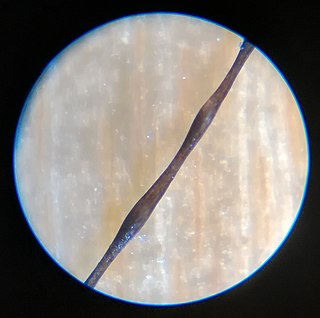
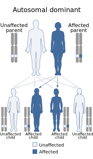
Monilethrix is a rare autosomal dominant hair disease that results in short, fragile, broken hair that appears beaded. It comes from the Latin word for necklace (monile) and the Greek word for hair (thrix). Hair becomes brittle, and breaks off at the thinner parts between the beads. It appears as a thinning or baldness of hair and was first described in 1897 by Walter Smith

<span class="mw-page-title-main">Keratin 6C</span>

Keratin 6C, is a type II cytokeratin, one of a number of isoforms of keratin 6 encoded by separate genes located within the type II keratin gene cluster on human chromosome 12q. This gene was uncovered recently by the Human Genome Project and its expression patterns in humans remains unknown.

<span class="mw-page-title-main">Genodermatosis</span> Medical condition

Genodermatosis is a hereditary skin disease with three inherited modes including single gene inheritance, multiple gene inheritance and chromosome inheritance. There are many different types of genodermatosis, the prevalence of genodermatosis ranges from 1 per 6000 people to 1 per 500,000 people. Genodermatosis has influence on the texture, color and structure of skin cuticle and connective tissue, specific lesion site and clinical manifestations on the body vary depending on the type. In the spite of the variety and complexity of genodermatosis, there are still some common methods that can help people diagnose. After diagnosis, different types of genodermatosis require different levels of therapy including interventions, nursing interventions and treatments. Among that, research of therapy for some new, complex and rare types are still in the developing stage. The impact of genodermatosis not only can be seen in body but also can be seen in all aspects of patients' life, including but not limited to psychological, family life, economic conditions and social activities. Accordingly, the patients need treatment, support and help in these areas.

<span class="mw-page-title-main">Steatocystoma multiplex</span> Medical condition

Steatocystoma multiplex, is a benign, autosomal dominant congenital condition resulting in multiple cysts on a person's body. Steatocystoma simplex is the solitary counterpart to steatocystoma multiplex.

<span class="mw-page-title-main">Ichthyosis bullosa of Siemens</span> Medical condition

Ichthyosis bullosa of Siemens is a type of familial, autosomal dominant ichthyosis, a rare skin disorder. It is also known as bullous congenital ichthyosiform erythroderma of Siemens or ichthyosis exfoliativa. It is a genetic disorder with no known cure which is estimated to affect about 1 in 500,000 people.

Peeling skin syndrome is an autosomal recessive disorder characterized by lifelong peeling of the stratum corneum, and may be associated with pruritus, short stature, and easily removed anagen hair.

Schöpf–Schulz–Passarge syndrome is an autosomal recessive condition with punctate symmetric palmoplantar keratoderma, with the keratoderma and fragility of the nails beginning around age 12. In addition to palmoplantar keratoderma, other symptoms include hypodontia, hypotrichosis, nail dystrophies, and eyelid cysts. Patients may also develop syringofibroadenoma and squamous cell carcinomas.

Howel–Evans syndrome is an extremely rare condition involving thickening of the skin in the palms of the hands and the soles of the feet (hyperkeratosis). This familial disease is associated with a high lifetime risk of esophageal cancer. For this reason, it is sometimes known as tylosis with oesophageal cancer (TOC).

Haim–Munk syndrome is a skin disease caused, like Papillon–Lefèvre syndrome, by a mutation in the cathepsin C gene. One of its features is thick curved finger and toenails.

Palmoplantar keratoderma with deafness, also known as Palmoplantar keratoderma-deafness syndrome is a rare genetic disorder which is characterized by either focal or diffuse early-onset palmoplantar keratoderma and sensorineural deafness. Transmission is autosomal dominant with incomplete penetrance.

References

  1. 1 2 3 4 5 6 7 8 Smith, Frances JD; Hansen, C. David; Hull, Peter R.; Kaspar, Roger L.; McLean, WH Irwin; O’Toole, Edel; Sprecher, Eli (1993), Adam, Margaret P.; Ardinger, Holly H.; Pagon, Roberta A.; Wallace, Stephanie E. (eds.), "Pachyonychia Congenita", GeneReviews®, University of Washington, Seattle, PMID   20301457 , retrieved 2018-08-01
  2. Eliason, Mark J.; Leachman, Sancy A.; Feng, Bing-jian; Schwartz, Mary E.; Hansen, C. David (October 2012). "A review of the clinical phenotype of 254 patients with genetically confirmed pachyonychia congenita". Journal of the American Academy of Dermatology. 67 (4): 680–686. doi:10.1016/j.jaad.2011.12.009. ISSN   1097-6787. PMID   22264670.
  3. "What Is Pachyonychia Congenita?". www.pachyonychia.org. Retrieved 2018-08-01.
  4. McLean W, Rugg E, Lunny D, Morley S, Lane E, Swensson O, Dopping-Hepenstal P, Griffiths W, Eady R, Higgins C, Navsaria H, Leigh I, Strachan T, Kunkeler L, Munro C (1995). "Keratin 16 and keratin 17 mutations cause pachyonychia congenita". Nature Genetics. 9 (3): 273–8. doi:10.1038/ng0395-273. PMID   7539673. S2CID   1873772.
  5. Bowden PE, Haley JL, Kansky A, Rothnagel JA, Jones DO, Turner RJ (1995). "Mutation of a type II keratin gene (K6a) in pachyonychia congenita". Nature Genetics. 10 (3): 363–5. doi:10.1038/ng0795-363. PMID   7545493. S2CID   26060130.
  6. Smith FJ, Jonkman MF, Van Goor H, Coleman CM, Covello SP, Uitto J, McLean WH (1998). "A Mutation in Human Keratin K6b Produces a Phenocopy of the K17 Disorder Pachyonychia Congenita Type 2". Human Molecular Genetics. 7 (7): 1143–8. doi:10.1093/hmg/7.7.1143. PMID   9618173.
  7. 1 2 3 "Pachyonychia Congenita - NORD (National Organization for Rare Disorders)". NORD (National Organization for Rare Disorders). Retrieved 2018-08-02.
  8. 1 2 3 Freedberg, et al. (2003). Fitzpatrick's Dermatology in General Medicine. (6th ed.). McGraw-Hill. ISBN   0-07-138076-0.
  9. 1 2 Rapini, Ronald P.; Bolognia, Jean L.; Jorizzo, Joseph L. (2007). Dermatology: 2-Volume Set. St. Louis: Mosby. p. 740. ISBN   978-1-4160-2999-1.
  10. 1 2 James, William; Berger, Timothy; Elston, Dirk (2005). Andrews' Diseases of the Skin: Clinical Dermatology. (10th ed.). Saunders. ISBN   0-7216-2921-0.
  11. 1 2 "International PC Research Registry (IPCRR)". www.pachyonychia.org. Retrieved 2018-08-01.
  12. 1 2 Goldberg, I.; Fruchter, D.; Meilick, A.; Schwartz, M.E.; Sprecher, E. (2013-01-30). "Best treatment practices for pachyonychia congenita". Journal of the European Academy of Dermatology and Venereology. 28 (3): 279–285. doi:10.1111/jdv.12098. ISSN   0926-9959. PMID   23363249. S2CID   29684041.
  13. "Clinical Trials & Studies". www.pachyonychia.org. Retrieved 2018-08-01.