Related Research Articles

In cell biology, the cytoplasm is all of the material within a eukaryotic cell, enclosed by the cell membrane, except for the cell nucleus. The material inside the nucleus and contained within the nuclear membrane is termed the nucleoplasm. The main components of the cytoplasm are cytosol, the organelles, and various cytoplasmic inclusions. The cytoplasm is about 80% water and is usually colorless.
Peptidoglycan or murein is a unique large macromolecule, a polysaccharide, consisting of sugars and amino acids that forms a mesh-like peptidoglycan layer outside the plasma membrane, the rigid cell wall characteristic of most bacteria. The sugar component consists of alternating residues of β-(1,4) linked N-acetylglucosamine (NAG) and N-acetylmuramic acid (NAM). Attached to the N-acetylmuramic acid is a oligopeptide chain made of three to five amino acids. The peptide chain can be cross-linked to the peptide chain of another strand forming the 3D mesh-like layer. Peptidoglycan serves a structural role in the bacterial cell wall, giving structural strength, as well as counteracting the osmotic pressure of the cytoplasm. This repetitive linking results in a dense peptidoglycan layer which is critical for maintaining cell form and withstanding high osmotic pressures, and it is regularly replaced by peptidoglycan production. Peptidoglycan hydrolysis and synthesis are two processes that must occur in order for cells to grow and multiply, a technique carried out in three stages: clipping of current material, insertion of new material, and re-crosslinking of existing material to new material.
A vacuole is a membrane-bound organelle which is present in plant and fungal cells and some protist, animal, and bacterial cells. Vacuoles are essentially enclosed compartments which are filled with water containing inorganic and organic molecules including enzymes in solution, though in certain cases they may contain solids which have been engulfed. Vacuoles are formed by the fusion of multiple membrane vesicles and are effectively just larger forms of these. The organelle has no basic shape or size; its structure varies according to the requirements of the cell.

In cell biology, a vesicle is a structure within or outside a cell, consisting of liquid or cytoplasm enclosed by a lipid bilayer. Vesicles form naturally during the processes of secretion (exocytosis), uptake (endocytosis) and transport of materials within the plasma membrane. Alternatively, they may be prepared artificially, in which case they are called liposomes. If there is only one phospholipid bilayer, the vesicles are called unilamellar liposomes; otherwise they are called multilamellar liposomes. The membrane enclosing the vesicle is also a lamellar phase, similar to that of the plasma membrane, and intracellular vesicles can fuse with the plasma membrane to release their contents outside the cell. Vesicles can also fuse with other organelles within the cell. A vesicle released from the cell is known as an extracellular vesicle.

Endosomes are a collection of intracellular sorting organelles in eukaryotic cells. They are parts of endocytic membrane transport pathway originating from the trans Golgi network. Molecules or ligands internalized from the plasma membrane can follow this pathway all the way to lysosomes for degradation or can be recycled back to the cell membrane in the endocytic cycle. Molecules are also transported to endosomes from the trans Golgi network and either continue to lysosomes or recycle back to the Golgi apparatus.
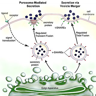
Secretion is the movement of material from one point to another, such as a secreted chemical substance from a cell or gland. In contrast, excretion is the removal of certain substances or waste products from a cell or organism. The classical mechanism of cell secretion is via secretory portals at the plasma membrane called porosomes. Porosomes are permanent cup-shaped lipoprotein structures embedded in the cell membrane, where secretory vesicles transiently dock and fuse to release intra-vesicular contents from the cell.
Turgor pressure is the force within the cell that pushes the plasma membrane against the cell wall.

Bacteria are ubiquitous, mostly free-living organisms often consisting of one biological cell. They constitute a large domain of prokaryotic microorganisms. Typically a few micrometres in length, bacteria were among the first life forms to appear on Earth, and are present in most of its habitats. Bacteria inhabit soil, water, acidic hot springs, radioactive waste, and the deep biosphere of Earth's crust. Bacteria are vital in many stages of the nutrient cycle by recycling nutrients such as the fixation of nitrogen from the atmosphere. The nutrient cycle includes the decomposition of dead bodies; bacteria are responsible for the putrefaction stage in this process. In the biological communities surrounding hydrothermal vents and cold seeps, extremophile bacteria provide the nutrients needed to sustain life by converting dissolved compounds, such as hydrogen sulphide and methane, to energy. Bacteria also live in symbiotic and parasitic relationships with plants and animals. Most bacteria have not been characterised and there are many species that cannot be grown in the laboratory. The study of bacteria is known as bacteriology, a branch of microbiology.
Exosomes are membrane-bound extracellular vesicles (EVs) that are produced in the endosomal compartment of most eukaryotic cells. The multivesicular body (MVB) is an endosome with intraluminal vesicles (ILVs) that bud inward into the endosomal lumen. If the MVB fuses with the cell surface, these ILVs are released as exosomes.

Microvesicles are a type of extracellular vesicle (EV) that are released from the cell membrane. In multicellular organisms, microvesicles and other EVs are found both in tissues and in many types of body fluids. Delimited by a phospholipid bilayer, microvesicles can be as small as the smallest EVs or as large as 1000 nm. They are considered to be larger, on average, than intracellularly-generated EVs known as exosomes. Microvesicles play a role in intercellular communication and can transport molecules such as mRNA, miRNA, and proteins between cells.

Archaea is a domain of single-celled organisms. These microorganisms lack cell nuclei and are therefore prokaryotes. Archaea were initially classified as bacteria, receiving the name archaebacteria, but this term has fallen out of use.
In biology, an organism is any living system that functions as an individual entity. All organisms are composed of cells. The idea of organism is based on the concept of minimal functional unit of life. Three traits has been proposed to play main role in qualification as an organism:

Eukaryota, whose members are known as eukaryotes, is a diverse domain of organisms whose cells have a nucleus. All animals, plants, fungi, and many unicellular organisms, are eukaryotes. They belong to the group of organisms Eukaryota or Eukarya, which is one of the three domains of life. Bacteria and Archaea make up the other two domains.
ExoCarta is a manually curated database of exosomal proteins, RNA and lipids.
Fission, in biology, is the division of a single entity into two or more parts and the regeneration of those parts to separate entities resembling the original. The object experiencing fission is usually a cell, but the term may also refer to how organisms, bodies, populations, or species split into discrete parts. The fission may be binary fission, in which a single organism produces two parts, or multiple fission, in which a single entity produces multiple parts.

An ectomycorrhiza is a form of symbiotic relationship that occurs between a fungal symbiont, or mycobiont, and the roots of various plant species. The mycobiont is often from the phyla Basidiomycota and Ascomycota, and more rarely from the Zygomycota. Ectomycorrhizas form on the roots of around 2% of plant species, usually woody plants, including species from the birch, dipterocarp, myrtle, beech, willow, pine and rose families. Research on ectomycorrhizas is increasingly important in areas such as ecosystem management and restoration, forestry and agriculture.
Membrane vesicle trafficking in eukaryotic animal cells involves movement of biochemical signal molecules from synthesis-and-packaging locations in the Golgi body to specific release locations on the inside of the plasma membrane of the secretory cell. It takes place in the form of Golgi membrane-bound micro-sized vesicles, termed membrane vesicles (MVs).
Extracellular vesicles (EVs) are lipid bilayer-delimited particles that are naturally released from almost all types of cell but, unlike a cell, cannot replicate. EVs range in diameter from near the size of the smallest physically possible unilamellar liposome to as large as 10 microns or more, although the vast majority of EVs are smaller than 200 nm. EVs can be divided according to size and synthesis route into Exosomes, microvesicles and apoptotic bodies. They carry a cargo of proteins, nucleic acids, lipids, metabolites, and even organelles from the parent cell. Most cells that have been studied to date are thought to release EVs, including some archaeal, bacterial, fungal, and plant cells that are surrounded by cell walls. A wide variety of EV subtypes have been proposed, defined variously by size, biogenesis pathway, cargo, cellular source, and function, leading to a historically heterogenous nomenclature including terms like exosomes and ectosomes.
Malassezia sympodialis is a species in the genus Malassezia. It is characterized by a pronounced lipophily, unilateral, percurrent or sympodial budding and an irregular, corrugated cell wall ultrastructure. It is one of the most common species found on the skin of healthy and diseased individuals. It is considered to be part of the skin's normal human microbiota and begins to colonize the skin of humans shortly after birth. Malassezia sympodialis, often has a symbiotic or commensal relationship with its host, but it can act as a pathogen causing a number of different skin diseases, such as atopic dermatitis.
"Intercellular communication" refers to the varying ways and structures biological cells use to communicate with each other directly or through their environment. Not all cells use all of the proteins or mechanisms and there are likely to be more yet to be discovered. Components of each type of intercellular communication may be involved in more than one type of communication making attempts at clearly separating the types of communication listed somewhat futile. The sections are loosely compiled from various areas of research rather than by a systematic attempt of classification by functional or structural characteristics.
References
- ↑ Girbardt, Manfred (1957). "Uber die Substruktur von Polystictus versicolor L.". Archives of Microbiology. 28 (3): 255–269. doi:10.1007/BF00411497. S2CID 38092176.
- ↑ Girbardt, M. (1961). "Licht- und elektronenmikroskopische Untersuchungen an Polystictus versicolor". Archives of Microbiology. 39 (4): 351–359. doi:10.1007/BF00411774. S2CID 31105712.
- ↑ Dillon, Lawrence S. (1981). Ultrastructure, Macromolecules, and Evolution. Boston, MA: Springer US. ISBN 9781461331476.
- ↑ Lackie, J.M. (2013). The dictionary of cell and molecular biology (5th ed.). London: Academic Press. p. 382. ISBN 978-0-12-384931-1.
- 1 2 An, Qianli; van Bel, Aart; Hückelhoven, Ralph (2007). "Do Plant Cells Secrete Exosomes Derived from Multivesicular Bodies?". Plant Signaling & Behavior. 2 (1): 4–7. doi:10.4161/psb.2.1.3596. PMC 2633885 . PMID 19704795.
| | This cell biology article is a stub. You can help Wikipedia by expanding it. |