Parelaphostrongylus tenuis

Last updated

Contents

Parelaphostrongylus tenuis
Scientific classification OOjs UI icon edit-ltr.svg
Domain: Eukaryota
Kingdom: Animalia
Phylum: Nematoda
Class: Chromadorea
Order: Rhabditida
Family: Protostrongylidae
Genus: Parelaphostrongylus
Species:
P. tenuis
Binomial name
Parelaphostrongylus tenuis
Dougherty, 1945

Parelaphostrongylus tenuis (also known as meningeal worm or brainworm) is a neurotropic nematode parasite common to white-tailed deer, Odocoileus virginianus, which causes damage to the central nervous system. [1] [2] [3] Moose (Alces alces), elk (Cervus canadensis), caribou (Rangifer tarandus), mule deer (Odocoileus hemionus), and others are also susceptible to the parasite, but are aberrant hosts and are infected in neurological instead of meningeal tissue. The frequency of infection in these species increases dramatically when their ranges overlap high densities of white-tailed deer. [4]

The lifecycle begins in infected meningeal tissues in the central nervous system (CNS) where adult brainworms lay eggs. The eggs are dislodged from the CNS and pass into the lungs, where they hatch. The larvae are then coughed up, swallowed, and proceed through the gastrointestinal tract. Snails and slugs then serve as intermediate hosts, which are later eaten by ungulates, allowing the process to continue. [3] [5] [ page needed ] Changes in climate and habitat beginning in the early 1900s have expanded range overlap between white-tailed deer and moose, increasing the frequency of infection within the moose population. [6]

Lifecycle

The lifecycle of P. tenuis is complex and multistaged.

Adults lay eggs on the dura mater (the outer layer of the meninges) of the brain or directly into the bloodstream of an infected host. [4] The eggs hatch into first-stage larvae, which travel in the bloodstream to the lungs, where they travel up the respiratory tract, are swallowed, and then pass out of the body in the mucus coating of fecal pellets. [4] [7]

Gastropods such as snails and slugs feed upon the mucus coating of the fecal pellets and ingest the larvae. [4] [7] While in the gastropod, the larvae develop into second- and third-stage larvae, which are capable of infection. [7]

Gastropods carrying second- and third-stage larvae may be accidentally ingested with plants, which results in the larvae being transmitted to a new host. The larvae then move into the new host's stomach wall and make their way to the CNS, as in white-tailed deer, or the brain as in other ungulates. Once in these tissues, they develop into their adult third stage of life and lay eggs to begin the cycle again. [4] [7]

Transmission

Adults of P. tenuis can persist for many years in a single host, which allows for many first-stage larvae to be shed in feces. [1] [8] It is quite common in many populations of white-tailed deer, which have built up a strong resistance. [4] After gastropod ingestion, moose or other deer may be hosts of the second- and third-stage worms. Moose resistance to P. tenuis is much lower than white-tailed deer, which results in a higher mortality rate. [9]

Infected deer density, temperature, climate conditions, and length of transmission periods all affect transmission levels.

Symptoms/clinical signs

Brainworm affects neurological and behavioral responses. Deer rarely show any external symptoms of P. tenius infection due to their high acquired resistance. Moose, however, have low resistance, and may show a number of symptoms. Though infrequent, cases of moose recovering from brainworm infection have been reported. In both deer and moose, symptom severity does not necessarily vary with severity of infection.

Several other ungulates are susceptible to brainworm infection, including elk, caribou, mule deer, sheep, goats, llamas, alpacas, rarely cattle, and rarely horses. Severe neurological damage similar to that of infected moose is shown to occur in these species.

Diagnosis

Presently, a commercial antibody test that can detect P. tenuis antemortem does not exist. [12] [13] Diagnosis in deer can be conducted by analyzing fecal pellets for larval P. tenuis, or post mortem necropsy to detect presence of adult P. tenuis in the brain cavity or second- and third-stage worms along the spinal cord. [1] [10] [11] However, brainworm larvae are difficult to distinguish from other parasitic worm species that can also be found in fecal pellets, [14] so detection of adult worms through necropsy is recommended. Diagnosis in moose is conducted with necropsy to detect worms in the brain or spinal cord. [7] [14] Diagnosis in horses can be conducted with postmortem samples used for polymerase chain reaction (PCR) testing. [12] [15] Positive PCR results show evidence of P.tenuis infecting equines. [12] [15] In veterinary patients, such as goats, an eosinophilic pleocytosis can be suggestive of P. tenuis infection. [16] [17] [18]

History

In 1912, an unknown neurological disease affecting moose was first reported in Minnesota. [19] Major declines in the moose population were reported, 1925–27 and 1933–34, following the discovery of this unknown disease. In 1963, a meningeal brainworm, Pneumostrongylus tenuis, was determined as the etiological agent causing neurological disease in moose. Around 1971, taxonomists reclassified it as Parelaphostrongylus tenuis. [20] [21] [22]

White-tailed deer populations in the eastern United States are now known to be commonly infected with meningeal worm. Infection in the southeastern and western United States is less common. This disease has also been found in the Canadian provinces of Ontario, Saskatchewan, Manitoba, and Nova Scotia. [23] The grassland biomes of the United States and Canada apparently act as a barrier to the movement of the disease. Evidence from Canada seems to support this, since the western distribution of meningeal worm has changed little since the 1960s. [11] [24]

Epidemiology

The geographic ranges of moose and white-tailed deer were historically separate prior to the 20th century. [1] [10] Moose are well adapted to winter survival, whereas deer are not. They could not withstand the harsh winters in these regions of the northeastern United States and the southeastern provinces of Canada. [9] Deer populations began to move into the southern portions of moose range in the early 1900s following changes in climate, [11] logging, mining, forest fires, and increasing human development. [1] [10] High-quality deer habitat associated with these changes has led to dramatic increases in deer abundance in these regions. [1] [10]

White-tailed deer are the normal host of the P. tenuis parasite and are immunologically adapted to its presence. Deer and P. tenuis have coadapted in an evolutionary arms race over time. Deer remain largely unaffected by the presence of P. tenuis because of the immunity they have built as a result of coadaptation. [4] [25] The prevalence and infection rate of P. tenuis in deer is density dependent; increased rates of infection by the parasite are the result of higher deer densities. [1] [10]

Moose populations on the southern fringes of their range have recently experienced dramatic declines. As deer encroached upon the southern fringes of moose range, they introduced the parasite to a naïve host, the moose. [14] Upon transmission of the pathogen into moose, the worm causes cerebrospinal nematodiasis, a disease of the nervous system that often results in death. [4] P. tenuis may be one factor that has contributed to moose declines in southern portions of their range. This disruption of host-pathogen dynamics due to habitat alterations has promoted the spread of this parasite to a naïve host and has potentially contributed to declines in moose abundance and productivity in regions with high deer densities. [9] Continued increase in deer abundance and density in the southern fringes of moose range are predicted to facilitate sustained transmission of P. tenuis to moose. Continued transmission of deer-related parasites, coupled with low productivity, habitat degradation, and a northward shift in the moose thermoneutral zone, leads to a troubling prognosis for southern moose populations. [14] Murray et al. (2006) predict, if current trends continue, moose populations in these regions will not be viable and population declines will persist.

Treatment

Currently, there is no definitive treatment for P. tenuis in mammals, though research is still being conducted. The use of anthelmintics (ivermectin and fenbendazole) have been attempted in white-tailed deer. The results indicate, however, that ivermectin was ineffective against larvae that had already reached the spinal cord. [26] Fenbendazole and ivermectin, combined with anti-inflammatory therapy have been used to manage infections in goats. [16] [18] [17]

Current and past research

The majority of moose brainworm research in North America has been conducted in northern Minnesota, where a historical moose population of 4,000 to 5,000 moose had declined to an estimated 1,200 individuals by 1997. [25] In this region, wildlife managers are challenged to predict and attempt to mitigate moose declines resulting from deer-related pathogens. Because no effective methods to prevent the transmission or infection of brainworm in moose have been found, managers have focused their efforts on decreasing deer densities in these regions. [10] The Minnesota DNR identified that population densities greater than 12 deer/square mile result in increased moose mortality as a result of brainworm. Managers in these areas are responsible for evaluating suitable habitat for moose and deer, as well as setting management priorities and population goals to decrease the transmission of Parelaphostrongylus tenuis in moose. [25] Though the main focus of research has been on white-tailed deer and moose, research has found that the guinea pig can be used as an experimental model of P. tenuis infection. [27]

Related Research Articles

<span class="mw-page-title-main">Trichuriasis</span> Infection by Trichuris trichiura (whipworm)

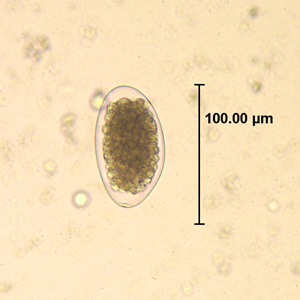
Trichuriasis, also known as whipworm infection, is an infection by the parasitic worm Trichuris trichiura (whipworm). If infection is only with a few worms, there are often no symptoms. In those who are infected with many worms, there may be abdominal pain, fatigue and diarrhea. The diarrhea sometimes contains blood. Infections in children may cause poor intellectual and physical development. Low red blood cell levels may occur due to loss of blood.

<i>Loa loa</i> filariasis Medical condition

Loa loa filariasis is a skin and eye disease caused by the nematode worm Loa loa. Humans contract this disease through the bite of a deer fly or mango fly, the vectors for Loa loa. The adult Loa loa filarial worm migrates throughout the subcutaneous tissues of humans, occasionally crossing into subconjunctival tissues of the eye where it can be easily observed. Loa loa does not normally affect one's vision but can be painful when moving about the eyeball or across the bridge of the nose. The disease can cause red itchy swellings below the skin called "Calabar swellings". The disease is treated with the drug diethylcarbamazine (DEC), and when appropriate, surgical methods may be employed to remove adult worms from the conjunctiva. Loiasis belongs to the so-called neglected diseases.

<i>Loa loa</i> Species of roundworm

Loa loa is a filarial (arthropod-borne) nematode (roundworm) that causes Loa loa filariasis. Loa loa actually means "worm worm", but is commonly known as the "eye worm", as it localizes to the conjunctiva of the eye. Loa loa is commonly found in Africa. It mainly inhabits rain forests in West Africa and has native origins in Ethiopia. The disease caused by Loa loa is called loiasis and is one of the neglected tropical diseases.

<span class="mw-page-title-main">Chronic wasting disease</span> Prion disease affecting the deer family

Chronic wasting disease (CWD), sometimes called zombie deer disease, is a transmissible spongiform encephalopathy (TSE) affecting deer. TSEs are a family of diseases thought to be caused by misfolded proteins called prions and include similar diseases such as BSE in cattle, Creutzfeldt–Jakob disease (CJD) in humans and scrapie in sheep. Natural infection causing CWD affects members of the deer family. In the United States, CWD affects mule deer, white-tailed deer, red deer, sika deer, elk, caribou, and moose. Experimental transmission of CWD to other species such as squirrel monkeys and genetically modified (humanized) mice has been shown.

<span class="mw-page-title-main">Onchocerciasis</span> Human helminthiasis (infection by parasite)

Onchocerciasis, also known as river blindness, is a disease caused by infection with the parasitic worm Onchocerca volvulus. Symptoms include severe itching, bumps under the skin, and blindness. It is the second-most common cause of blindness due to infection, after trachoma.

<i>Dirofilaria immitis</i> Species of worm that causes parasitic disease in animals

Dirofilaria immitis, also known as heartworm or dog heartworm, is a parasitic roundworm that is a type of filarial worm, a small thread-like worm, and which causes dirofilariasis. It is spread from host to host through the bites of mosquitoes. Four genera of mosquitoes transmit dirofilariasis, Aedes, Culex, Anopheles, and Mansonia. The definitive host is the dog, but it can also infect cats, wolves, coyotes, jackals, foxes, ferrets, bears, seals, sea lions and, under rare circumstances, humans.

<i>Trichinella</i> Genus of worms

Trichinella is the genus of parasitic roundworms of the phylum Nematoda that cause trichinosis. Members of this genus are often called trichinella or trichina worms. A characteristic of Nematoda is the one-way digestive tract, with a pseudocoelom.

<i>Dictyocaulus</i> Genus of nematode parasites of the bronchial tree of horses, sheep, goats, deer, and cattle

Dictyocaulus is a genus of nematode parasites of the bronchial tree of horses, sheep, goats, deer, and cattle. Dictyocaulus arnfieldi is the lungworm of horses, and Dictyocaulus viviparus is the lungworm affecting ruminants.

Elaeophora schneideri is a nematode which infests several mammalian hosts in North America. It is transmitted by horse-flies. Infection in the normal definitive hosts, mule deer or black-tailed deer, seldom produces clinical symptoms. In other hosts, such as sheep, elk, moose, and goats, infection with E. schneideri leads to elaeophorosis. Symptoms of elaeophorosis include necrosis of the muzzle, ears, and optic nerves; lack of coordination (ataxia); facial or lower limb dermatitis; horn deformities; blindness; and death.

<span class="mw-page-title-main">Moose sickness</span>

Moose sickness is a neurological condition seen in the northern mixed-wood forests of central and eastern North America where moose distribution overlaps with that of white-tailed deer. The disease is characterized by an unsteady gait, stumbling, head held to one side, circling, staying in one location, and in severe cases, being unable to get up.

Muelleries capillaris, also known as the hair or goat lungworm, is one of the most economically important nematodes of small ruminants. Although normally non-pathogenic in sheep, the parasite causes a disease condition called muelleriosis in goats. Sheep and goats commonly become infected after accidentally ingesting M. capillaris infected snails or slugs, and the parasite produces eggs in the lungs of its host, causing life-threatening bronchopneumonia in serious cases.

Lungworms are parasitic nematode worms of the order Strongylida that infest the lungs of vertebrates. The name is used for a variety of different groups of nematodes, some of which also have other common names; what they have in common is that they migrate to their hosts' lungs or respiratory tracts, and cause bronchitis or pneumonia. The lungworm will gradually damage the airways or lung tissue by inciting an inflammatory reaction inside the tissue. Ultimately, the parasites survive and reproduce in the respiratory tissues. The category is thus more a descriptive than a precisely taxonomic one.

<i>Toxocara cati</i> Species of worm

Toxocara cati, also known as the feline roundworm, is a parasite of cats and other felids. It is one of the most common nematodes of cats, infecting both wild and domestic felids worldwide. Adult worms are localised in the gut of the host. In adult cats, the infection – which is called toxocariasis – is usually asymptomatic. However, massive infection in juvenile cats can be fatal.

<span class="mw-page-title-main">Thelaziasis</span> Medical condition

Thelaziasis is the term for infestation with parasitic nematodes of the genus Thelazia. The adults of all Thelazia species discovered so far inhabit the eyes and associated tissues of various mammal and bird hosts, including humans. Thelazia nematodes are often referred to as "eyeworms".

<i>Trichostrongylus</i> Genus of roundworms

Trichostrongylus species are nematodes, which are ubiquitous among herbivores worldwide, including cattle, sheep, donkeys, goats, deer, and rabbits. At least 10 Trichostrongylus species have been associated with human infections. Infections occur via ingestion of infective larvae from contaminated vegetables or water. Epidemiological studies indicate a worldwide distribution of Trichostrongylus infections in humans, with the highest prevalence rates observed in individuals from regions with poor sanitary conditions, in rural areas, or who are farmers / herders. Human infections are most prevalent in the Middle East and Asia, with a worldwide estimated prevalence of 5.5 million people.

<i>Baylisascaris procyonis</i> Species of roundworm

Baylisascaris procyonis, also known by the common name raccoon roundworm, is a roundworm nematode, found ubiquitously in raccoons, the definitive hosts. It is named after H. A. Baylis, who studied them in the 1920s–30s, and Greek askaris. Baylisascaris larvae in paratenic hosts can migrate, causing larva migrans. Baylisascariasis as the zoonotic infection of humans is rare, though extremely dangerous due to the ability of the parasite's larvae to migrate into brain tissue and cause damage. Concern for human infection has been increasing over the years due to the urbanization of rural areas, resulting in the increase in proximity and potential human interaction with raccoons.

<i>Taenia hydatigena</i> Species of flatworm

Taenia hydatigena is one of the adult forms of the canine and feline tapeworm. This infection has a worldwide geographic distribution. Humans with taeniasis can infect other humans or animal intermediate hosts by eggs and gravid proglottids passed in the feces.

<span class="mw-page-title-main">Deer cutaneous fibroma</span> Disease of deer

Cutaneous fibromas are common neoplasms occurring in wild and domestic deer of many species and are caused by host-specific viral infections. The fibromas occur most frequently in animals under 2 years of age, with cases in older deer reported occasionally or rarely.

<i>Dermacentor albipictus</i> Species of tick

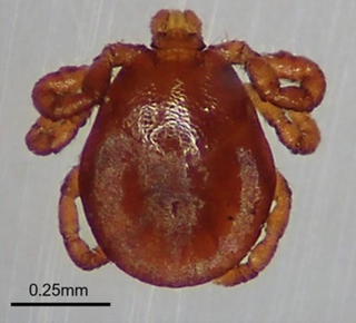
Dermacentor albipictus, the winter tick, is a species of hard tick that parasitizes many different mammal species in North America. It is commonly associated with cervid species such as elk, white-tailed deer, mule deer and caribou but is primarily known as a serious pest of moose. As early as 1909, Ernest Thompson Seton described the winter tick as a greater enemy of the moose than were "wolves, bears, and cougars."

Setaria cervi is a species of parasitic roundworms belonging to the genus Setaria. It infects cattle, bison, yak, reindeer, buffalo, moose, and sheep all over the world. It is most prevalent in Europe and Asia. Different species of Aedes mosquito can transmit the filarial worm. Stable fly Haematobia stimulans is the major vector. The mature roundworms are primarily present in the abdominal (peritoneal) cavity, but are capable of migrating to central nervous system causing serious neurological disease.

References

  1. 1 2 3 4 5 6 7 Duffy MS, Greaves TA, Keppie NJ, Burt MD (April 2002). "Meningeal worm is a long-lived parasitic nematode in white-tailed deer". Journal of Wildlife Diseases. 38 (2): 448–52. doi:10.7589/0090-3558-38.2.448. PMID   12038147. S2CID   39879199.
  2. Smith MC, et al. (1994). Goat Medicine. Vol. 150. Lea & Febiger.
  3. 1 2 ""Brain Worm" (Meningeal Worm) Infestation in Llamas and Alpacas". University of Tennessee. Retrieved 2013-11-14.
  4. 1 2 3 4 5 6 7 8 "DNR - Brainworm". Michigan DNR. Retrieved 2013-10-24.
  5. Fowler ME (1989). Medicine and Surgery of South American Camelids: Llama, Alpaca, Vicuna, Guanaco (First ed.). Ames, IA: Iowa State University Press. ISBN   978-0813803937 . Retrieved January 18, 2017.[ page needed ]
  6. DEBOW, JACOB; BLOUIN, JOSHUA; ROSENBLATT, ELIAS. "Effects of Winter Ticks and Internal Parasites on Moose Survival in Vermont, USA". The Journal of Wildlife Management.
  7. 1 2 3 4 5 "Brain worm". New York State Department of Environmental Conservation. Retrieved 2013-10-24.
  8. Karns PD, Jordan PA (1969). "Pneumostrongylus tenuis in moose on a deer-free island". The Journal of Wildlife Management. 33 (2): 431–433. doi:10.2307/3799850. JSTOR   3799850.
  9. 1 2 3 4 5 Lankester MW (2010). "Understanding the Impact of Meningeal Worm, Parelaphostrongylus tenuis, on Moose Populations". Alces. 46: 53–70. Retrieved January 18, 2017.
  10. 1 2 3 4 5 6 7 Karns, P.D. (1967). "Pneumostrongylus tenuis in deer in Minnesota and implications for moose". Journal of Wildlife Management. 31 (2): 299–303. doi:10.2307/3798320. JSTOR   3798320.
  11. 1 2 3 4 Wasel SM, Samuel WM, Crichton V (April 2003). "Distribution and ecology of meningeal worm, Parelaphostrongylus tenuis (Nematoda), in northcentral North America". Journal of Wildlife Diseases. 39 (2): 338–46. doi:10.7589/0090-3558-39.2.338. PMID   12910761. S2CID   23444907.
  12. 1 2 3 Mittelman NS, Divers TJ, Engiles JB, Gerhold R, Ness S, Scrivani PV, Southard T, Johnson AL (May 2017). "Parelaphostrongylus tenuis Cerebrospinal Nematodiasis in a Horse with Cervical Scoliosis and Meningomyelitis". Journal of Veterinary Internal Medicine. 31 (3): 890–893. doi:10.1111/jvim.14691. PMC   5435076 . PMID   28317172.
  13. Dobey CL, Grunenwald C, Newman SJ, Muller L, Gerhold RW (November 2014). "Retrospective study of central nervous system lesions and association with Parelaphostrongylus species by histology and specific nested polymerase chain reaction in domestic camelids and wild ungulates". Journal of Veterinary Diagnostic Investigation. 26 (6): 748–54. doi: 10.1177/1040638714553427 . PMID   25274743.
  14. 1 2 3 4 Murray DL, Cox EW, Ballard WB, Whitlaw HA, Lenarz MS, Custer TW, Barnett T, Fuller TK (2006). "Pathogens, nutritional deficiency, and climate influences on a declining moose population". Wildlife Monographs. 166 (1): 1–30. doi:10.2193/0084-0173(2006)166[1:PNDACI]2.0.CO;2. S2CID   17800980.
  15. 1 2 Tanabe M, Gerhold RW, Beckstead RB, de Lahunta A, Wade SE (July 2010). "Molecular confirmation of Parelaphostrongylus tenuis Infection in a horse with verminous encephalitis". Veterinary Pathology. 47 (4): 759. doi:10.1177/0300985810363488. PMID   20581347.
  16. 1 2 Garcia, Jessica; Smith, Joe; Fry, Michael; Mulon, Pierre-Yves (May 2023). "Clinical presentation, diagnostics, treatment, and outcome of goats diagnosed with presumptive cerebrospinal nematodiasis at a veterinary teaching hospital". The Canadian Veterinary Journal. 64 (5): 419–425. ISSN   0008-5286. PMC   10150576 . PMID   37138711.
  17. 1 2 Breuer, Ryan; Merkatoris, Paul; Tepley, Samantha; Dierks, Caitlyn; Klostermann, Cassandra; Flaherty, Heather; Smith, Joseph (2019-01-01). "Treatment of cerebrospinal nematodiasis in a Boer Buck". Veterinary Record Case Reports. 7 (1): e000706. doi:10.1136/vetreccr-2018-000706. ISSN   2052-6121. S2CID   86538320.
  18. 1 2 Smith, Joe; Kreuder, Amanda; Breuer, Ryan; Still-Brooks, Kelly (2019-08-12). "Meningeal Worm Infection in Central Iowa Goat Herds II: Individual Cases and Treatment Using a Camelid Therapeutic Protocol". Animal Industry Report. 2019 (1).
  19. Fenstermacher R, Jellison WL (1933). "Diseases affecting moose". University of Minnesota Agriculture Exponent State Bulletin. 6.
  20. Anderson RC (1964). "Neurologic disease in moose infected experimentally with Pneumostrongylus tenuis from white-tailed deer". Pathology. 1 (4): 289–322. doi: 10.1177/030098586400100402 .
  21. Loken KI, Schlotthauer JC, Kurtz HJ, Karns PD (1965). "Pneumostrongylus tenuis in Minnesota moose (Alces alces)". Bulletin Wildlife Disease Association. 1 (2): 7.
  22. Botzler RG, Brown RW (2014). Foundations of Wildlife Diseases. Univ. of Calif. Press. p. 62.
  23. Benson DA, Dodds GD (1997). The deer of Nova Scotia. Province of Nova Scotia: Department of Lands and Forests. Cited in: Telfer ES (2004). "Continuing environmental change-An example from Nova Scotia". The Canadian Field-Naturalist. 118 (1): 39–44. doi: 10.22621/cfn.v118i1.880 .
  24. Bindernagel JA, Anderson RC (1972). "Distribution of the meningeal worm in white-tailed deer in Canada". The Journal of Wildlife Management. 36 (4): 1349–1353. doi:10.2307/3799283. JSTOR   3799283.
  25. 1 2 3 Peterson B (1997). "Moose Walking in Circles" (online pdf of print issue). The Minnesota Volunteer (September–October): 24–31. Retrieved January 18, 2017.
  26. Kocan AA (October 1985). "The Use of Ivermectin in the Treatment and Prevention of Infection with Parelaphostrongylus tenuis (Dougherty) (Nematoda: Metastrongyloidea) in White-tailed Deer (Odocoileus virginianus Zimmermann)". Journal of Wildlife Diseases. 21 (4): 454–455. doi: 10.7589/0090-3558-21.4.454 . PMID   3841163.
  27. Haigh JC, Mackintosh C, Griffin F (2002-08-01). "Viral, parasitic and prion diseases of farmed deer and bison". Revue Scientifique et Technique de l'OIE. 21 (2): 219–248. doi: 10.20506/rst.21.2.1331 . PMID   11974612.