Photoactive yellow protein

Last updated
PYP
Identifiers
SymbolPYP
Pfam PF00989
InterPro IPR012130
SMART SM00091
PROSITE PS50112
SCOP2 55786 / SCOPe / SUPFAM
CDD cd00130
Available protein structures:
Pfam   structures / ECOD  
PDB RCSB PDB; PDBe; PDBj
PDBsum structure summary

In molecular biology, the PYP domain (photoactive yellow protein) is a p-coumaric acid-binding protein domain. They are present in various proteins in bacteria.

Contents

PYP is a highly soluble globular protein with an alpha/beta fold structure. It is a member of the PAS domain superfamily, which also contains a variety of other kinds of photosensory proteins.

PYP was first discovered in 1985. [1]

A recently (2016) developed chemogenetic system named FAST (Fluorescence-Activating and absorption Shifting Tag) was engineered from PYP to specifically and reversibly bind a series of hydroxybenzylidene rhodanine (HBR) derivatives for their fluorogenic properties. Upon interaction with FAST, the fluorogen is locked into a fluorescent conformation unlike when in solution. This new protein labelling system is used in a variety of microscopy and cytometry setups. [2]

p-Coumaric acid

p-Coumaric acid is a cofactor of Photoactive yellow protein|photoactive yellow proteins. [3] Adducts of p-coumaric acid bound to PYP form crystals that diffract well for x-ray crystallography experiments. These structural studies have provided insight into photosensitive proteins, e.g. the role of hydrogen bonding, molecular isomerization and photoactivity. [4] [5] [6] [7]

Photochemical transitions

It was originally believed that due to light emissions resembling that of retinal bound rhodopsin, the photosensor molecule bound to PYP should resemble the structure of retinal bound rhodopsin, the photosensor molecule bound to PYP should resemble the structure of retinal. [8] Scientists were therefore amazed when the PYP Cys 69 was bound by a thiol ester linkage as the light sensitive prosthetic group p-coumaric acid. [3] During the photoreactive mechanism: [3] [8]

  1. Light absorption yields the native protein to absorb a maximum wavelength of 446 nm, ε = 45500 M−1 cm−1.
  2. Within a nanosecond the absorbed maximum wavelength is shifted to 465 nm.
  3. Then on a sub-millisecond timescale is excited to a 355 nm state.
p-Coumaric acid exhibiting trans-cis isomerization due to absorption of light causing photochemical transitions. To the right demonstrates the molecular structure of p-coumaric acid in the ligand binding site and hydrogen bonding interactions involved in the innate oxyanion hole. To the left is a crystallographic image of p-coumaric acid in the ligand binding site based on PDB: 2PYP PyMOL rendering. 2019-03-11 Pcoumaric acid figures.pdf
p-Coumaric acid exhibiting transcis isomerization due to absorption of light causing photochemical transitions. To the right demonstrates the molecular structure of p-coumaric acid in the ligand binding site and hydrogen bonding interactions involved in the innate oxyanion hole. To the left is a crystallographic image of p-coumaric acid in the ligand binding site based on PDB: 2PYP PyMOL rendering.

These observed phenomena are due to the transcis isomerization of the vinyl trans double bond in the p-coumaric acid. [3] [7] [6] Scientists noted by observing the crystal structure of p-coumaric acid bound by PYP that the hydroxyl group connected to the C4 carbon of the phenyl ring appeared to be deprotonated – effectively a phenolate functional group. [7] [10] This was due to abnormally short hydrogen bonding lengths observed in the protein crystal structure. [9]

Role of hydrogen bonding

Hydrogen bonds in proteins such as PYP take part in interrelated networks, where at the center of p-coumaric acid's phenolate O4 atom, there is an oxyanion hole that is crucial for photosensory function. [6] [11] [12] Oxyanion holes exist in enzymes to stabilize transitions states of reaction intermediates, thus stabilizing the transcis isomerization of p-coumaric acid. [5] [13] During the transition state it is believed that the p-coumaric acid phenolate O4 takes part in a hydrogen bond network between Glu46, Tyr42 and Thr50 of PYP. [13] [5] These interactions are apart from the thiol ester linkage to Cys 69 keeping p-coumaric acid in the ligand binding site. [3] Upon transitioning to the cis-isomeric form of p-coumaric acid the favorable hydrogen bonds are no longer in close interaction.

Related Research Articles

<span class="mw-page-title-main">Denaturation (biochemistry)</span> Loss of structure in proteins and nucleic acids due to external stress

In biochemistry, denaturation is a process in which proteins or nucleic acids lose the quaternary structure, tertiary structure, and secondary structure which is present in their native state, by application of some external stress or compound such as a strong acid or base, a concentrated inorganic salt, an organic solvent, agitation and radiation or heat. If proteins in a living cell are denatured, this results in disruption of cell activity and possibly cell death. Protein denaturation is also a consequence of cell death. Denatured proteins can exhibit a wide range of characteristics, from conformational change and loss of solubility to aggregation due to the exposure of hydrophobic groups. The loss of solubility as a result of denaturation is called coagulation. Denatured proteins lose their 3D structure and therefore cannot function.

<span class="mw-page-title-main">Peptide bond</span> Covalent chemical bond between amino acids in a peptide or protein chain

In organic chemistry, a peptide bond is an amide type of covalent chemical bond linking two consecutive alpha-amino acids from C1 of one alpha-amino acid and N2 of another, along a peptide or protein chain.

In chemistry, a zwitterion, also called an inner salt or dipolar ion, is a molecule that contains an equal number of positively- and negatively-charged functional groups. With amino acids, for example, in solution a chemical equilibrium will be established between the "parent" molecule and the zwitterion.

Proline (symbol Pro or P) is an organic acid classed as a proteinogenic amino acid (used in the biosynthesis of proteins), although it does not contain the amino group -NH
2
but is rather a secondary amine. The secondary amine nitrogen is in the protonated form (NH2+) under biological conditions, while the carboxyl group is in the deprotonated −COO form. The "side chain" from the α carbon connects to the nitrogen forming a pyrrolidine loop, classifying it as a aliphatic amino acid. It is non-essential in humans, meaning the body can synthesize it from the non-essential amino acid L-glutamate. It is encoded by all the codons starting with CC (CCU, CCC, CCA, and CCG).

In biochemistry, a disulfide refers to a functional group with the structure R−S−S−R′. The linkage is also called an SS-bond or sometimes a disulfide bridge and is usually derived by the coupling of two thiol groups. In biology, disulfide bridges formed between thiol groups in two cysteine residues are an important component of the secondary and tertiary structure of proteins. Persulfide usually refers to R−S−S−H compounds.

<span class="mw-page-title-main">Flavin group</span> Group of chemical compounds

Flavins refers generally to the class of organic compounds containing the tricyclic heterocycle isoalloxazine or its isomer alloxazine, and derivatives thereof. The biochemical source of flavin is the vitamin riboflavin. The flavin moiety is often attached with an adenosine diphosphate to form flavin adenine dinucleotide (FAD), and, in other circumstances, is found as flavin mononucleotide, a phosphorylated form of riboflavin. It is in one or the other of these forms that flavin is present as a prosthetic group in flavoproteins.

<span class="mw-page-title-main">Rhodopsin</span> Light-sensitive receptor protein

Rhodopsin, also known as visual purple, is a protein encoded by the RHO gene and a G-protein-coupled receptor (GPCR). It is the opsin of the rod cells in the retina and a light-sensitive receptor protein that triggers visual phototransduction in rods. Rhodopsin mediates dim light vision and thus is extremely sensitive to light. When rhodopsin is exposed to light, it immediately photobleaches. In humans, it is regenerated fully in about 30 minutes, after which the rods are more sensitive. Defects in the rhodopsin gene cause eye diseases such as retinitis pigmentosa and congenital stationary night blindness.

<span class="mw-page-title-main">Transducin</span>

Transducin (Gt) is a protein naturally expressed in vertebrate retina rods and cones and it is very important in vertebrate phototransduction. It is a type of heterotrimeric G-protein with different α subunits in rod and cone photoreceptors.

<span class="mw-page-title-main">Bacteriorhodopsin</span> Protein used by single-celled organisms

Bacteriorhodopsin (Bop) is a protein used by Archaea, most notably by haloarchaea, a class of the Euryarchaeota. It acts as a proton pump; that is, it captures light energy and uses it to move protons across the membrane out of the cell. The resulting proton gradient is subsequently converted into chemical energy.

<span class="mw-page-title-main">Retinal</span> Chemical compound

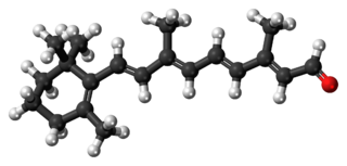
Retinal is a polyene chromophore. Retinal, bound to proteins called opsins, is the chemical basis of visual phototransduction, the light-detection stage of visual perception (vision).

<span class="mw-page-title-main">Proteorhodopsin</span> Family of transmembrane proteins

Proteorhodopsin is a family of transmembrane proteins that use retinal as a chromophore for light-mediated functionality, in this case, a proton pump. pRhodopsin is found in marine planktonic bacteria, archaea and eukaryotes (protae), but was first discovered in bacteria.

<span class="mw-page-title-main">Aconitase</span> Class of enzymes

Aconitase is an enzyme that catalyses the stereo-specific isomerization of citrate to isocitrate via cis-aconitate in the tricarboxylic acid cycle, a non-redox-active process.

<i>p</i>-Coumaric acid Chemical compound

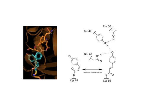
p-Coumaric acid is an organic compound with the formula HOC6H4CH=CHCO2H. It is one of the three isomers of hydroxycinnamic acid. It is a white solid that is only slightly soluble in water but very soluble in ethanol and diethyl ether.

In chemistry, persulfide refers to the functional group R-S-S-H. Persulfides are intermediates in the biosynthesis of iron-sulfur proteins and are invoked as precursors to hydrogen sulfide, a signaling molecule.

Retinylidene proteins, or rhodopsins in a broad sense, are proteins that use retinal as a chromophore for light reception. They are the molecular basis for a variety of light-sensing systems from phototaxis in flagellates to eyesight in animals. Retinylidene proteins include all forms of opsin and rhodopsin. While rhodopsin in the narrow sense refers to a dim-light visual pigment found in vertebrates, usually on rod cells, rhodopsin in the broad sense refers to any molecule consisting of an opsin and a retinal chromophore in the ground state. When activated by light, the chromophore is isomerized, at which point the molecule as a whole is no longer rhodopsin, but a related molecule such as metarhodopsin. However, it remains a retinylidene protein. The chromophore then separates from the opsin, at which point the bare opsin is a retinylidene protein. Thus, the molecule remains a retinylidene protein throughout the phototransduction cycle.

<span class="mw-page-title-main">Cystathionine beta-lyase</span> Enzyme

Cystathionine beta-lyase, also commonly referred to as CBL or β-cystathionase, is an enzyme that primarily catalyzes the following α,β-elimination reaction

<span class="mw-page-title-main">Retinal G protein coupled receptor</span> Protein-coding gene in the species Homo sapiens

RPE-retinal G protein-coupled receptor also known as RGR-opsin is a protein that in humans is encoded by the RGR gene. RGR-opsin is a member of the rhodopsin-like receptor subfamily of GPCR. Like other opsins which bind retinaldehyde, it contains a conserved lysine residue in the seventh transmembrane domain. RGR-opsin comes in different isoforms produced by alternative splicing.

<span class="mw-page-title-main">DsbA</span>

DsbA is a bacterial thiol disulfide oxidoreductase (TDOR). DsbA is a key component of the Dsb family of enzymes. DsbA catalyzes intrachain disulfide bond formation as peptides emerge into the cell's periplasm.

Halorhodospira halophila is a species of Halorhodospira distinguished by its ability to grow optimally in an environment of 15–20% salinity. It was formerly called Ectothiorhodospira halophila. It is an anaerobic, rod-shaped Gram-negative bacterium. H. halophila has a flagellum.

Archaerhodopsin proteins are a family of retinal-containing photoreceptors found in the archaea genera Halobacterium and Halorubrum. Like the homologous bacteriorhodopsin (bR) protein, archaerhodopsins harvest energy from sunlight to pump H+ ions out of the cell, establishing a proton motive force that is used for ATP synthesis. They have some structural similarities to the mammalian GPCR protein rhodopsin, but are not true homologs.

References

  1. Meyer TE (January 1985). "Isolation and characterization of soluble cytochromes, ferredoxins and other chromophoric proteins from the halophilic phototrophic bacterium Ectothiorhodospira halophila". Biochimica et Biophysica Acta (BBA) - Bioenergetics. 806 (1): 175–83. doi:10.1016/0005-2728(85)90094-5. PMID   2981543.
  2. Plamont MA, Billon-Denis E, Maurin S, Gauron C, Pimenta FM, Specht CG, et al. (January 2016). "Small fluorescence-activating and absorption-shifting tag for tunable protein imaging in vivo". Proceedings of the National Academy of Sciences of the United States of America. 113 (3): 497–502. doi: 10.1073/pnas.1513094113 . PMC   4725535 . PMID   26711992.
  3. 1 2 3 4 5 Hoff WD, Düx P, Hård K, Devreese B, Nugteren-Roodzant IM, Crielaard W, Boelens R, Kaptein R, van Beeumen J, Hellingwerf KJ (November 1994). "Thiol ester-linked p-coumaric acid as a new photoactive prosthetic group in a protein with rhodopsin-like photochemistry". Biochemistry. 33 (47): 13959–62. doi:10.1021/bi00251a001. PMID   7947803.
  4. "PDB101: Molecule of the Month: Photoactive Yellow Protein". RCSB: PDB-101. Retrieved 2019-03-12.
  5. 1 2 3 Pinney MM, Natarajan A, Yabukarski F, Sanchez DM, Liu F, Liang R, Doukov T, Schwans JP, Martinez TJ, Herschlag D (August 2018). "Structural Coupling Throughout the Active Site Hydrogen Bond Networks of Ketosteroid Isomerase and Photoactive Yellow Protein". Journal of the American Chemical Society. 140 (31): 9827–9843. doi:10.1021/jacs.8b01596. OSTI   1476146. PMID   29990421. S2CID   207190006.
  6. 1 2 3 Hellingwerf KJ (February 2000). "Key issues in the photochemistry and signalling-state formation of photosensor proteins". Journal of Photochemistry and Photobiology B: Biology. 54 (2–3): 94–102. doi:10.1016/S1011-1344(00)00004-X. PMID   10836537.
  7. 1 2 3 Premvardhan LL, Buda F, Van Der Horst MA, Lührs DC, Hellingwerf KJ, Van Grondelle R (2004-01-30). "Impact of Photon Absorption on the Electronic Properties of p -Coumaric Acid Derivatives of the Photoactive Yellow Protein Chromophore". The Journal of Physical Chemistry B. 108 (16): 5138–5148. doi:10.1021/jp037469b. hdl: 1887/3480076 .
  8. 1 2 Meyer TE, Yakali E, Cusanovich MA, Tollin G (January 1987). "Properties of a water-soluble, yellow protein isolated from a halophilic phototrophic bacterium that has photochemical activity analogous to sensory rhodopsin". Biochemistry. 26 (2): 418–23. doi:10.1021/bi00376a012. PMID   3828315.
  9. 1 2 Genick UK, Borgstahl GE, Ng K, Ren Z, Pradervand C, Burke PM, Srajer V, Teng TY, Schildkamp W, McRee DE, Moffat K, Getzoff ED (March 1997). "Structure of a protein photocycle intermediate by millisecond time-resolved crystallography". Science. 275 (5305): 1471–5. doi:10.1126/science.275.5305.1471. PMID   9045611. S2CID   20434371.
  10. Yamaguchi S, Kamikubo H, Kurihara K, Kuroki R, Niimura N, Shimizu N, Yamazaki Y, Kataoka M (January 2009). "Low-barrier hydrogen bond in photoactive yellow protein". Proceedings of the National Academy of Sciences of the United States of America. 106 (2): 440–4. Bibcode:2009PNAS..106..440Y. doi: 10.1073/pnas.0811882106 . PMC   2626721 . PMID   19122140.
  11. Borgstahl GE, Williams DR, Getzoff ED (May 1995). "1.4 Å structure of photoactive yellow protein, a cytosolic photoreceptor: unusual fold, active site, and chromophore". Biochemistry. 34 (19): 6278–87. doi:10.1021/bi00019a004. PMID   7756254.
  12. Stahl AD, Hospes M, Singhal K, van Stokkum I, van Grondelle R, Groot ML, Hellingwerf KJ (September 2011). "On the involvement of single-bond rotation in the primary photochemistry of photoactive yellow protein". Biophysical Journal. 101 (5): 1184–92. Bibcode:2011BpJ...101.1184S. doi:10.1016/j.bpj.2011.06.065. PMC   3164125 . PMID   21889456.
  13. 1 2 Herschlag D, Pinney MM (June 2018). "Hydrogen Bonds: Simple after All?". Biochemistry. 57 (24): 3338–3352. doi: 10.1021/acs.biochem.8b00217 . PMID   29678112.

Further reading

This article incorporates text from the public domain Pfam and InterPro: IPR012130