Photon-counting mammography

Last updated

Photon-counting mammography was introduced commercially in 2003 and was the first widely available application of photon-counting detector technology in medical x-ray imaging. [1] Photon-counting mammography improves dose efficiency compared to conventional technologies, [2] and enables spectral imaging.

Contents

Background

Conventional detectors for x-ray imaging are energy integrating, i.e., all photon interactions over a certain time interval are integrated. Photon-counting detectors, on the other hand, are fast enough to register single photon events. Photon-counting detectors have been used in nuclear medicine for decades, but their introduction to transmission imaging was relatively late, mainly as a result of the higher flux that leads to an unwanted condition called pulse pileup, which is one of the main challenges for photon-counting detectors. Partly for this reason, the first, and to date (2020) only, widely available photon-counting x-ray imaging modality is a mammography system; mammography requires high spatial resolution, which results in small detector elements and therefore relatively low count rates. [1] The MicroDose mammography system was introduced by Sectra Mamea in 2003 and acquired by Philips in 2011.

The MicroDose system is based on an array of silicon strip detectors in a multi-slit configuration that is scanned across the object to acquire an image. [1] When a photon interacts in the detector, charge is released and collected by electrodes on the sensor. The electrodes are connected to parallel channels in an application-specific integrated circuit (ASIC). Each channel comprises an amplifier and a shaper, which convert the charge to a pulse with an amplitude proportional to the energy of the impinging photon. This pulse height is measured by comparators, generally referred to as energy thresholds, which are followed by corresponding counters. The counters register the sum of all events within a specific energy window and are generally referred to as energy bins. The lower-most threshold is put below the expected incident spectrum to prevent electronic noise from being counted. The energy resolution of the MicroDose detector ranges from 2.0–2.3 keV standard deviation in the range 20–40 keV. [1]

The advantages of silicon as a detector material include high charge-collection efficiency, ready availability of high-quality high-purity material, and established methods for test and assembly driven by the semiconductor industry. The main challenge is the relatively low detection efficiency of silicon, which in the MicroDose system is addressed by arranging the silicon wafers edge on. [1] Several research groups and commercial companies are investigating cadmium–zinc telluride (CZT) as sensor material. [3] The higher atomic number of CZT results in higher absorption than silicon, but the higher K-fluorescent yield degrades the spectral response and leads to cross-talk. [4] Also, manufacturing of macro-sized crystals of these materials poses practical challenges, and the crystals generally suffer from lattice defects and impurities that lead to charge trapping, which limits charge-collection efficiency, [5] and may cause long-term polarization effects. [6] Other solid-state materials, such as gallium arsenide, [7] and gas detectors, [8] are currently quite far from clinical implementation.

Clinical applications

Improved dose efficiency

Photon-counting mammography allows for a reduction of patient dose while keeping image quality on par with conventional technologies, or, equivalently, improving image quality at equal dose. A study that compared photon-counting mammography to the state-wide average of the North Rhine-Westphalian mammography screening program in Germany reported a slightly improved diagnostic performance at a dose that was 40% of conventional technologies. [2]

Improved dose efficiency in photon-counting mammography compared to conventional technologies is mainly enabled by:

Energy weighting

Even though equal weighting of photons, intrinsic to photon-counting detectors, improves dose efficiency compared to energy-integrating detectors, a higher weighting of low-energy photons is generally optimal because x-ray contrast drops with increasing photon energy when the photo-electric effect dominates and away from any absorption edges, which holds true for mammography without contrast agents. Photon-counting detectors allow for measuring the energy of impinging photons and therefore enable optimal weighting for a given imaging case. This technique, generally referred to as energy weighting, was pioneered for mammography applications by Cahn et al. [9] At the limit of infinite energy resolution, energy weighting results in a CNR improvement of approximately 10% compared to equal weighting of photons, [9] whereas studies with realistic energy resolution report CNR improvements of a few percent. [11] The first results in clinical application were reported by Berglund et al. who was able to improve the CNR of clinical images by 2.2–5.2%, which translates to a potential dose reduction at a constant CNR in the range of 4.5%–11%. [12]

Material decomposition and K-edge imaging

Photon-counting detectors can measure the energy spectrum of the incident photons, a technique broadly referred to as spectral imaging. Spectral information can be used to extract quantitative information about the composition of the object because the two dominant x-ray interaction effects at mammographic energies, the photo-electric effect and Compton scattering, have different energy and material dependencies. So-called material decomposition has been investigated to measure breast density, [13] improve lesion visibility, [11] characterize lesions, [14] and to measure the attenuation of breast constituents. [15] [16]

High-atomic-number contrast agents add additional energy dependencies to the object and can be separated from soft tissue, often referred to as K-edge imaging. In photon-counting mammography, contrast-enhanced imaging has been focused on iodine imaging. [17] [18]

Tomosynthesis

Photon-counting breast tomosynthesis has been developed to a prototype state. [19] [20] [21] Tomosynthesis relies on a number of low-dose projections, which makes the influence of electronic noise higher than for conventional mammography, and photon-counting detectors with rejection of electronic noise are therefore beneficial with potentially higher improvements in dose efficiency than for conventional mammography. Further, the slit-scanning technique is expected to provide additional benefit because scatter rejection based on conventional anti-scatter grids is challenging to implement at a range of projection angles. However, slit-scanning tomosynthesis requires larger modifications of existing systems, and the technique has so far not been applied in wide-spread clinical use. Applications of spectral imaging that have been investigated for photon-counting tomosynthesis include breast-density measurement, [22] lesion characterization, [23] and iodine K-edge imaging. [24] [25] [26]

Phase-contrast imaging

The slit-scanning geometry of some implementations of photon-counting mammography offers advantages for implementation of grating-based phase-contrast imaging. [27] Phantom studies [28] and pre-clinical imaging [29] has been conducted with this technique and initial promising results have been reported. The spectral resolution of photon-counting detectors can potentially be used to reduce the coherence requirements of phase-contrast imaging setups. [30]

Related Research Articles

<span class="mw-page-title-main">X-ray</span> Form of short-wavelength electromagnetic radiation

X-ray radiation, or, much less commonly, X-radiation, is a penetrating form of high-energy electromagnetic radiation. Most X-rays have a wavelength ranging from 10 nanometers to 10 picometers, corresponding to frequencies in the range 30 petahertz to 30 exahertz (3×10¹⁶ Hz to 3×10¹⁹ Hz) and energies in the range 124 keV to 145 eV, respectively. X-ray wavelengths are shorter than those of UV rays and typically longer than those of gamma rays. In many languages, X-radiation is referred to as Röntgen radiation, after the German scientist Wilhelm Conrad Röntgen, who discovered it on November 8, 1895. He named it X-radiation to signify an unknown type of radiation. Spellings of X-ray(s) in English include the variants x-ray(s), xray(s), and X ray(s).
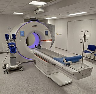
<span class="mw-page-title-main">CT scan</span> Medical imaging procedure using X-rays to produce cross-sectional images

A computed tomography scan is a medical imaging technique used to obtain detailed internal images of the body. The personnel that perform CT scans are called radiographers or radiology technologists.
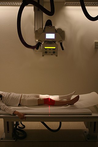
<span class="mw-page-title-main">Radiography</span> Imaging technique using ionizing and non-ionizing radiation

Radiography is an imaging technique using X-rays, gamma rays, or similar ionizing radiation and non-ionizing radiation to view the internal form of an object. Applications of radiography include medical radiography and industrial radiography. Similar techniques are used in airport security. To create an image in conventional radiography, a beam of X-rays is produced by an X-ray generator and is projected toward the object. A certain amount of the X-rays or other radiation is absorbed by the object, dependent on the object's density and structural composition. The X-rays that pass through the object are captured behind the object by a detector. The generation of flat two dimensional images by this technique is called projectional radiography. In computed tomography an X-ray source and its associated detectors rotate around the subject which itself moves through the conical X-ray beam produced. Any given point within the subject is crossed from many directions by many different beams at different times. Information regarding attenuation of these beams is collated and subjected to computation to generate two dimensional images in three planes which can be further processed to produce a three dimensional image.

<span class="mw-page-title-main">Mammography</span> Process of using low-energy X-rays to examine the human breast for diagnosis and screening

Mammography is the process of using low-energy X-rays to examine the human breast for diagnosis and screening. The goal of mammography is the early detection of breast cancer, typically through detection of characteristic masses or microcalcifications.

<span class="mw-page-title-main">Gamma camera</span> Camera to record gamma radiation

A gamma camera (γ-camera), also called a scintillation camera or Anger camera, is a device used to image gamma radiation emitting radioisotopes, a technique known as scintigraphy. The applications of scintigraphy include early drug development and nuclear medical imaging to view and analyse images of the human body or the distribution of medically injected, inhaled, or ingested radionuclides emitting gamma rays.

Technetium (<sup>99m</sup>Tc) sestamibi Pharmaceutical drug

Technetium (99mTc) sestamibi (INN) is a pharmaceutical agent used in nuclear medicine imaging. The drug is a coordination complex consisting of the radioisotope technetium-99m bound to six (sesta=6) methoxyisobutylisonitrile (MIBI) ligands. The anion is not defined. The generic drug became available late September 2008. A scan of a patient using MIBI is commonly known as a "MIBI scan".

Charge sharing is an effect of signal degradation through transfer of charges from one electronic domain to another.

X-ray optics is the branch of optics that manipulates X-rays instead of visible light. It deals with focusing and other ways of manipulating the X-ray beams for research techniques such as X-ray crystallography, X-ray fluorescence, small-angle X-ray scattering, X-ray microscopy, X-ray phase-contrast imaging, and X-ray astronomy.

<span class="mw-page-title-main">Tomosynthesis</span>

Tomosynthesis, also digital tomosynthesis (DTS), is a method for performing high-resolution limited-angle tomography at radiation dose levels comparable with projectional radiography. It has been studied for a variety of clinical applications, including vascular imaging, dental imaging, orthopedic imaging, mammographic imaging, musculoskeletal imaging, and chest imaging.

A Compound refractive lens (CRL) is a series of individual lenses arranged in a linear array in order to achieve focusing of X-rays in the energy range of 5-40 keV. They are an alternative to the KB mirror.

<span class="mw-page-title-main">Flat-panel detector</span> Class of solid-state x-ray digital radiography devices

Flat-panel detectors are a class of solid-state x-ray digital radiography devices similar in principle to the image sensors used in digital photography and video. They are used in both projectional radiography and as an alternative to x-ray image intensifiers (IIs) in fluoroscopy equipment.

<span class="mw-page-title-main">Positron emission mammography</span> Imaging procedure used to detect breast cancer

Positron emission mammography (PEM) is a nuclear medicine imaging modality used to detect or characterise breast cancer. Mammography typically refers to x-ray imaging of the breast, while PEM uses an injected positron emitting isotope and a dedicated scanner to locate breast tumors. Scintimammography is another nuclear medicine breast imaging technique, however it is performed using a gamma camera. Breasts can be imaged on standard whole-body PET scanners, however dedicated PEM scanners offer advantages including improved resolution.
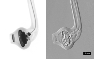
<span class="mw-page-title-main">Phase-contrast X-ray imaging</span> Imaging systems using changes in phase

Phase-contrast X-ray imaging or phase-sensitive X-ray imaging is a general term for different technical methods that use information concerning changes in the phase of an X-ray beam that passes through an object in order to create its images. Standard X-ray imaging techniques like radiography or computed tomography (CT) rely on a decrease of the X-ray beam's intensity (attenuation) when traversing the sample, which can be measured directly with the assistance of an X-ray detector. However, in phase contrast X-ray imaging, the beam's phase shift caused by the sample is not measured directly, but is transformed into variations in intensity, which then can be recorded by the detector.

<span class="mw-page-title-main">X-ray detector</span> Instrument that can measure properties of X-rays

X-ray detectors are devices used to measure the flux, spatial distribution, spectrum, and/or other properties of X-rays.

<span class="mw-page-title-main">Photon counting</span> Counting photons using a single-photon detector

Photon counting is a technique in which individual photons are counted using a single-photon detector (SPD). A single-photon detector emits a pulse of signal for each detected photon. The counting efficiency is determined by the quantum efficiency and the system's electronic losses.
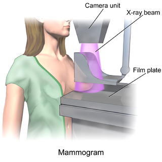
<span class="mw-page-title-main">Breast imaging</span>

In medicine, breast imaging is a sub-speciality of diagnostic radiology that involves imaging of the breasts for screening or diagnostic purposes. There are various methods of breast imaging using a variety of technologies as described in detail below. Traditional screening and diagnostic mammography uses x-ray technology and has been the mainstay of breast imaging for many decades. Breast tomosynthesis is a relatively new digital x-ray mammography technique that produces multiple image slices of the breast similar to, but distinct from, computed tomography (CT). Xeromammography and galactography are somewhat outdated technologies that also use x-ray technology and are now used infrequently in the detection of breast cancer. Breast ultrasound is another technology employed in diagnosis and screening that can help differentiate between fluid filled and solid lesions, an important factor to determine if a lesion may be cancerous. Breast MRI is a technology typically reserved for high-risk patients and patients recently diagnosed with breast cancer. Lastly, scintimammography is used in a subgroup of patients who have abnormal mammograms or whose screening is not reliable on the basis of using traditional mammography or ultrasound.

Jeffrey Harold Siewerdsen is an American physicist and biomedical engineer who is a Professor of Biomedical Engineering, Computer Science, Radiology, and Neurosurgery at Johns Hopkins University. He is Co-Director of the Carnegie Center for Surgical Innovation at Johns Hopkins School of Medicine and is a member of the Malone Center for Engineering in Healthcare. He is among the original inventors of cone-beam CT-guided radiotherapy as well as weight-bearing cone-beam CT for musculoskeletal radiology and orthopedic surgery. His work also includes the early development of flat-panel detectors on mobile C-arms for intraoperative cone-beam CT in image-guided surgery. He developed early models for the signal and noise performance of flat-panel detectors and later extended such analysis to dual-energy imaging and 3D imaging performance in cone-beam CT. His core laboratory at Johns Hopkins University is the ISTAR Lab in the Department of Biomedical Engineering at the Johns Hopkins Hospital.

Photon-counting computed tomography (PCCT) is a form of X-ray computed tomography (CT) in which X-rays are detected using a photon-counting detector (PCD) which registers the interactions of individual photons. By keeping track of the deposited energy in each interaction, the detector pixels of a PCD each record an approximate energy spectrum, making it a spectral or energy-resolved CT technique. In contrast, more conventional CT scanners use energy-integrating detectors (EIDs), where the total energy deposited in a pixel during a fixed period of time is registered. These EIDs thus register only photon intensity, comparable to black-and-white photography, whereas PCDs register also spectral information, similar to color photography.

Spectral imaging is an umbrella term for energy-resolved X-ray imaging in medicine. The technique makes use of the energy dependence of X-ray attenuation to either increase the contrast-to-noise ratio, or to provide quantitative image data and reduce image artefacts by so-called material decomposition. Dual-energy imaging, i.e. imaging at two energy levels, is a special case of spectral imaging and is still the most widely used terminology, but the terms "spectral imaging" and "spectral CT" have been coined to acknowledge the fact that photon-counting detectors have the potential for measurements at a larger number of energy levels.

Diffuse optical mammography, or simply optical mammography, is an emerging imaging technique that enables the investigation of the breast composition through spectral analysis. It combines in a single non-invasive tool the capability to implement breast cancer risk assessment, lesion characterization, therapy monitoring and prediction of therapy outcome. It is an application of diffuse optics, which studies light propagation in strongly diffusive media, such as biological tissues, working in the red and near-infrared spectral range, between 600 and 1100 nm.

References

  1. 1 2 3 4 5 6 Fredenberg E, Lundqvist M, Cederström B, Åslund M, Danielsson M (2010). "Energy resolution of a photon-counting silicon strip detector". Nuclear Instruments and Methods in Physics Research Section A: Accelerators, Spectrometers, Detectors and Associated Equipment. 613 (1): 156–162. arXiv: 2101.07789 . Bibcode:2010NIMPA.613..156F. doi:10.1016/j.nima.2009.10.152. ISSN   0168-9002. S2CID   121348971.
  2. 1 2 Weigel S, Berkemeyer S, Girnus R, Sommer A, Lenzen H, Heindel W (May 2014). "Digital mammography screening with photon-counting technique: can a high diagnostic performance be realized at low mean glandular dose?". Radiology. 271 (2): 345–55. doi: 10.1148/radiol.13131181 . PMID   24495234.
  3. Iwanczyk JS, Nygård E, Meirav O, Arenson J, Barber WC, Hartsough NE, et al. (2009). "Photon Counting Energy Dispersive Detector Arrays for X-ray Imaging". IEEE Transactions on Nuclear Science. 56 (3): 535–542. Bibcode:2009ITNS...56..535I. doi:10.1109/TNS.2009.2013709. PMC   2777741 . PMID   19920884.
  4. Shikhaliev PM, Fritz SG, Chapman JW (November 2009). "Photon counting multienergy x-ray imaging: effect of the characteristic x rays on detector performance". Medical Physics. 36 (11): 5107–19. Bibcode:2009MedPh..36.5107S. doi:10.1118/1.3245875. PMID   19994521.
  5. Knoll GF (2000). Radiation detection and measurement. John Wiley & Sons. ISBN   978-0-471-07338-3.
  6. Szeles C, Soldner SA, Vydrin S, Graves J, Bale DS (2008). "CdZnTe Semiconductor Detectors for Spectroscopic X-ray Imaging". IEEE Transactions on Nuclear Science. 55 (1): 572–582. Bibcode:2008ITNS...55..572S. doi:10.1109/TNS.2007.914034. ISSN   0018-9499. S2CID   43453671.
  7. Amendolia SR, Bisogni MG, Delogu P, Fantacci ME, Paternoster G, Rosso V, Stefanini A (April 2009). "Characterization of a mammographic system based on single photon counting pixel arrays coupled to GaAs x-ray detectors". Medical Physics. 36 (4): 1330–9. Bibcode:2009MedPh..36.1330A. doi:10.1118/1.3097284. PMID   19472640.
  8. Thunberg SJ, Adelow L, Blom O, Coster A, Egerstrom J, Eklund M, et al. (2004-05-06). Yaffe MJ, Flynn MJ (eds.). "Dose reduction in mammography with photon counting imaging". Medical Imaging 2004: Physics of Medical Imaging. International Society for Optics and Photonics. 5368: 457–465. Bibcode:2004SPIE.5368..457T. doi:10.1117/12.530649. S2CID   72756191.
  9. 1 2 3 Cahn RN, Cederström B, Danielsson M, Hall A, Lundqvist M, Nygren D (December 1999). "Detective quantum efficiency dependence on x-ray energy weighting in mammography". Medical Physics. 26 (12): 2680–3. Bibcode:1999MedPh..26.2680C. doi:10.1118/1.598807. PMID   10619253.
  10. Aslund M, Cederström B, Lundqvist M, Danielsson M (April 2006). "Scatter rejection in multislit digital mammography". Medical Physics. 33 (4): 933–40. Bibcode:2006MedPh..33..933A. doi:10.1118/1.2179122. PMID   16696469.
  11. 1 2 Fredenberg E, Åslund M, Cederström B, Lundqvist M, Danielsson M (2010). "Observer model optimization of a spectral mammography system". Medical Imaging 2010: Physics of Medical Imaging. International Society for Optics and Photonics. 7622: 762210. arXiv: 2101.09440 . Bibcode:2010SPIE.7622E..10F. doi:10.1117/12.845480. S2CID   121950145.
  12. Berglund J, Johansson H, Lundqvist M, Cederström B, Fredenberg E (October 2014). "Energy weighting improves dose efficiency in clinical practice: implementation on a spectral photon-counting mammography system". Journal of Medical Imaging. 1 (3): 031003. doi:10.1117/1.JMI.1.3.031003. PMC   4478791 . PMID   26158045.
  13. Ding H, Molloi S (August 2012). "Quantification of breast density with spectral mammography based on a scanned multi-slit photon-counting detector: a feasibility study". Physics in Medicine and Biology. 57 (15): 4719–4738. Bibcode:2012PMB....57.4719D. doi:10.1088/0031-9155/57/15/4719. PMC   3478949 . PMID   22771941.
  14. Ghammraoui B, Glick SJ (June 2017). "Investigating the feasibility of classifying breast microcalcifications using photon-counting spectral mammography: A simulation study". Medical Physics. 44 (6): 2304–2311. Bibcode:2017MedPh..44.2304G. doi:10.1002/mp.12230. PMID   28332199. S2CID   38228845.
  15. Berggren K, Eriksson M, Hall P, Wallis MG, Fredenberg E (October 2018). "In vivo measurement of the effective atomic number of breast skin using spectral mammography". Physics in Medicine and Biology. 63 (21): 215023. Bibcode:2018PMB....63u5023B. doi: 10.1088/1361-6560/aae78c . PMID   30375362.
  16. Fredenberg E, Willsher P, Moa E, Dance DR, Young KC, Wallis MG (November 2018). "Measurement of breast-tissue x-ray attenuation by spectral imaging: fresh and fixed normal and malignant tissue". Physics in Medicine and Biology. 63 (23): 235003. arXiv: 2101.02755 . Bibcode:2018PMB....63w5003F. doi:10.1088/1361-6560/aaea83. PMID   30465547. S2CID   53717425.
  17. Ding H, Molloi S (August 2017). "Quantitative contrast-enhanced spectral mammography based on photon-counting detectors: A feasibility study". Medical Physics. 44 (8): 3939–3951. Bibcode:2017MedPh..44.3939D. doi:10.1002/mp.12296. PMC   5553693 . PMID   28432828.
  18. Fredenberg E, Hemmendorff M, Cederström B, Aslund M, Danielsson M (May 2010). "Contrast-enhanced spectral mammography with a photon-counting detector". Medical Physics. 37 (5): 2017–2029. arXiv: 2101.07787 . Bibcode:2010MedPh..37.2017F. doi:10.1118/1.3371689. PMID   20527535. S2CID   31601055.
  19. Wallis MG, Moa E, Zanca F, Leifland K, Danielsson M (March 2012). "Two-view and single-view tomosynthesis versus full-field digital mammography: high-resolution X-ray imaging observer study". Radiology. 262 (3): 788–796. doi:10.1148/radiol.11103514. PMID   22274840.
  20. Berggren K, Cederström B, Lundqvist M, Fredenberg E (October 2018). "Cascaded systems analysis of shift-variant image quality in slit-scanning breast tomosynthesis". Medical Physics. 45 (10): 4392–4401. Bibcode:2018MedPh..45.4392B. doi:10.1002/mp.13116. PMID   30091470. S2CID   51941096.
  21. Maidment AD, Ullberg C, Lindman K, Adelöw L, Egerström J, Eklund M, et al. (2006-03-02). Flynn MJ, Hsieh J (eds.). "Evaluation of a photon-counting breast tomosynthesis imaging system". Medical Imaging 2006: Physics of Medical Imaging. SPIE. 6142: 89–99. Bibcode:2006SPIE.6142...89M. doi:10.1117/12.654651. S2CID   173179592.
  22. Fredenberg E, Berggren K, Bartels M, Erhard K (2016). "Volumetric Breast-Density Measurement Using Spectral Photon-Counting Tomosynthesis: First Clinical Results". In Tingberg A, Lång K, Timberg P (eds.). Breast Imaging. Lecture Notes in Computer Science. Vol. 9699. Cham: Springer International Publishing. pp. 576–584. arXiv: 2101.02758 . doi:10.1007/978-3-319-41546-8_72. ISBN   978-3-319-41546-8. S2CID   1257696.
  23. Cederström B, Fredenberg E, Berggren K, Erhard K, Danielsson M, Wallis M (2017). Flohr TG, Lo JY, Schmidt GT (eds.). "Lesion characterization in spectral photon-counting tomosynthesis". Medical Imaging 2017: Physics of Medical Imaging. International Society for Optics and Photonics. 10132: 1013205. arXiv: 2102.00175 . Bibcode:2017SPIE10132E..05C. doi:10.1117/12.2253966. S2CID   125903314.
  24. Schmitzberger FF, Fallenberg EM, Lawaczeck R, Hemmendorff M, Moa E, Danielsson M, et al. (May 2011). "Development of low-dose photon-counting contrast-enhanced tomosynthesis with spectral imaging". Radiology. 259 (2): 558–564. doi:10.1148/radiol.11101682. PMID   21330558.
  25. Carton AK, Ullberg C, Lindman K, Acciavatti R, Francke T, Maidment AD (November 2010). "Optimization of a dual-energy contrast-enhanced technique for a photon-counting digital breast tomosynthesis system: I. A theoretical model". Medical Physics. 37 (11): 5896–5907. Bibcode:2010MedPh..37.5896C. doi:10.1118/1.3490556. PMC   3188980 . PMID   21158302.
  26. Carton AK, Ullberg C, Maidment AD (November 2010). "Optimization of a dual-energy contrast-enhanced technique for a photon-counting digital breast tomosynthesis system: II. An experimental validation". Medical Physics. 37 (11): 5908–5913. Bibcode:2010MedPh..37.5908C. doi:10.1118/1.3488889. PMC   3188981 . PMID   21158303.
  27. Fredenberg E, Danielsson M, Stayman JW, Siewerdsen JH, Aslund M (September 2012). "Ideal-observer detectability in photon-counting differential phase-contrast imaging using a linear-systems approach". Medical Physics. 39 (9): 5317–35. Bibcode:2012MedPh..39.5317F. doi:10.1118/1.4739195. PMC   3427340 . PMID   22957600.
  28. Koehler T, Daerr H, Martens G, Kuhn N, Löscher S, van Stevendaal U, Roessl E (April 2015). "Slit-scanning differential x-ray phase-contrast mammography: proof-of-concept experimental studies". Medical Physics. 42 (4): 1959–65. Bibcode:2015MedPh..42.1959K. doi: 10.1118/1.4914420 . PMID   25832086.
  29. Arboleda C, Wang Z, Jefimovs K, Koehler T, Van Stevendaal U, Kuhn N, et al. (March 2020). "Towards clinical grating-interferometry mammography". European Radiology. 30 (3): 1419–1425. doi:10.1007/s00330-019-06362-x. PMC   7033145 . PMID   31440834.
  30. Fredenberg E, Roessl E, Koehler T, van Stevendaal U, Schulze-Wenck I, Wieberneit N, et al. (2012-02-23). "Photon-counting spectral phase-contrast mammography". In Pelc NJ, Nishikawa RM, Whiting BR (eds.). Medical Imaging 2012: Physics of Medical Imaging. Vol. 8313. San Diego, California, USA. pp. 83130F. arXiv: 2101.09660 . doi:10.1117/12.910615. S2CID   121130207.{{cite book}}: CS1 maint: location missing publisher (link)