Photosynthetic reaction centre protein family

Last updated
Type II reaction centre protein
Photosynthetic Reaction Center Drawing.png
Structure of the photosynthetic reaction centre from Rhodopseudomonas viridis ( PDB: 1PRC ). Middle transmembrane section is the two subunits in this family; green blocks represent chlorophyll. Top section is the 4-heme (red) cytochrome c subunit (infobox below). The bottom section along with its connected TM helices is the H subunit.
Identifiers
SymbolPhoto_RC
Pfam PF00124
InterPro IPR000484
PROSITE PDOC00217
SCOP2 1prc / SCOPe / SUPFAM
TCDB 3.E.2
OPM superfamily 2
OPM protein 1dxr
Available protein structures:
Pfam   structures / ECOD  
PDB RCSB PDB; PDBe; PDBj
PDBsum structure summary
PDB 1clt M:37-306 4prc M:37-306 1r2c M:37-306

1prc M:37-306 5prc M:37-306 1vrn M:37-306 3prc M:37-306 2prc M:37-306 6prc M:37-306 7prc M:37-306 1dxr M:37-306 1dop A:105-296

Contents

2axt A:28-330 1s5l D:28-327
Type I reaction centre protein
1jb0 opm.png
Side view of Cyanobacterial photosystem I. Large near-symmetrical proteins in the center, colored blue and pink, are the two subunits of this family.
Identifiers
SymbolPsaA_PsaB
Pfam PF00223
InterPro IPR001280
PROSITE PDOC00347
SCOP2 1jb0 / SCOPe / SUPFAM
TCDB 5.B.4
OPM superfamily 2
OPM protein 1jb0
Membranome 535
Available protein structures:
Pfam   structures / ECOD  
PDB RCSB PDB; PDBe; PDBj
PDBsum structure summary
Bacterial type II reaction centre, cytochrome c subunit
Identifiers
SymbolCytoC_RC
Pfam PF02276
Pfam clan CL0317
InterPro IPR003158
SCOP2 1prc / SCOPe / SUPFAM
Available protein structures:
Pfam   structures / ECOD  
PDB RCSB PDB; PDBe; PDBj
PDBsum structure summary

Photosynthetic reaction centre proteins are main protein components of photosynthetic reaction centres (RCs) of bacteria and plants. They are transmembrane proteins embedded in the chloroplast thylakoid or bacterial cell membrane.

Plants, algae, and cyanobacteria have one type of PRC for each of its two photosystems. Non-oxygenic bacteria, on the other hand, have an RC resembling either the Photosystem I centre (Type I) or the Photosystem II centre (Type II). In either case, PRCs have two related proteins (L/M; D1/D2; PsaA/PsaB) making up a quasi-symmetrical 5-helical core complex with pockets for pigment binding. The two types are structurally related and share a common ancestor. [1] [2] Each type have different pockets for ligands to accommodate their specific reactions: while Type I RCs use iron sulfur clusters to accept electrons, Type II RCs use quinones. The centre units of Type I RCs also have six extra transmembrane helices for gathering energy. [2]

In bacteria

The Type II photosynthetic apparatus in non-oxygenic bacteria consists of light-harvesting protein-pigment complexes LH1 and LH2, which use carotenoid and bacteriochlorophyll as primary donors. [3] LH1 acts as the energy collection hub, temporarily storing it before its transfer to the photosynthetic reaction centre (RC). [4] Electrons are transferred from the primary donor via an intermediate acceptor (bacteriophaeophytin) to the primary acceptor (quinine Qa), and finally to the secondary acceptor (quinone Qb), resulting in the formation of ubiquinol QbH2. RC uses the excitation energy to shuffle electrons across the membrane, transferring them via ubiquinol to the cytochrome bc1 complex in order to establish a proton gradient across the membrane, which is used by ATP synthetase to form ATP. [5] [6] [7]

The core complex is anchored in the cell membrane, consisting of one unit of RC surrounded by LH1; in some species there may be additional subunits. [8] A type II RC consists of three subunits: L (light), M (medium), and H (heavy; InterPro :  IPR005652 ). Subunits L and M provide the scaffolding for the chromophore, while subunit H contains a cytoplasmic domain. [9] In Rhodopseudomonas viridis , there is also a non-membranous tetrahaem cytochrome (4Hcyt) subunit on the periplasmic surface.

The structure for a type I system in the anaerobe Heliobacterium modesticaldum was resolved in 2017 ( PDB: 5V8K ). As a homodimer consisting of only one type of protein in the core complex, it is considered a closer example to what an ancestral unit before the Type I/II split is like compared to all heterodimeric systems. [2]

Oxygenic systems

The D1 (PsbA) and D2 (PsbD) photosystem II (PSII) reaction centre proteins from cyanobacteria, algae and plants only show approximately 15% sequence homology with the L and M subunits, however the conserved amino acids correspond to the binding sites of the photochemically active cofactors. As a result, the reaction centres (RCs) of purple photosynthetic bacteria and PSII display considerable structural similarity in terms of cofactor organisation.

The D1 and D2 proteins occur as a heterodimer that form the reaction core of PSII, a multisubunit protein-pigment complex containing over forty different cofactors, which are anchored in the cell membrane in cyanobacteria, and in the thylakoid membrane in algae and plants. Upon absorption of light energy, the D1/D2 heterodimer undergoes charge separation, and the electrons are transferred from the primary donor (chlorophyll a) via phaeophytin to the primary acceptor quinone Qa, then to the secondary acceptor Qb, which like the bacterial system, culminates in the production of ATP. However, PSII has an additional function over the bacterial system. At the oxidising side of PSII, a redox-active residue in the D1 protein reduces P680, the oxidised tyrosine then withdrawing electrons from a manganese cluster, which in turn withdraw electrons from water, leading to the splitting of water and the formation of molecular oxygen. PSII thus provides a source of electrons that can be used by photosystem I to produce the reducing power (NADPH) required to convert CO2 to glucose. [10] [11]

Instead of assigning specialized roles to quinones, the PsaA-PsaB photosystem I centre evolved to make both quinones immobile. It also recruited the iron-sulphur PsaC subunit to further mitigate the risk of oxidative stress. [2]

In viruses

Photosynthetic reaction centre genes from PSII (PsbA, PsbD) have been discovered within marine bacteriophage. [12] [13] [14] Though it is widely accepted dogma that arbitrary pieces of DNA can be borne by phage between hosts (transduction), one would hardly expect to find transduced DNA within a large number of viruses. Transduction is presumed to be common in general, but for any single piece of DNA to be routinely transduced would be highly unexpected. Instead, conceptually, a gene routinely found in surveys of viral DNA would have to be a functional element of the virus itself (this does not imply that the gene would not be transferred among hosts - which the photosystem within viruses is [15] - but instead that there is a viral function for the gene, that it is not merely hitchhiking with the virus). However, free viruses lack the machinery needed to support metabolism, let alone photosynthesis. As a result, photosystem genes are not likely to be a functional component of the virus like a capsid protein or tail fibre. Instead, it is expressed within an infected host cell. [16] [17] Most virus genes that are expressed in the host context are useful for hijacking the host machinery to produce viruses or for replication of the viral genome. These can include reverse transcriptases, integrases, nucleases or other enzymes. Photosystem components do not fit this mould either.

The production of an active photosystem during viral infection provides active photosynthesis to dying cells. This is not viral altruism towards the host, however. The problem with viral infections tends to be that they disable the host relatively rapidly. As protein expression is shunted from the host genome to the viral genome, the photosystem degrades relatively rapidly (due in part to the interaction with light, which is highly corrosive), cutting off the supply of nutrients to the replicating virus. [18] A solution to this problem is to add rapidly degraded photosystem genes to the virus, such that the nutrient flow is uninhibited and more viruses are produced. One would expect that this discovery will lead to other discoveries of a similar nature; that elements of the host metabolism key to viral production and easily damaged during infection are actively replaced or supported by the virus during infection. Indeed, recently, PSI gene cassettes containing whole gene suites [(psaJF, C, A, B, K, E and D) and (psaD, C, A and B)] were also reported to exist in marine cyanophages from the Pacific and Indian Oceans [19] [20] [21]

Subfamilies

See also

Notes

  1. Sadekar S, Raymond J, Blankenship RE (November 2006). "Conservation of distantly related membrane proteins: photosynthetic reaction centers share a common structural core". Molecular Biology and Evolution. 23 (11): 2001–7. doi: 10.1093/molbev/msl079 . PMID   16887904.
  2. 1 2 3 4 Orf GS, Gisriel C, Redding KE (October 2018). "Evolution of photosynthetic reaction centers: insights from the structure of the heliobacterial reaction center". Photosynthesis Research. 138 (1): 11–37. doi:10.1007/s11120-018-0503-2. OSTI   1494566. PMID   29603081. S2CID   4473759.
  3. Lancaster CR, Bibikova MV, Sabatino P, Oesterhelt D, Michel H (December 2000). "Structural basis of the drastically increased initial electron transfer rate in the reaction center from a Rhodopseudomonas viridis mutant described at 2.00-A resolution". The Journal of Biological Chemistry. 275 (50): 39364–8. doi: 10.1074/jbc.M008225200 . PMID   11005826.
  4. Bahatyrova S, Frese RN, Siebert CA, Olsen JD, Van Der Werf KO, Van Grondelle R, Niederman RA, Bullough PA, Otto C, Hunter CN (August 2004). "The native architecture of a photosynthetic membrane" (PDF). Nature. 430 (7003): 1058–62. Bibcode:2004Natur.430.1058B. doi:10.1038/nature02823. PMID   15329728. S2CID   486505.
  5. Scheuring S (October 2006). "AFM studies of the supramolecular assembly of bacterial photosynthetic core-complexes". Current Opinion in Chemical Biology. 10 (5): 387–93. doi:10.1016/j.cbpa.2006.08.007. PMID   16931113.
  6. Remy A, Gerwert K (August 2003). "Coupling of light-induced electron transfer to proton uptake in photosynthesis". Nature Structural Biology. 10 (8): 637–44. doi:10.1038/nsb954. PMID   12872158. S2CID   20008703.
  7. Deisenhofer J, Michel H (August 1989). "Nobel lecture. The photosynthetic reaction centre from the purple bacterium Rhodopseudomonas viridis". The EMBO Journal. 8 (8): 2149–70. doi:10.1002/j.1460-2075.1989.tb08338.x. PMC   401143 . PMID   2676514.
  8. Miki K, Kobayashi M, Nogi T, Fathir I, Nozawa T (2000). "Crystal structures of photosynthetic reaction center and high-potential iron-sulfur protein from Thermochromatium tepidum: thermostability and electron transfer". Proc. Natl. Acad. Sci. U.S.A. 97 (25): 13561–13566. Bibcode:2000PNAS...9713561N. doi: 10.1073/pnas.240224997 . PMC   17615 . PMID   11095707.
  9. Michel H, Ermler U, Schiffer M (1994). "Structure and function of the photosynthetic reaction center from Rhodobacter sphaeroides". J. Bioenerg. Biomembr. 26 (1): 5–15. doi:10.1007/BF00763216. PMID   8027023. S2CID   84295064.
  10. Kamiya N, Shen JR (2003). "Crystal structure of oxygen-evolving photosystem II from Thermosynechococcus vulcanus at 3.7-A resolution". Proc. Natl. Acad. Sci. U.S.A. 100 (1): 98–103. Bibcode:2003PNAS..100...98K. doi: 10.1073/pnas.0135651100 . PMC   140893 . PMID   12518057.
  11. Schroder WP, Shi LX (2004). "The low molecular mass subunits of the photosynthetic supracomplex, photosystem II". Biochim. Biophys. Acta. 1608 (2–3): 75–96. doi: 10.1016/j.bbabio.2003.12.004 . PMID   14871485.
  12. Sharon I, Tzahor S, Williamson S, Shmoish M, Man-Aharonovich D, Rusch DB, Yooseph S, Zeidner G, Golden SS, Mackey SR, Adir N, Weingart U, Horn D, Venter JC, Mandel-Gutfreund Y, Béjà O (2007). "Viral photosynthetic reaction center genes and transcripts in the marine environment". ISME J . 1 (6): 492–501. doi: 10.1038/ismej.2007.67 . PMID   18043651.
  13. Millard A, Clokie MR, Shub DA, Mann NH (2004). "Genetic organization of the psbAD region in phages infecting marine Synechococcus strains". Proc. Natl. Acad. Sci. U.S.A. 101 (30): 11007–12. Bibcode:2004PNAS..10111007M. doi: 10.1073/pnas.0401478101 . PMC   503734 . PMID   15263091.
  14. Sullivan MB, Lindell D, Lee JA, Thompson LR, Bielawski JP, Chisholm SW (2006). "Prevalence and evolution of core photosystem II genes in marine cyanobacterial viruses and their hosts". PLoS Biol. 4 (8): e234. doi:10.1371/journal.pbio.0040234. PMC   1484495 . PMID   16802857. Open Access logo PLoS transparent.svg
  15. Lindell D, Sullivan MB, Johnson ZI, Tolonen AC, Rohwer F, Chisholm SW (2004). "Transfer of photosynthesis genes to and from Prochlorococcus viruses". Proc. Natl. Acad. Sci. U.S.A. 101 (30): 11013–8. Bibcode:2004PNAS..10111013L. doi: 10.1073/pnas.0401526101 . PMC   503735 . PMID   15256601.
  16. Lindell D, Jaffe JD, Johnson ZI, Church GM, Chisholm SW (2005). "Photosynthesis genes in marine viruses yield proteins during host infection". Nature . 438 (7064): 86–9. Bibcode:2005Natur.438...86L. doi:10.1038/nature04111. PMID   16222247. S2CID   4347406.
  17. Clokie MR, Shan J, Bailey S, Jia Y, Krisch HM, West S, Mann NH (2006). "Transcription of a 'photosynthetic' T4-type phage during infection of a marine cyanobacterium". Environ. Microbiol. 8 (5): 827–35. doi:10.1111/j.1462-2920.2005.00969.x. PMID   16623740.
  18. Bailey S, Clokie MR, Millard A, Mann NH (2004). "Cyanophage infection and photoinhibition in marine cyanobacteria". Res. Microbiol. 155 (9): 720–5. doi:10.1016/j.resmic.2004.06.002. PMID   15501648.
  19. Sharon I, Alperovitch A, Rohwer F, Haynes M, Glaser F, Atamna-Ismaeel N, Pinter RY, Partensky F, Koonin EV, Wolf YI, Nelson N, Béjà O (2009). "Photosystem-I gene cassettes are present in marine virus genomes". Nature. 461 (7261): 258–262. Bibcode:2009Natur.461..258S. doi:10.1038/nature08284. PMC   4605144 . PMID   19710652.
  20. Alperovitch-Lavy A, Sharon I, Rohwer F, Aro EM, Glaser F, Milo R, Nelson N, Béjà O (2011). "Reconstructing a puzzle: existence of cyanophages containing both photosystem-I and photosystem-II gene suites inferred from oceanic metagenomic datasets". Environ. Microbiol. 13 (1): 24–32. doi:10.1111/j.1462-2920.2010.02304.x. PMID   20649642.
  21. Béjà O, Fridman S, Glaser F (2012). "Viral clones from the GOS expedition with an unusual photosystem-I gene cassette organization". ISME J. 6 (8): 1617–20. doi:10.1038/ismej.2012.23. PMC   3400403 . PMID   22456446.

Related Research Articles

<span class="mw-page-title-main">Photosynthesis</span> Biological process to convert light into chemical energy

Photosynthesis is a process used by plants and other organisms to convert light energy into chemical energy that, through cellular respiration, can later be released to fuel the organism's activities. Some of this chemical energy is stored in carbohydrate molecules, such as sugars and starches, which are synthesized from carbon dioxide and water – hence the name photosynthesis, from the Greek phōs, "light", and synthesis, "putting together". Most plants, algae, and cyanobacteria perform photosynthesis; such organisms are called photoautotrophs. Photosynthesis is largely responsible for producing and maintaining the oxygen content of the Earth's atmosphere, and supplies most of the energy necessary for life on Earth.

<span class="mw-page-title-main">Thylakoid</span> Membrane enclosed compartments in chloroplasts and cyanobacteria

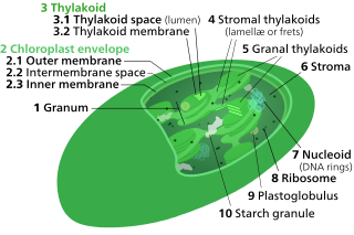
Thylakoids are membrane-bound compartments inside chloroplasts and cyanobacteria. They are the site of the light-dependent reactions of photosynthesis. Thylakoids consist of a thylakoid membrane surrounding a thylakoid lumen. Chloroplast thylakoids frequently form stacks of disks referred to as grana. Grana are connected by intergranal/stromal thylakoids, which join granum stacks together as a single functional compartment.

<span class="mw-page-title-main">Photosystem</span>

Photosystems are functional and structural units of protein complexes involved in photosynthesis. Together they carry out the primary photochemistry of photosynthesis: the absorption of light and the transfer of energy and electrons. Photosystems are found in the thylakoid membranes of plants, algae, and cyanobacteria. These membranes are located inside the chloroplasts of plants and algae, and in the cytoplasmic membrane of photosynthetic bacteria. There are two kinds of photosystems: PSI and PSII.

<span class="mw-page-title-main">Photosystem II</span> First protein complex in light-dependent reactions of oxygenic photosynthesis

Photosystem II is the first protein complex in the light-dependent reactions of oxygenic photosynthesis. It is located in the thylakoid membrane of plants, algae, and cyanobacteria. Within the photosystem, enzymes capture photons of light to energize electrons that are then transferred through a variety of coenzymes and cofactors to reduce plastoquinone to plastoquinol. The energized electrons are replaced by oxidizing water to form hydrogen ions and molecular oxygen.

<span class="mw-page-title-main">Photosystem I</span> Second protein complex in photosynthetic light reactions

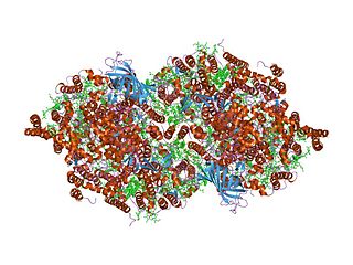
Photosystem I is one of two photosystems in the photosynthetic light reactions of algae, plants, and cyanobacteria. Photosystem I is an integral membrane protein complex that uses light energy to catalyze the transfer of electrons across the thylakoid membrane from plastocyanin to ferredoxin. Ultimately, the electrons that are transferred by Photosystem I are used to produce the moderate-energy hydrogen carrier NADPH. The photon energy absorbed by Photosystem I also produces a proton-motive force that is used to generate ATP. PSI is composed of more than 110 cofactors, significantly more than Photosystem II.

Chlorophyll <i>a</i> Chemical compound

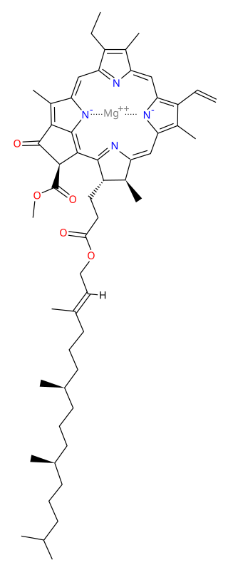
Chlorophyll a is a specific form of chlorophyll used in oxygenic photosynthesis. It absorbs most energy from wavelengths of violet-blue and orange-red light, and it is a poor absorber of green and near-green portions of the spectrum. Chlorophyll does not reflect light but chlorophyll-containing tissues appear green because green light, diffusively reflected by structures like cell walls, becomes enriched in the reflected light. This photosynthetic pigment is essential for photosynthesis in eukaryotes, cyanobacteria and prochlorophytes because of its role as primary electron donor in the electron transport chain. Chlorophyll a also transfers resonance energy in the antenna complex, ending in the reaction center where specific chlorophylls P680 and P700 are located.

Site-directed spin labeling (SDSL) is a technique for investigating the structure and local dynamics of proteins using electron spin resonance. The theory of SDSL is based on the specific reaction of spin labels with amino acids. A spin label's built-in protein structure can be detected by EPR spectroscopy. SDSL is also a useful tool in examinations of the protein folding process.

The oxygen-evolving complex (OEC), also known as the water-splitting complex, is the portion of photosystem II where photo-oxidation of water occurs during the light reactions of photosynthesis. The OEC is surrounded by four core protein subunits of photosystem II at the membrane-lumen interface.

Cytochrome b<sub>6</sub>f complex Enzyme

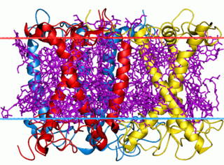
The cytochrome b6f complex is an enzyme found in the thylakoid membrane in chloroplasts of plants, cyanobacteria, and green algae, that catalyzes the transfer of electrons from plastoquinol to plastocyanin. The reaction is analogous to the reaction catalyzed by cytochrome bc1 of the mitochondrial electron transport chain. During photosynthesis, the cytochrome b6f complex is one step along the chain that transfers electrons from Photosystem II to Photosystem I, and at the same time pumps protons into the thylakoid space, contributing to the generation of an electrochemical (energy) gradient that is later used to synthesize ATP from ADP.

<span class="mw-page-title-main">Light-harvesting complexes of green plants</span>

The light-harvesting complex is an array of protein and chlorophyll molecules embedded in the thylakoid membrane of plants and cyanobacteria, which transfer light energy to one chlorophyll a molecule at the reaction center of a photosystem.

<span class="mw-page-title-main">Photosynthetic reaction centre</span>

A photosynthetic reaction center is a complex of several proteins, pigments and other co-factors that together execute the primary energy conversion reactions of photosynthesis. Molecular excitations, either originating directly from sunlight or transferred as excitation energy via light-harvesting antenna systems, give rise to electron transfer reactions along the path of a series of protein-bound co-factors. These co-factors are light-absorbing molecules (also named chromophores or pigments) such as chlorophyll and pheophytin, as well as quinones. The energy of the photon is used to excite an electron of a pigment. The free energy created is then used, via a chain of nearby electron acceptors, for a transfer of hydrogen atoms (as protons and electrons) from H2O or hydrogen sulfide towards carbon dioxide, eventually producing glucose. These electron transfer steps ultimately result in the conversion of the energy of photons to chemical energy.

A light-harvesting complex consists of a number of chromophores which are complex subunit proteins that may be part of a larger super complex of a photosystem, the functional unit in photosynthesis. It is used by plants and photosynthetic bacteria to collect more of the incoming light than would be captured by the photosynthetic reaction center alone. The light which is captured by the chromophores is capable of exciting molecules from their ground state to a higher energy state, known as the excited state. This excited state does not last very long and is known to be short-lived.

<span class="mw-page-title-main">Photoinhibition</span>

Photoinhibition is light-induced reduction in the photosynthetic capacity of a plant, alga, or cyanobacterium. Photosystem II (PSII) is more sensitive to light than the rest of the photosynthetic machinery, and most researchers define the term as light-induced damage to PSII. In living organisms, photoinhibited PSII centres are continuously repaired via degradation and synthesis of the D1 protein of the photosynthetic reaction center of PSII. Photoinhibition is also used in a wider sense, as dynamic photoinhibition, to describe all reactions that decrease the efficiency of photosynthesis when plants are exposed to light.

In enzymology, a ferredoxin-NADP+ reductase (EC 1.18.1.2) abbreviated FNR, is an enzyme that catalyzes the chemical reaction

<span class="mw-page-title-main">Cytochrome b559</span>

Cytochrome b559 is an important component of Photosystem II.

<span class="mw-page-title-main">Photosystem II light-harvesting protein</span>

Photosystem II light-harvesting proteins are the intrinsic transmembrane proteins CP43 (PsbC) and CP47 (PsbB) occurring in the reaction centre of photosystem II (PSII). These polypeptides bind to chlorophyll a and β-Carotene and pass the excitation energy on to the reaction centre.

<span class="mw-page-title-main">Light-dependent reactions</span> Photosynthetic reactions

Light-dependent reactions is jargon for certain photochemical reactions that are involved in photosynthesis, the main process by which plants acquire energy. There are two light dependent reactions, the first occurs at photosystem II (PSII) and the second occurs at photosystem I (PSI),

<span class="mw-page-title-main">Anoxygenic photosynthesis</span> Process used by obligate anaerobes

Bacterial anoxygenic photosynthesis differs from the better known oxygenic photosynthesis in plants by the reductant used and the byproduct generated.

<span class="mw-page-title-main">Ycf9 protein domain</span> Plastid protein involved in photosynthesis

In molecular biology, the PsbZ (Ycf9) is a protein domain, which is low in molecular weight. It is a transmembrane protein and therefore is located in the thylakoid membrane of chloroplasts in cyanobacteria and plants. More specifically, it is located in Photosystem II (PSII) and in the light-harvesting complex II (LHCII). Ycf9 acts as a structural linker, that stabilises the PSII-LHCII supercomplexes. Moreover, the supercomplex fails to form in PsbZ-deficient mutants, providing further evidence to suggest Ycf9's role as a structural linker. This may be caused by a marked decrease in two LHCII antenna proteins, CP26 and CP29, found in PsbZ-deficient mutants, which result in structural changes, as well as functional modifications in PSII.

<span class="mw-page-title-main">Bill Rutherford</span>

Alfred William Rutherford FRS is Professor and Chair in Biochemistry of Solar energy in the Department of Life sciences at Imperial College London.

References