Plasma cell granuloma

Last updated

Plasma cell granulomas (PCGs) are uncommon, non-neoplastic lesions of unknown etiology and are considered an entity of IgG4-related diseases. [1] [2] [3] [4]

Contents

History/Introduction

PCGs were first discovered and described in 1973 by Bahadori and Liebow. [1] PCGs are characterized by the proliferation and infiltration of different inflammatory cells with the main cell observed in highest concentrations being plasma cells. [1] [2] More specifically, PCGs are a type of mass-forming lesion arising from the accumulation of polyclonal plasma cells surrounded in a swirling storiform orientation of fibrosis and spindle cell proliferation. [3] [4] It has been noted that these granulomas have the propensity to manifest on any organ or soft tissue. [3] Plasma cell granulomas are generally found to be benign, but in some cases, the granulomas have the ability to initiate malignancy and become symptomatic regardless of location or size. [3] Common sites of plasma cell granulomas are in the oral gingiva, lungs, vagina, larynx, orbit, spinal cord meninges, breast, pelvic soft tissue, bladder, mesentery, retroperitoneum, kidney, lymph nodes, spleen, pancreas, liver, stomach, heart, thyroid, and trachea. [1] [4] [5] [6] Microscopically, plasma cell granulomas, demonstrate a lesional pattern of inflammatory pseudotumor. [1] The term inflammatory pseudotumor has previously been used to classify plasma cell granulomas. However, this term has become more uncommon in recent years due to its lack of specificity. [3] Today, scientist use more up to date diagnostic and medical terminology to avoid classifying lesions in the same group that are likely to have different etiologies. [3] Other names associated with plasma cell granulomas are inflammatory myofibroblastic tumor, inflammatory myofibrohistiocytic tumor, benign myofibroblastoma, pseudosarcoma, fibrous histocytoma, fibroxanthoma, xanthomatous pseudotumor, xanthogranuloma, myxoid hamartoma, and lymphoid hamartoma. [1] [2] [4] [7] [8] [9]

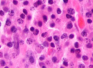
General image of plasma cell granuloma that was classified in a broader category of inflammatory pseudotumor. Original author of image was: Yale Rosen from USA Inflammatory pseudotumor - Plasma cell granuloma Case 176 (5601450952).jpg
General image of plasma cell granuloma that was classified in a broader category of inflammatory pseudotumor. Original author of image was: Yale Rosen from USA

Etiology

The etiology of plasma cell granulomas is widely unknown, however, there are a few ideas on what causes the condition to develop. [4] Studies suggest that one possible causative factor is the presence of a foreign body. A foreign body is likely to give off an antigenic cue causing the accumulation of polyclonal IgG4 positive plasma cells. [2] [3] [4] Others suggest that plasma cell granulomas have an autoimmune origin. [4] PCGs have also been found to be drug and/or hormone induced. [4]

Pathology

Diagnosis/Differentiation

Since plasma cell granulomas have the ability to occur at any site, even though they are uncommon, it should be included in differential diagnostics in regard to plasma cell neoplasms. [3] With granulomas that arise in the mouth, PCGs are commonly misclassified considering the malignancy of the disease due to its aggressive behavior and clinical presentation. [4] Plasma cell granulomas located in the oral cavity have been identified with disruption and damage of surrounding tissues. [4] In comparison, PCGs manifesting in other internal organs are usually secondary findings on radiographic images. [4] Occasionally, plasma cell granulomas are misdiagnosed as malignant lymphoma or malignant plasmacytoma during initial examination due to radiological evidence of its ability to erode and infiltrate bone. [4] Typically, under microscopic analysis, a plasma cell granuloma will display a dense population of morphologically similar plasma cells sporadically mixed together with other inflammatory lymphocytes seen in a storiform orientation of fibrotic connective tissue. [3] [4] To avoid the misdiagnosis of a PCG as a plasmacytoma, it should be noted PCGs are formed from typical plasma cells while plasmacytomas consist of both atypical and typical plasma cells. [4] It is imperative that such differentiation between plasma cell granuloma and plasmacytomas are completed prior to informing the patient undergoing clinical examination. [4] The importance of an accurate diagnosis between the two is due to the different prognosis and progression of the mass-forming lesions. [4] While plasma cell granulomas are often found to be benign, plasmacytomas have been associated with a more aggressive and invasive behavior in which they have the ability to transform or evolve into multiple myeloma. [4] One of the most reliable distinguishing factors from plasmacytomas and plasma cell granulomas, is the polyclonality of the plasma cells involved with plasma cell granulomas. [3] PCGs that manifest in the mouth, specifically the gingiva, have been seen to exhibit similar physical characteristics of other conditions such as epulis, fibroma, pyogenic granuloma, and peripheral giant cell granuloma. [4] Histopathological analysis of plasma cell granulomas have been found to be the most precise confirmative diagnosis to distinguish PCGs from other lesional tumors of plasma cell origins. [4]

Histopathology

Analyzing the changes that arise in the tissue associated with the mass-forming lesion has been found to be the most accurate and precise method for confirming the diagnosis of plasma cell granulomas. This can be achieved by taking excision biopsies of the unknown mass for examination. [4] Typically, plasma cell granulomas appear microscopically as a lesional mass consisting of an abundance of plasma cells intermingled among different inflammatory cells set in fibrous connective tissue displayed in a spiral appearance. [3] [4]  Depending on the location of the plasma cell granuloma, vasculitis can be present but is not considered a confirmatory factor for diagnosis. [3] Kappa/lambda in-situ hybridization is another diagnostic test that needs to be done when trying to characterize the lesion for further confirmation. [3] Common results associated with kappa/lambda in-situ hybridization studies of PCGs are an abundant population of CD138 positive polyclonal plasma cells. [3] If instead, results show monoclonality of plasma cells it is indicative of plasmacytoma. [3]

Blech et al. - Biophysical J 2019 116 1637: "Structure of a Therapeutic Full-Length Anti-NPRA IgG4 Antibody: Dissecting Conformational Diversity" were describing that IgG4 has an unusual preferred configuration that is different to the classical Y-shape described for many other antibodies. See there for details. IgG1 vs IgG4 configuration.png
Blech et al. – Biophysical J 2019 116 1637: "Structure of a Therapeutic Full-Length Anti-NPRA IgG4 Antibody: Dissecting Conformational Diversity" were describing that IgG4 has an unusual preferred configuration that is different to the classical Y-shape described for many other antibodies. See there for details.

Determining Plasma Cell Clonality

The clonality of plasma cells is most accurately determined by two different methods. [3] One method is flow cytometry and the other is kappa/lambda in-situ hybridization and/or immunohistochemical staining (IHC). To obtain the most precise results of clonality it is recommended that both methods be used. [12] Flow cytometry is generally carried out with plasma cell granulomas to obtain the ratio of kappa light chains to lambda light chains. [3] Immunohistochemical staining of plasma cell granulomas is an important diagnostic method to show PCG lesions are also an entity within the IgG4-related diseases. [3] IHC staining is especially helpful because it allows for the assessment of the quantitative number of IgG4 positive plasma cells. [3] Immunohistochemical staining will also provide the overall ratio of IgG4 secreting plasma cells to the total number of IgG secreting plasma cells within a given mass. [3] Plasma cell granulomas have been characterized showing a ratio of IgG4:IgG of greater than 40 percent. [3] A ratio above 40 percent indicates abnormal elevated levels of IgG4 positive plasma cells. [3] Even though IgG levels can be measured in serum, histopathological analysis has been concluded to be the most accurate characterization method of PCG lesions. [3] Using serum IgG4 levels can be a misleading diagnostic, considering that up to 40 percent of patients diagnosed with plasma cell granuloma have IgG4 serum levels within normal reference ranges. [3]

Treatment for PCG

The typical treatment for plasma cell granuloma is a complete surgical excision of the lesional mass. [3] [4] Generally when the mass is removed, patients will display an absolute reversal of symptoms. [3] When surgical removal of the granuloma is not applicable due to size or location, the lesions have been found to respond well to radiation therapy as well as glucocorticoids or steroids. [3] While recurrence rate for plasma cell granulomas is very low, they have been reported; therefore, it is recommended that patients come back for yearly follow up visits. [3] [4]

Localization

See also

Related Research Articles

<span class="mw-page-title-main">Plasmacytoma</span> Growth of a plasma cell tumour within soft tissue or the axial skeleton

Plasmacytoma is a plasma cell dyscrasia in which a plasma cell tumour grows within soft tissue or within the axial skeleton.

Epulis fissuratum is a benign hyperplasia of fibrous connective tissue which develops as a reactive lesion to chronic mechanical irritation produced by the flange of a poorly fitting denture. More simply, epulis fissuratum is where excess folds of firm tissue form inside the mouth, as a result of rubbing on the edge of dentures that do not fit well. It is a harmless condition and does not represent oral cancer. Treatment is by simple surgical removal of the lesion, and also by adjustment of the denture or provision of a new denture.
<span class="mw-page-title-main">Pyogenic granuloma</span> Vascular tumor on both mucosa and skin

A pyogenic granuloma or lobular capillary hemangioma is a vascular tumor that occurs on both mucosa and skin, and appears as an overgrowth of tissue due to irritation, physical trauma, or hormonal factors. It is often found to involve the gums, skin, or nasal septum, and has also been found far from the head, such as in the thigh.

<span class="mw-page-title-main">Immunoglobulin light chain</span> Small antibody polypeptide subunit (immunoglobin)

The immunoglobulin light chain is the small polypeptide subunit of an antibody (immunoglobulin).
<span class="mw-page-title-main">Sperm granuloma</span> Lump of extravasated sperm found in some vasectomized men

A sperm granuloma is a lump of leaked sperm that appears along the vasa deferentia or epididymides in vasectomized individuals. While the majority of sperm granulomas are present along the vas deferens, the rest of them form at the epididymis. Sperm granulomas range in size, from one millimeter to one centimeter. They consist of a central mass of degenerating sperm surrounded by tissue containing blood vessels and immune system cells. Sperm granulomas may also have a yellow, white, or cream colored center when cut open. While some sperm granulomas can be painful, most of them are painless and asymptomatic. Sperm granulomas can appear as a result of surgery, trauma, or an infection. They can appear as early as four days after surgery and fully formed ones can appear as late as 208 days later.

In hematology, plasma cell dyscrasias are a spectrum of progressively more severe monoclonal gammopathies in which a clone or multiple clones of pre-malignant or malignant plasma cells over-produce and secrete into the blood stream a myeloma protein, i.e. an abnormal monoclonal antibody or portion thereof. The exception to this rule is the disorder termed non-secretory multiple myeloma; this disorder is a form of plasma cell dyscrasia in which no myeloma protein is detected in serum or urine of individuals who have clear evidence of an increase in clonal bone marrow plasma cells and/or evidence of clonal plasma cell-mediated tissue injury. Here, a clone of plasma cells refers to group of plasma cells that are abnormal in that they have an identical genetic identity and therefore are descendants of a single genetically distinct ancestor cell.

Fibromatosis colli (FMC), also termed sternocleidomastoid tumor of infancy, pseudotumor of infancy, and infancy sternocleidomastoid pseudotumor, is an uncommon, congenital tumor in one of the two sternocleidomastoid neck muscles although rare cases have presented with a FMC tumor in both sternocleidomastoid muscles. A tumor is here defined as a growth of tissue that is not coordinated with the normal surrounding tissue and persists in growing even if the original trigger for its growth is removed. FMC tumors are benign growths that may cause disfigurements but are not cancers and do not metastasize to distant tissues.

Epulis is any tumor like enlargement situated on the gingival or alveolar mucosa. The word literally means "(growth) on the gingiva", and describes only the location of the mass and has no further implications on the nature of the lesion. There are three types: fibromatous, ossifying and acanthomatous. The related term parulis refers to a mass of inflamed granulation tissue at the opening of a draining sinus on the alveolus over the root of an infected tooth. Another closely related term is gingival enlargement, which tends to be used where the enlargement is more generalized over the whole gingiva rather than a localized mass.

<span class="mw-page-title-main">Idiopathic orbital inflammatory disease</span> Medical condition

Idiopathic orbital inflammatory (IOI) disease refers to a marginated mass-like enhancing soft tissue involving any area of the orbit. It is the most common painful orbital mass in the adult population, and is associated with proptosis, cranial nerve palsy, uveitis, and retinal detachment. Idiopathic orbital inflammatory syndrome, also known as orbital pseudotumor, was first described by Gleason in 1903 and by Busse and Hochheim. It was then characterized as a distinct entity in 1905 by Birch-Hirschfeld. It is a benign, nongranulomatous orbital inflammatory process characterized by extraocular orbital and adnexal inflammation with no known local or systemic cause. Its diagnosis is of exclusion once neoplasm, primary infection and systemic disorders have been ruled out. Once diagnosed, it is characterized by its chronicity, anatomic location or histologic subtype.

<span class="mw-page-title-main">IgG4-related disease</span> Medical condition

IgG4-related disease (IgG4-RD), formerly known as IgG4-related systemic disease, is a chronic inflammatory condition characterized by tissue infiltration with lymphocytes and IgG4-secreting plasma cells, various degrees of fibrosis (scarring) and a usually prompt response to oral steroids. In approximately 51–70% of people with this disease, serum IgG4 concentrations are elevated during an acute phase.

<span class="mw-page-title-main">Inflammatory myofibroblastic tumour</span> Medical condition

Inflammatory myofibroblastic tumor (IMT) is a rare neoplasm of the mesodermal cells that form the connective tissues which support virtually all of the organs and tissues of the body. IMT was formerly termed inflammatory pseudotumor. Currently, however, inflammatory pseudotumor designates a large and heterogeneous group of soft tissue tumors that includes inflammatory myofibroblastic tumor, plasma cell granuloma, xanthomatous pseudotumor, solitary mast cell granuloma, inflammatory fibrosarcoma, pseudosarcomatous myofibroblastic proliferation, myofibroblastoma, inflammatory myofibrohistiocytic proliferation, and other tumors that develop from connective tissue cells. Inflammatory pseudotumour is a generic term applied to various neoplastic and non-neoplastic tissue lesions which share a common microscopic appearance consisting of spindle cells and a prominent presence of the white blood cells that populate chronic or, less commonly, acute inflamed tissues.

According to the WHO classification, three lesional patterns can be observed

<span class="mw-page-title-main">Inflammatory fibroid polyp</span> Medical condition

Inflammatory fibroid polyp (IFP) is a benign abnormal growth of tissue projecting into the lumen of the gastrointestinal tract.

The xanthogranulomatous process (XP), is a form of acute and chronic inflammation characterized by an exuberant clustering of foamy macrophages among other inflammatory cells. Localization in the kidney and renal pelvis has been the most frequent and better known occurrence followed by that in the gallbladder but many others have been subsequently recorded. The pathological findings of the process and etiopathogenetic and clinical observations have been reviewed by Cozzutto and Carbone.

<span class="mw-page-title-main">Otic polyp</span> Medical condition

An otic polyp is a benign proliferation of chronic inflammatory cells associated with granulation tissue, in response to a longstanding inflammatory process of the middle ear.

Chronic sclerosing sialadenitis is a chronic (long-lasting) inflammatory condition affecting the salivary gland. Relatively rare in occurrence, this condition is benign, but presents as hard, indurated and enlarged masses that are clinically indistinguishable from salivary gland neoplasms or tumors. It is now regarded as a manifestation of IgG4-related disease.

Acral myxoinflammatory fibroblastic sarcoma (AMSF), also termed myxoinflammatory fibroblastic sarcoma (MSF), is a rare, low-grade, soft tissue tumor that the World Health Organization (2020) classified as in the category of rarely metastasizing fibroblastic and myofibroblastic tumors. It is a locally aggressive neoplasm that often recurs at the site of its surgical removal. However, it usually grows slowly and in only 1–2% of cases spreads to distant tissues.

<span class="mw-page-title-main">Proliferative fasciitis and proliferative myositis</span> Medical condition

Proliferative fasciitis and proliferative myositis (PF/PM) are rare benign soft tissue lesions that increase in size over several weeks and often regress over the ensuing 1–3 months. The lesions in PF/PM are typically obvious tumors or swellings. Historically, many studies had grouped the two descriptive forms of PF/PM as similar disorders with the exception that proliferative fasciitis occurs in subcutaneous tissues while proliferative myositis occurs in muscle tissues. In 2020, the World Health Organization agreed with this view and defined these lesions as virtually identical disorders termed proliferative fasciitis/proliferative myositis or proliferative fasciitis and proliferative myositis. The Organization also classified them as one of the various forms of the fibroblastic and myofibroblastic tumors.

Fibroblastic and myofibroblastic tumors (FMTs) develop from the mesenchymal stem cells which differentiate into fibroblasts and/or the myocytes/myoblasts that differentiate into muscle cells. FMTs are a heterogeneous group of soft tissue neoplasms. The World Health Organization (2020) defined tumors as being FMTs based on their morphology and, more importantly, newly discovered abnormalities in the expression levels of key gene products made by these tumors' neoplastic cells. Histopathologically, FMTs consist of neoplastic connective tissue cells which have differented into cells that have microscopic appearances resembling fibroblasts and/or myofibroblasts. The fibroblastic cells are characterized as spindle-shaped cells with inconspicuous nucleoli that express vimentin, an intracellular protein typically found in mesenchymal cells, and CD34, a cell surface membrane glycoprotein. Myofibroblastic cells are plumper with more abundant cytoplasm and more prominent nucleoli; they express smooth muscle marker proteins such as smooth muscle actins, desmin, and caldesmon. The World Health Organization further classified FMTs into four tumor forms based on their varying levels of aggressiveness: benign, intermediate, intermediate, and malignant.

Cellular angiofibroma (CAF) is a rare, benign tumor of superficial soft tissues that was first described by M. R. Nucci et al. in 1997. These tumors occur predominantly in the distal parts of the female and male reproductive systems, i.e. in the vulva-vaginal and inguinal-scrotal areas, respectively, or, less commonly, in various other superficial soft tissue areas throughout the body. CAF tumors develop exclusively in adults who typically are more than 30 years old.

References

  1. 1 2 3 4 5 6 7 Jhingta, Pravesh Kumar; Mardi, Kavita; Sharma, Deepak; Bhardwaj, Vinay Kumar; Bhardwaj, Ashu; Saroch, Nitin; Negi, Nishant (2018). "An Enigmatic Clinical Presentation of Plasma Cell Granuloma of the Oral Cavity". Contemporary Clinical Dentistry. 9 (1): 132–136. doi: 10.4103/ccd.ccd_816_17 . ISSN   0976-237X. PMC   5863397 . PMID   29599600.
  2. 1 2 3 4 Manohar, Balaji; Bhuvaneshwari, S (2011). "Plasma cell granuloma of gingiva". Journal of Indian Society of Periodontology. 15 (1): 64–66. doi: 10.4103/0972-124X.82275 . ISSN   0972-124X. PMC   3134051 . PMID   21772725.
  3. 1 2 3 4 5 6 7 8 9 10 11 12 13 14 15 16 17 18 19 20 21 22 23 24 25 26 27 28 Forcucci, Jessica; Butler-Williams, Shannon; Miller, Nicole; Lazarchick, John (2015-05-01). "Plasma Cell Granuloma: An Entity within the Spectrum of IgG4-Related Disease". Annals of Clinical & Laboratory Science. 45 (3): 340–343. ISSN   0091-7370. PMID   26116600.
  4. 1 2 3 4 5 6 7 8 9 10 11 12 13 14 15 16 17 18 19 20 21 22 23 24 Dyna, Albert; Muthusekhar, M. R.; Kumar, Santhosh P.; Murugesan, Krishnan (2022). "A Rare Case of Hormone-Induced Plasma Cell Granuloma of the Gingiva". Cureus. 14 (4): e23897. doi: 10.7759/cureus.23897 . PMC   9076496 . PMID   35530856. ProQuest   2671878822.
  5. 1 2 Hammar, Samuel P. (2006-01-01), Dabbs, David J. (ed.), "Chapter 11 - Immunohistology of Lung and Pleural Neoplasms", Diagnostic Immunohistochemistry (Second Edition), Churchill Livingstone, pp. 329–403, ISBN   978-0-443-06652-8 , retrieved 2022-12-05
  6. 1 2 "Plasma Cell Granuloma - an overview | ScienceDirect Topics". www.sciencedirect.com. Retrieved 2022-12-05.
  7. Braham, Yosra; Migaou, Asma; Njima, Manel; Achour, Asma; Ben Saad, Ahmed; Cheikh Mhamed, Saoussen; Fahem, Nesrine; Rouatbi, Naceur; Joobeur, Samah (2020-01-01). "Inflammatory myofibroblastic tumor of the lung: A rare entity". Respiratory Medicine Case Reports. 31: 101287. doi: 10.1016/j.rmcr.2020.101287 . ISSN   2213-0071. PMC   7683262 . PMID   33251105.
  8. Weinberger, Steven E, MD (December 4, 2022). Flaherty, Kevin R. MD, MS; Dieffenbach, Paul, MD (eds.). "Inflammatory myofibroblastic tumor (plasma cell granuloma) of the lung". www.uptodate.com. Retrieved 2022-12-05.{{cite web}}: CS1 maint: multiple names: authors list (link)
  9. Zen, Yoh; Kitagawa, Satoshi; Minato, Hiroshi; Kurumaya, Hiroshi; Katayanagi, Kazuyoshi; Masuda, Shinji; Niwa, Hideki; Fujimura, Masaki; Nakanuma, Yasuni (2005-07-01). "IgG4-positive plasma cells in inflammatory pseudotumor (plasma cell granuloma) of the lung". Human Pathology. 36 (7): 710–717. doi:10.1016/j.humpath.2005.05.011. ISSN   0046-8177. PMID   16084938.
  10. Pathology, Atlas of Pulmonary (2011-04-04), Inflammatory pseudotumor - Plasma cell granuloma Case 176 , retrieved 2022-12-07
  11. "File:IgG1 vs IgG4 configuration.png - Wikipedia". commons.wikimedia.org. 11 November 2020. Retrieved 2022-12-08.
  12. Hristov, Alexandra C.; Comfere, Nneka I.; Vidal, Claudia I.; Sundram, Uma (November 2020). "Kappa and lambda immunohistochemistry and in situ hybridization in the evaluation of atypical cutaneous lymphoid infiltrates". Journal of Cutaneous Pathology. 47 (11): 1103–1110. doi: 10.1111/cup.13858 . hdl: 2027.42/163451 . ISSN   0303-6987. PMID   32870521.
  13. Nonomura, A; Mizukami, Y; Matsubara, F; Shimizu, J; Oda, M; Watanabe, Y; Kamimura, R; Takashima, T; Kitagawa, M (1992). "Seven patients with plasma cell granuloma (inflammatory pseudotumor) of the lung, including two with intrabronchial growth: An immunohistochemical and electron microscopic study". Internal Medicine (Tokyo, Japan). 31 (6): 756–65. doi: 10.2169/internalmedicine.31.756 . PMID   1392177.
  14. Balachandran, A; Shivbalan, S (2004). "Plasma cell granuloma of the lung". Indian Pediatrics. 41 (3): 292–3. PMID   15064526.