Related Research Articles

Magnetic resonance imaging (MRI) is a medical imaging technique used in radiology to form pictures of the anatomy and the physiological processes of the body. MRI scanners use strong magnetic fields, magnetic field gradients, and radio waves to generate images of the organs in the body. MRI does not involve X-rays or the use of ionizing radiation, which distinguishes it from computed tomography (CT) and positron emission tomography (PET) scans. MRI is a medical application of nuclear magnetic resonance (NMR) which can also be used for imaging in other NMR applications, such as NMR spectroscopy.

In a multicellular organism, an organ is a collection of tissues joined in a structural unit to serve a common function. In the hierarchy of life, an organ lies between tissue and an organ system. Tissues are formed from same type cells to act together in a function. Tissues of different types combine to form an organ which has a specific function. The intestinal wall for example is formed by epithelial tissue and smooth muscle tissue. Two or more organs working together in the execution of a specific body function form an organ system, also called a biological system or body system.

The bile duct is a part of the biliary tract. It is formed by the union of the common hepatic duct and cystic duct. It ends by uniting with the pancreatic duct to form the hepatopancreatic ampulla. It possesses its own sphincter to enable regulation of bile flow.
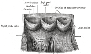
An aortic sinus, also known as a sinus of Valsalva, is one of the anatomic dilations of the ascending aorta, which occurs just above the aortic valve. These widenings are between the wall of the aorta and each of the three cusps of the aortic valve.

The nasal septum separates the left and right airways of the nasal cavity, dividing the two nostrils.

The chordae tendineae or tendinous cords, colloquially known as the heart strings, are inelastic cords of fibrous connective tissue that connect the papillary muscles to the tricuspid valve and the mitral valve in the heart.
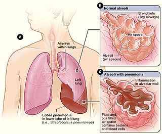
Lobar pneumonia is a form of pneumonia characterized by inflammatory exudate within the intra-alveolar space resulting in consolidation that affects a large and continuous area of the lobe of a lung.

The dural venous sinuses are venous sinuses (channels) found between the endosteal and meningeal layers of dura mater in the brain. They receive blood from the cerebral veins, and cerebrospinal fluid (CSF) from the subarachnoid space via arachnoid granulations. They mainly empty into the internal jugular vein.

In the anatomy of the human digestive tract, there are two colic flexures, or curvatures in the transverse colon. The right colic flexure is also known as the hepatic flexure, and the left colic flexure is also known as the splenic flexure. Note that "right" refers to the patient's anatomical right, which may be depicted on the left of a diagram.
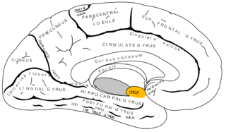
The uncus is an anterior extremity of the parahippocampal gyrus. It is separated from the apex of the temporal lobe by a slight fissure called the incisura temporalis.
A portacaval anastomosis or portocaval anastomosis is a specific type of circulatory anastomosis that occurs between the veins of the portal circulation and the vena cava, thus forming one of the principal types of portasystemic anastomosis or portosystemic anastomosis, as it connects the portal circulation to the systemic circulation, providing an alternative pathway for the blood. When there is a blockage of the portal system, portocaval anastomosis enables the blood to still reach the systemic venous circulation. The inferior end of the esophagus and the superior part of the rectum are potential sites of a harmful portocaval anastomosis.

A flexion teardrop fracture is a fracture of the anteroinferior aspect of a cervical vertebral body due to flexion of the spine along with vertical axial compression. The fracture continues sagittally through the vertebral body, and is associated with deformity of the body and subluxation or dislocation of the facet joints at the injured level. A flexion teardrop fracture is usually associated with a spinal cord injury, often a result of displacement of the posterior portion of the vertebral body into the spinal canal.
Radiopaedia is a wiki-based international collaborative educational web resource containing a radiology encyclopedia and imaging case repository. It is currently the largest freely available radiology related resource in the world with more than 50,000 patient cases and over 16,000 reference articles on radiology-related topics. The open edit nature of articles allows radiologists, radiology trainees, radiographers, sonographers, and other healthcare professionals interested in medical imaging to refine most content through time. An editorial board peer reviews all contributions.

An ALPSAlesion is an injury at the front of the shoulder associated with shoulder dislocation.

Adenocarcinoma of the lung is the most common type of lung cancer, and like other forms of lung cancer, it is characterized by distinct cellular and molecular features. It is classified as one of several non-small cell lung cancers (NSCLC), to distinguish it from small cell lung cancer which has a different behavior and prognosis. Lung adenocarcinoma is further classified into several subtypes and variants. The signs and symptoms of this specific type of lung cancer are similar to other forms of lung cancer, and patients most commonly complain of persistent cough and shortness of breath.
Fascia iliaca blocks is a local anesthetic nerve block, a type of regional anesthesia technique, used to provide analgesia or anaesthesia to the hip and thigh. FICB can performed by using ultrasound or with a loss of resistance technique, the latter sometimes referred to as the "two-pop-method". FICB works by affecting the femoral, obturator and the lateral cutaneous nerves with a local anesthetic.

Emphysema is any air-filled enlargement in the body's tissues. Most commonly emphysema refers to the enlargement of air spaces (alveoli) in the lungs, and is also known as pulmonary emphysema.
An anatomical variation, anatomical variant, or anatomical variability is a presentation of body structure with morphological features different from those that are typically described in the majority of individuals. Anatomical variations are categorized into three types including morphometric, consistency, and spatial.

An MRI sequence in magnetic resonance imaging (MRI) is a particular setting of pulse sequences and pulsed field gradients, resulting in a particular image appearance.
A bone sarcoma is a primary malignant bone tumour, a type of sarcoma that starts in the bones. This is in contrast to most bone cancers that are secondary having developed as a metastasis from another cancer. Bone sarcomas are rare, and mostly affect the legs. The other type of sarcoma is a soft-tissue sarcoma.
References
- ↑ Jones, Jeremy. "Pneumoretroperitoneum | Radiology Reference Article | Radiopaedia.org". Radiopaedia. Retrieved 2019-04-09.
| | This article about a medical condition affecting the respiratory system is a stub. You can help Wikipedia by expanding it. |