Popliteal artery entrapment syndrome

Last updated
Popliteal artery entrapment syndrome
Popliteal artery.jpg
Normal course of the popliteal artery at the back of the knee
Symptoms Numbness, discoloration, absent pulses, coolness
Complications Distal artery stenosis, limb amputation, arterial thromboembolism
DurationChronic
CausesCongenital or functional
Risk factors Young athlete males.
TreatmentOpen surgical decompression
Prognosis Favorable

The popliteal artery entrapment syndrome (PAES) is an uncommon pathology that occurs when the popliteal artery is compressed by the surrounding popliteal fossa myofascial structures. [1] This results in claudication and chronic leg ischemia. This condition mainly occurs more in young athletes than in the elderlies. [2] Elderlies, who present with similar symptoms, are more likely to be diagnosed with peripheral artery disease with associated atherosclerosis. [2] Patients with PAES mainly present with intermittent feet and calf pain associated with exercises and relieved with rest. [3] PAES can be diagnosed with a combination of medical history, physical examination, and advanced imaging modalities such as duplex ultrasound, computer tomography, or magnetic resonance angiography. Management can range from non-intervention to open surgical decompression with a generally good prognosis. [4] Complications of untreated PAES can include stenotic artery degeneration, complete popliteal artery occlusion, distal arterial thromboembolism, or even formation of an aneurysm.

Contents

History

In 1879, the syndrome was first described in a 64 years old male by Anderson Stuart, a medical student. [1] In 1959, Hamming and Vink first described the management of the PAES in a 12-year-old patient. The patient was treated with myotomy of the medial head of the gastrocnemius muscle and concomitant endarterectomy of the popliteal artery. They later reported four more cases and claimed that the incidence of this pathology in patients younger than 30 years old with claudication was 40%. Servello was the first to draw attention to diminished distal pulses observed with forced plantar- or dorsiflexion in patients with this syndrome. [5] In 1981, Bouhoutsos and Daskalakis reported 45 cases of this syndrome in a population of 20,000 Greek soldiers. [6] Over the last few decades, the increasing frequency with which popliteal artery entrapment is reported, strongly suggests greater awareness of the syndrome. [7]

Epidemiology

In the general population, popliteal artery entrapment syndrome (PAES) has an estimated prevalence of 0.16%. [8] It is most commonly found in young, physically active males. [2] In fact, sixty percent of all cases of this syndrome occur in athletically active males under the age of 30. [9] The predilection of this syndrome presents in a male to female ratio of 15:1. [8] This discrepancy in prevalence may be overestimated due the findings that males are generally found to be more physically active than females or because a large portion of the data is from military hospitals that treat mostly male populations. [9] People, who participate in running, soccer, football, basketball, or rugby, are at increased risk. [10]

Newborns and young children are also at increased PAES risk due to congenital causes. During embryonic development, the medial head of gastrocnemius migrates medially and superiorly. This migration can cause structural abnormalities, such as irregular positioning of the popliteal artery, and can account for the rare instances of entrapment caused by the popliteus muscle. [9] Less than 3% of all people are born with this anatomical defect that progresses into PAES, and of those who are born with the anatomical defect, the majority never develop symptoms. [10] Bilateral presentation of PAES is found in approximately 30% of cases. [11]

Pathophysiology and classification

PAES can be classified as either congenital or functional. [3] Analysis of human embryological development has shown that the popliteal artery and the medial head of the gastrocnemius muscle arise at approximately the same time. Because of that, abnormal development of muscle's position in relation to the nearby vessels can result in potential vascular compromise. [3] The varying types of PAES can be classified based on aberrant migration and resultant attachments of the medial head of the gastrocnemius muscle. Type VI PAES (functional PAES) describes a subtype that is due to repeated microtrauma resulting in the destruction of the internal elastic lamina and damage to the smooth muscles resulting in fibrosis and scar formation.

Types of Popliteal Artery Entrapment Syndrome
Type IThe popliteal artery courses more medially around a normally positioned medial head of the gastrocnemius muscle. [3]
Type IIThe medial head of the gastrocnemius muscle attaches more laterally to the femur. [3]
Type IIIAberrant additional tendon of the gastrocnemius muscle encircles a normally positioned popliteal artery. [3]
Type IVThe popliteal artery is compressed by the popliteus muscle or a fibrous brand. [3]
Type VThe compression of both the popliteal artery and vein by any of the above causes. [3]
Type VIThe normally positioned popliteal artery is entrapped by the gastrocnemius muscle hypertrophy. [3]

Additionally, a more practical classification system was introduced by Heidelberg et al. [12] This system classifies PAES into three main types:

Medical history and physical examination

Patients with PAES are typically healthy young males without previous history of cardiovascular risk factors such as smoking, hypertension, hypercholesterolemia, or diabetes. [13] Typically, patients present with intermittent claudication that is worsened with exercise and relieved with rest. [3] Associated symptoms include numbness, discoloration, pallor, and coolness in the affected lower extremity. [13] Physical examination of suspected PAES may show hypertrophy of the calf muscles, as well as diminished, unequal, or absent pulses in the lower extremity upon plantar- or dorsiflexion. [14]

Diagnosis

Differential diagnosis

Evaluation

Angiograms of a patient diagnosed with popliteal artery entrapment syndrome of the left lower extremity. Image A shows a neutral popliteal artery before provocative maneuvers. Images B and C show the obstruction (orange arrows) enhanced with provocative maneuvers of plantar flexion and dorsiflexion, respectively. Angiogram of a patient diagnosed with popliteal artery entrapment syndromes.png
Angiograms of a patient diagnosed with popliteal artery entrapment syndrome of the left lower extremity. Image A shows a neutral popliteal artery before provocative maneuvers. Images B and C show the obstruction (orange arrows) enhanced with provocative maneuvers of plantar flexion and dorsiflexion, respectively.

PAES should be suspected in young healthy male patients with clinical symptoms consistent with compression of the vascular structures and without significant cardiovascular risk factors such as smoking. [13] Multiple imaging modalities are used to confirm the diagnosis of PAES. [23] Based on a systemic review by Sinha et al, digital subtraction angiography (DSA) is the most common imaging used for PAES diagnosis, followed by ankle–brachial index (18 percent), computed tomography angiography (CTA) (12 percent), magnetic resonance angiography (MRA) (12 percent), duplex ultrasonography (DU) (10 percent), exercise ankle-brachial index (4 percent), and other modalities (4 percent). [23] According to a recent study by Willimas et al, a combination of DU and MRA is far superior in diagnosing PAES. [24]

Provocative maneuvers can be used to improve visualization of PAES on the imagines. [25] The patient is initially positioned supine with the legs straight, and then instructed to forcefully plantar-flex. A plantarflexion force of 0 to 70 percent maximum has been shown to maximize the sensitivity and specificity for PAES diagnosis. [25] The DU can be a quick, inexpensive, and noninvasive initial screening for PAES. Flow velocities in the popliteal artery will increase, as the popliteal artery is compressed, which is reflected on the DU. If DU is negative but there is still strong suspicion for PAES, MRA or CTA with provocative maneuvers are needed as follow-up imaging. MRA would demonstrate a focal occlusion or narrowing of the mid-popliteal artery, post-stenotic dilatation, or aneurysm of the distal popliteal artery. If MRA or CTA is [13] non-conclusive, DSA may be used as a further option with a high sensitivity (> 97%) for PAES diagnosis. [23]

Additionally, functional PAES in which the gastrocnemius hypertrophy causes arterial compression during exercise can be best evaluated with dynamic CT. [23] For dynamic CT, initial images are taken with the patient still. Further images are taken following a series of provocative maneuvers.

Management

The outcome following the surgery is usually favorable. Successful resolution of PAES occurs in 77 percent of cases. [23] Surgical complications include deep vein thrombosis, hematoma, wound infection, or seroma. [23] After the surgery, patient is usually monitored using arterial duplex ultrasonography 1, 3, 6, and 12 months, and annually after that. [3]

Complications of untreated PAES

Early detection and management of PAES can lead to a favorable outcome. [23] However, prolonged compression of popliteal artery can lead to extensive arterial damage and permanent claudication or limb loss. [28] Complications of PAES may include: [13]

Nonetheless, the course of PAES is often slow and takes time, thus, limbs loss is rarely seen, even in PAES patients. [13]

Related Research Articles

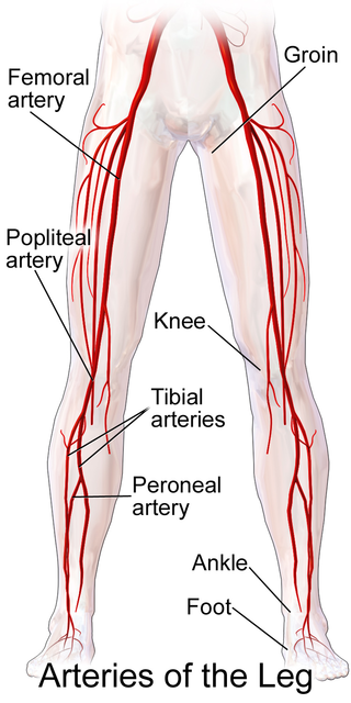
In medicine, a pulse represents the tactile arterial palpation of the cardiac cycle (heartbeat) by trained fingertips. The pulse may be palpated in any place that allows an artery to be compressed near the surface of the body, such as at the neck, wrist, at the groin, behind the knee, near the ankle joint, and on foot. Pulse is equivalent to measuring the heart rate. The heart rate can also be measured by listening to the heart beat by auscultation, traditionally using a stethoscope and counting it for a minute. The radial pulse is commonly measured using three fingers. This has a reason: the finger closest to the heart is used to occlude the pulse pressure, the middle finger is used get a crude estimate of the blood pressure, and the finger most distal to the heart is used to nullify the effect of the ulnar pulse as the two arteries are connected via the palmar arches. The study of the pulse is known as sphygmology.

<span class="mw-page-title-main">Interventional radiology</span> Medical subspecialty

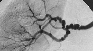
Interventional radiology (IR) is a medical specialty that performs various minimally-invasive procedures using medical imaging guidance, such as x-ray fluoroscopy, computed tomography, magnetic resonance imaging, or ultrasound. IR performs both diagnostic and therapeutic procedures through very small incisions or body orifices. Diagnostic IR procedures are those intended to help make a diagnosis or guide further medical treatment, and include image-guided biopsy of a tumor or injection of an imaging contrast agent into a hollow structure, such as a blood vessel or a duct. By contrast, therapeutic IR procedures provide direct treatment—they include catheter-based medicine delivery, medical device placement, and angioplasty of narrowed structures.

<span class="mw-page-title-main">Peripheral artery disease</span> Medical condition

Peripheral artery disease (PAD) is an abnormal narrowing of arteries other than those that supply the heart or brain. When narrowing occurs in the heart, it is called coronary artery disease, and in the brain, it is called cerebrovascular disease. Peripheral artery disease most commonly affects the legs, but other arteries may also be involved – such as those of the arms, neck, or kidneys. The classic symptom is leg pain when walking which resolves with rest, known as intermittent claudication. Other symptoms include skin ulcers, bluish skin, cold skin, or abnormal nail and hair growth in the affected leg. Complications may include an infection or tissue death which may require amputation; coronary artery disease, or stroke. Up to 50% of people with PAD do not have symptoms.

<span class="mw-page-title-main">Vascular surgery</span> Medical specialty, operative procedures for the treatment of vascular disorders

Vascular surgery is a surgical subspecialty in which vascular diseases involving the arteries, veins, or lymphatic vessels, are managed by medical therapy, minimally-invasive catheter procedures and surgical reconstruction. The specialty evolved from general and cardiovascular surgery where it refined the management of just the vessels, no longer treating the heart or other organs. Modern vascular surgery includes open surgery techniques, endovascular techniques and medical management of vascular diseases - unlike the parent specialities. The vascular surgeon is trained in the diagnosis and management of diseases affecting all parts of the vascular system excluding the coronaries and intracranial vasculature. Vascular surgeons also are called to assist other physicians to carry out surgery near vessels, or to salvage vascular injuries that include hemorrhage control, dissection, occlusion or simply for safe exposure of vascular structures.

<span class="mw-page-title-main">Baker's cyst</span> Medical condition

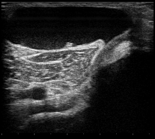
A Baker's cyst, also known as a popliteal cyst, is a type of fluid collection behind the knee. Often there are no symptoms. If symptoms do occur these may include swelling and pain behind the knee, or knee stiffness. If the cyst breaks open, pain may significantly increase with swelling of the calf. Rarely complications such as deep vein thrombosis, peripheral neuropathy, ischemia, or compartment syndrome may occur.

<span class="mw-page-title-main">Popliteal artery</span> Continuation of the femoral artery that supplies the lower leg

The popliteal artery is a deeply placed continuation of the femoral artery opening in the distal portion of the adductor magnus muscle. It courses through the popliteal fossa and ends at the lower border of the popliteus muscle, where it branches into the anterior and posterior tibial arteries.

<span class="mw-page-title-main">Aortoiliac occlusive disease</span> Medical condition

In medicine, aortoiliac occlusive disease is a form of central artery disease involving the blockage of the abdominal aorta as it transitions into the common iliac arteries.

<span class="mw-page-title-main">Takayasu's arteritis</span> Medical condition

Takayasu's arteritis (TA), also known as aortic arch syndrome, nonspecific aortoarteritis, and pulseless disease, is a form of large vessel granulomatous vasculitis with massive intimal fibrosis and vascular narrowing, most commonly affecting young or middle-aged women of Asian descent, though anyone can be affected. It mainly affects the aorta and its branches, as well as the pulmonary arteries. Females are about 8–9 times more likely to be affected than males.

<span class="mw-page-title-main">Arteritis</span> Medical condition

Arteritis is the inflammation of the walls of arteries, usually as a result of infection or autoimmune response. Arteritis, a complex disorder, is still not entirely understood. Arteritis may be distinguished by its different types, based on the organ systems affected by the disease. A complication of arteritis is thrombosis, which can be fatal. Arteritis and phlebitis are forms of vasculitis.

Intermittent claudication, also known as vascular claudication, is a symptom that describes muscle pain on mild exertion, classically in the calf muscle, which occurs during exercise, such as walking, and is relieved by a short period of rest. It is classically associated with early-stage peripheral artery disease, and can progress to critical limb ischemia unless treated or risk factors are modified and maintained.

<span class="mw-page-title-main">Shin splints</span> Medical condition

A shin splint, also known as medial tibial stress syndrome, is pain along the inside edge of the shinbone (tibia) due to inflammation of tissue in the area. Generally this is between the middle of the lower leg and the ankle. The pain may be dull or sharp, and is generally brought on by high-impact exercise that overloads the tibia. It generally resolves during periods of rest. Complications may include stress fractures.

Claudication is a medical term usually referring to impairment in walking, or pain, discomfort, numbness, or tiredness in the legs that occurs during walking or standing and is relieved by rest. The perceived level of pain from claudication can be mild to extremely severe. Claudication is most common in the calves but it can also affect the feet, thighs, hips, buttocks, or arms. The word claudication comes from the Latin claudicare meaning 'to limp'.

<span class="mw-page-title-main">Popliteal vein</span> Large leg vein

The popliteal vein is a vein of the lower limb. It is formed from the anterior tibial vein and the posterior tibial vein. It travels medial to the popliteal artery, and becomes the femoral vein. It drains blood from the leg. It can be assessed using medical ultrasound. It can be affected by popliteal vein entrapment.

<span class="mw-page-title-main">Plantaris muscle</span> One of the superficial muscles of the superficial posterior compartment of the leg,

The plantaris is one of the superficial muscles of the superficial posterior compartment of the leg, one of the fascial compartments of the leg.

<span class="mw-page-title-main">Fibromuscular dysplasia</span> Human arterial disease

Fibromuscular dysplasia (FMD) is a non-atherosclerotic, non-inflammatory disease of the blood vessels that causes abnormal growth within the wall of an artery. FMD has been found in nearly every arterial bed in the body although the most common arteries affected are the renal and carotid arteries.

<span class="mw-page-title-main">Acute limb ischaemia</span> Occurs when there is a sudden lack of blood flow to a limb

Acute limb ischaemia (ALI) occurs when there is a sudden lack of blood flow to a limb.

<span class="mw-page-title-main">Knee dislocation</span> Medical condition

A knee dislocation is an injury in which there is disruption of the knee joint between the tibia and the femur. Symptoms include pain and instability of the knee. Complications may include injury to an artery, most commonly the popliteal artery behind the knee, or compartment syndrome.

<span class="mw-page-title-main">Femoropopliteal bypass</span> Leg artery surgical procedure

Popliteal bypass surgery, more commonly known as femoropopliteal bypass or more generally as lower extremity bypass surgery, is a surgical procedure used to treat diseased leg arteries above or below the knee. It is used as a medical intervention to salvage limbs that are at risk of amputation and to improve walking ability in people with severe intermittent claudication and ischemic rest pain.

Adventitial cystic disease is a rare type of non-atherosclerotic peripheral artery disease. It can present as claudication, critical limb ischemia or acute limb ischemia. The most commonly affected vessel is the popliteal artery. The cause is unknown.

<span class="mw-page-title-main">Arterial occlusion</span>

Arterial occlusion is a condition involving partial or complete blockage of blood flow through an artery. Arteries are blood vessels that carry oxygenated blood to body tissues. An occlusion of arteries disrupts oxygen and blood supply to tissues, leading to ischemia. Depending on the extent of ischemia, symptoms of arterial occlusion range from simple soreness and pain that can be relieved with rest, to a lack of sensation or paralysis that could require amputation.

References

  1. 1 2 Gokkus, Kemal; Sagtas, Ergin; Bakalim, Tamer; Taskaya, Ertugrul; Aydin, Ahmet Turan (2014-07-14). "Popliteal entrapment syndrome. A systematic review of the literature and case presentation". Muscles, Ligaments and Tendons Journal. 4 (2): 141–148. doi:10.32098/mltj.02.2014.09. ISSN   2240-4554. PMC   4187583 . PMID   25332925.
  2. 1 2 3 Sharma, Aditya (2014). "Conditions Presenting with Symptoms of Peripheral Arterial Disease". Seminars in Interventional Radiology. 31 (4): 281–291. doi:10.1055/s-0034-1393963. PMC   4232436 . PMID   25435652.
  3. 1 2 3 4 5 6 7 8 9 10 11 Júnior, Francisco Cialdine Frota Carneiro; Carrijo, Eduardo Nazareno dos Anjos; Araújo, Samuel Tomaz; Nakano, Luis Carlos Uta; de Amorim, Jorge Eduardo; Cacione, Daniel Guimarães (2018-01-09). "Popliteal Artery Entrapment Syndrome: A Case Report and Review of the Literature". The American Journal of Case Reports. 19: 29–34. doi:10.12659/AJCR.905170. ISSN   1941-5923. PMC   5769514 . PMID   29311538.
  4. 1 2 Gourgiotis, Stavros; Aggelakas, John; Salemis, Nikolaos; Elias, Charalabos; Georgiou, Charalabos (February 2008). "Diagnosis and surgical approach of popliteal artery entrapment syndrome: a retrospective study". Vascular Health and Risk Management. 4 (1): 83–88. doi:10.2147/vhrm.2008.04.01.83. ISSN   1176-6344. PMC   2464757 . PMID   18629362.
  5. Servello, Manfredi (1962-11-01). "Clinical Syndrome of Anomalous Position of the Popliteal Artery". Circulation. 26 (5): 885–890. doi: 10.1161/01.CIR.26.5.885 . PMID   13988163. S2CID   261140.
  6. Bouhoutsos, J; Daskalakis, E (July 1981). "Muscular abnormalities affecting the popliteal vessels". British Journal of Surgery. 68 (7): 501–506. doi:10.1002/bjs.1800680720. PMID   7248723. S2CID   44852078.
  7. Stager, Andrew; Clement, Douglas (1999-07-01). "Popliteal Artery Entrapment Syndrome". Sports Medicine. 28 (1): 61–70. doi:10.2165/00007256-199928010-00006. ISSN   1179-2035. PMID   10461713. S2CID   26958095.
  8. 1 2 "Popliteal Artery Occlusive Disease: Background, Problem, Epidemiology". 2016-04-10.{{cite journal}}: Cite journal requires |journal= (help)
  9. 1 2 3 Stager, Andrew; Clement, Douglas (2012-09-23). "Popliteal Artery Entrapment Syndrome". Sports Medicine. 28 (1): 61–70. doi:10.2165/00007256-199928010-00006. ISSN   0112-1642. PMID   10461713. S2CID   26958095.
  10. 1 2 "Popliteal Artery Entrapment Syndrome (PAES) | Cleveland Clinic". Cleveland Clinic. Retrieved 2016-12-19.
  11. Drigny, Joffrey; Reboursière, Emmanuel; Desvergée, Antoine; Ruet, Alexis; Hulet, Christophe (June 2019). "Concurrent Exertional Compartment Syndrome and Functional Popliteal Artery Entrapment Syndrome: A Case Report". PM&R. 11 (6): 669–672. doi:10.1002/pmrj.12081. ISSN   1934-1482. PMID   30689303. S2CID   195661793.
  12. 1 2 3 4 Papaioannou, S; Tsitouridis, K; Giataganas, G; Rodokalakis, G; Kyriakou, V; Papastergiou, Ch; Arvaniti, M; Tsitouridis, I (2009). "Evaluation of popliteal arteries with CT angiography in popliteal artery entrapment syndrome". Hippokratia. 13 (1): 32–37. ISSN   1108-4189. PMC   2633250 . PMID   19240818.
  13. 1 2 3 4 5 6 7 Davis, Donald D.; Shaw, Palma M. (2022), "Popliteal Artery Entrapment Syndrome", StatPearls, Treasure Island (FL): StatPearls Publishing, PMID   28722994 , retrieved 2022-12-04
  14. "ClinicalKey". www.clinicalkey.com. Retrieved 2022-12-04.
  15. Bong MR, Polatsch DB, Jazrawi LM et al. Chronic exertional compartment syndrome: diagnosis and management.Bull Hosp Jt Dis. 2005;62(3-4):77-84.
  16. Noonan, Thomas J.; Garrett, William E. Jr (July 1999). "Muscle Strain Injury: Diagnosis and Treatment". JAAOS - Journal of the American Academy of Orthopaedic Surgeons. 7 (4): 262–269. doi:10.5435/00124635-199907000-00006. ISSN   1067-151X. PMID   10434080.
  17. McClure, Charles J.; Oh, Robert (2022), "Medial Tibial Stress Syndrome", StatPearls, Treasure Island (FL): StatPearls Publishing, PMID   30860714 , retrieved 2022-12-04
  18. "UpToDate". www.uptodate.com. Retrieved 2022-12-04.
  19. Dyson, Kathryn; Palan, Jeya; Mangwani, Jitendra (2019). "Bilateral non-traumatic lower leg fascial defects causing peroneal muscle herniation and novel use of a GraftJacket to repair the fascial defect". Journal of Clinical Orthopaedics and Trauma. 10 (5): 879–883. doi:10.1016/j.jcot.2018.09.009. ISSN   0976-5662. PMC   6739491 . PMID   31528061.
  20. Hicks, Brandon L.; Lam, Jason C.; Varacallo, Matthew (2022), "Piriformis Syndrome", StatPearls, Treasure Island (FL): StatPearls Publishing, PMID   28846222 , retrieved 2022-12-04
  21. "UpToDate". www.uptodate.com. Retrieved 2022-12-04.
  22. Al Qaraghli, Mustafa I.; De Jesus, Orlando (2022), "Lumbar Disc Herniation", StatPearls, Treasure Island (FL): StatPearls Publishing, PMID   32809713 , retrieved 2022-12-04
  23. 1 2 3 4 5 6 7 Sinha, Sidhartha; Houghton, Jon; Holt, Peter J.; Thompson, Matt M.; Loftus, Ian M.; Hinchliffe, Robert J. (2012-01-01). "Popliteal entrapment syndrome". Journal of Vascular Surgery. 55 (1): 252–262.e30. doi: 10.1016/j.jvs.2011.08.050 . ISSN   0741-5214. PMID   22116047.
  24. Williams, Charles; Kennedy, Dominic; Bastian-Jordan, Matthew; Hislop, Matthew; Cramp, Brendan; Dhupelia, Sanjay (September 2015). "A new diagnostic approach to popliteal artery entrapment syndrome". Journal of Medical Radiation Sciences. 62 (3): 226–229. doi:10.1002/jmrs.121. ISSN   2051-3895. PMC   4592677 . PMID   26451245.
  25. 1 2 Hislop, Matthew; Kennedy, Dominic; Cramp, Brendan; Dhupelia, Sanjay (2014). "Functional Popliteal Artery Entrapment Syndrome: Poorly Understood and Frequently Missed? A Review of Clinical Features, Appropriate Investigations, and Treatment Options". Journal of Sports Medicine. 2014: 105953. doi: 10.1155/2014/105953 . ISSN   2356-7651. PMC   4590902 . PMID   26464888.
  26. Dovell, G; Hinchliffe, RJ (January 2018). "Surgery for popliteal artery entrapment syndrome: use of an intraoperative tibial nerve stimulator and duplex ultrasound". Annals of the Royal College of Surgeons of England. 100 (1): 78–79. doi:10.1308/rcsann.2017.0099. ISSN   0035-8843. PMC   5838662 . PMID   29022807.
  27. 1 2 "ClinicalKey". www.clinicalkey.com. Retrieved 2022-12-04.
  28. Shahi, Niti; Arosemena, Mariano; Kwon, Jeontaik; Abai, Babak; Salvatore, Dawn; DiMuzio, Paul (2019-08-01). "Functional Popliteal Artery Entrapment Syndrome: A Review of Diagnosis and Management". Annals of Vascular Surgery. 59: 259–267. doi:10.1016/j.avsg.2018.12.105. ISSN   0890-5096. PMID   31028851. S2CID   135466438.