
René-Théophile-Hyacinthe Laennec was a French physician and musician. His skill of carving his own wooden flutes led him to invent the stethoscope in 1816, while working at the Hôpital Necker. He pioneered its usage in diagnosing various chest conditions. He became a lecturer at the Collège de France in 1822 and professor of medicine in 1823. His final appointments were that of head of the medical clinic at the Hôpital de la Charité and professor at the Collège de France. He died of tuberculosis in 1826 at the age of 45.

Electrocardiography is the process of producing an electrocardiogram. It is a graph of voltage versus time of the electrical activity of the heart using electrodes placed on the skin. These electrodes detect the small electrical changes that are a consequence of cardiac muscle depolarization followed by repolarization during each cardiac cycle (heartbeat). Changes in the normal ECG pattern occur in numerous cardiac abnormalities, including cardiac rhythm disturbances, inadequate coronary artery blood flow, and electrolyte disturbances.

Heart sounds are the noises generated by the beating heart and the resultant flow of blood through it. Specifically, the sounds reflect the turbulence created when the heart valves snap shut. In cardiac auscultation, an examiner may use a stethoscope to listen for these unique and distinct sounds that provide important auditory data regarding the condition of the heart.

Heart murmurs are heart sounds produced when blood is pumped across a heart valve and creates a sound loud enough to be heard with a stethoscope. Murmurs are of various types and are important in the detection of cardiac and valvular pathologies.

Pleurisy, also known as pleuritis, is inflammation of the membranes that surround the lungs and line the chest cavity (pleurae). This can result in a sharp chest pain while breathing. Occasionally the pain may be a constant dull ache. Other symptoms may include shortness of breath, cough, fever or weight loss, depending on the underlying cause.

A pleural effusion is excess fluid that accumulates in the pleural cavity, the fluid-filled space that surrounds the lungs. Excess fluid can impair breathing by limiting the expansion of the lungs. Various kinds of pleural effusion, depending on the nature of the fluid and what caused its entry into the pleural space, are hydrothorax, hemothorax (blood), urinothorax (urine), chylothorax (chyle), or pyothorax (pus) commonly known as pleural empyema. In contrast, a pneumothorax is the accumulation of air in the pleural space, and is commonly called a "collapsed lung".

Chest pain is pain or discomfort in the chest, typically the front of the chest. It may be described as sharp, dull, pressure, heaviness or squeezing. Associated symptoms may include pain in the shoulder, arm, upper abdomen, or jaw, along with nausea, sweating, or shortness of breath. It can be divided into heart-related and non-heart-related pain. Pain due to insufficient blood flow to the heart is also called angina pectoris. Those with diabetes or the elderly may have less clear symptoms.
Precordial thump is a medical procedure used in the treatment of ventricular fibrillation or pulseless ventricular tachycardia under certain conditions. The procedure has a very low success rate, but may be used in those with witnessed, monitored onset of one of the "shockable" cardiac rhythms if a defibrillator is not immediately available. It should not delay cardiopulmonary resuscitation (CPR) and defibrillation, nor should it be used in those with unwitnessed out-of-hospital cardiac arrest.
Percussion is a method of tapping on a surface to determine the underlying structures, and is used in clinical examinations to assess the condition of the thorax or abdomen. It is one of the four methods of clinical examination, together with inspection, palpation, auscultation, and inquiry. It is done with the middle finger of one hand tapping on the middle finger of the other hand using a wrist action. The nonstriking finger is placed firmly on the body over tissue. When percussing boney areas such as the clavicle, the pleximeter can be omitted and the bone is tapped directly such as when percussing an apical cavitary lung lesion typical of TB.
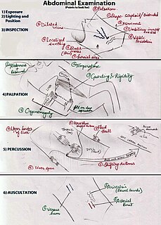
An abdominal examination is a portion of the physical examination which a physician or nurse uses to clinically observe the abdomen of a patient for signs of disease. The physical examination typically occurs after a thorough medical history is taken, that is, after the physician asks the patient the course of their symptoms. The abdominal examination is conventionally split into four different stages: first, inspection of the patient and the visible characteristics of their abdomen. Auscultation (listening) of the abdomen with a stethoscope. Palpation of the patient's abdomen. Finally, percussion (tapping) of the patient's abdomen and abdominal organs. Depending on the need to test for specific diseases such as ascites, special tests may be performed as a part of the physical examination. An abdominal examination may be performed because the physician suspects a disease of the organs inside the abdominal cavity, or simply as a part of a complete physical examination for other conditions. In a complete physical examination, the abdominal exam classically follows the respiratory examination and cardiovascular examination.
In medicine, the cardiac examination, also precordial exam, is performed as part of a physical examination, or when a patient presents with chest pain suggestive of a cardiovascular pathology. It would typically be modified depending on the indication and integrated with other examinations especially the respiratory examination.
Pulsus paradoxus, also paradoxic pulse or paradoxical pulse, is an abnormally large decrease in stroke volume, systolic blood pressure and pulse wave amplitude during inspiration. The normal fall in pressure is less than 10 mmHg. When the drop is more than 10 mmHg, it is referred to as pulsus paradoxus. Pulsus paradoxus is not related to pulse rate or heart rate, and it is not a paradoxical rise in systolic pressure. The normal variation of blood pressure during breathing/respiration is a decline in blood pressure during inhalation and an increase during exhalation. Pulsus paradoxus is a sign that is indicative of several conditions, including cardiac tamponade, chronic sleep apnea, croup, and obstructive lung disease.

A respiratory examination, or lung examination, is performed as part of a physical examination, in response to respiratory symptoms such as shortness of breath, cough, or chest pain, and is often carried out with a cardiac examination.
In medicine, shifting dullness refers to a sign elicited on physical examination for ascites.
Commotio cordis is an often lethal disruption of heart rhythm that occurs as a result of a blow to the area directly over the heart at a critical time during the cycle of a heart beat, producing what is termed an R-on-T phenomenon that leads to the condition. It is a form of ventricular fibrillation (V-Fib), not mechanical damage to the heart muscle or surrounding organs, and not the result of heart disease. The fatality rate is about 65 percent even with prompt CPR and defibrillation, and more than 80 percent without.

Precordial catch syndrome (PCS) is a non-serious condition in which there are sharp stabbing pains in the chest. These typically get worse with breathing in and occur within a small area. Spells of pain usually last less than a few minutes. Typically it begins at rest and other symptoms are absent. Concerns about the condition may result in anxiety.
A functional murmur is a heart murmur that is primarily due to physiologic conditions outside the heart, as opposed to structural defects in the heart itself. Serious conditions can arise even in the absence of a primary heart defect, and it is possible for peripheral conditions to generate abnormalities in the heart. Therefore, caution should be applied to use of the terms "innocent" or "benign" in this context.Use of the term dates to the mid 19th century.
Whispered pectoriloquy refers to an increased loudness of whispering noted during auscultation with a stethoscope on the lung fields on a patient's torso.
A parasternal heave, lift, or thrust is a precordial impulse that may be felt (palpated) in patients with cardiac or respiratory disease. Precordial impulses are visible or palpable pulsations of the chest wall, which originate on the heart or the great vessels.
The cardiovascular examination is a portion of the physical examination that involves evaluation of the cardiovascular system. The exact contents of the examination will vary depending on the presenting complaint but a complete examination will involve the heart, lungs, belly and the blood vessels.