Presynaptic inhibition

Last updated
A circuit diagram of postsynaptic inhibition (A, B) and presynaptic inhibition (C). Excitation is shown in green and inhibition is shown in red. Modified Axo-axonic synapse.svg
A circuit diagram of postsynaptic inhibition (A, B) and presynaptic inhibition (C). Excitation is shown in green and inhibition is shown in red.

Presynaptic inhibition is a phenomenon in which an inhibitory neuron provides synaptic input to the axon of another neuron (axo-axonal synapse) to make it less likely to fire an action potential. Presynaptic inhibition occurs when an inhibitory neurotransmitter, like GABA, acts on GABA receptors on the axon terminal. Or when endocannabinoids act as retrograde messengers by binding to presynaptic CB1 receptors, thereby indirectly modulating GABA and the excitability of dopamine neurons by reducing it and other presynaptic released neurotransmitters. [1] Presynaptic inhibition is ubiquitous among sensory neurons. [2]

Contents

Function

Sensory stimuli, such as pain, proprioception, and somatosensation, are sensed by primary afferent fibers. Somatosensory neurons encode information about the body's current state (e.g. temperature, pain, pressure, position, etc.). For vertebrate animals, these primary afferent fibers form synapses onto the spinal cord, specifically in the dorsal horn area, onto a variety of downstream targets including both excitatory neurons and inhibitory neurons. Synapses between primary afferent fibers and their targets are the first opportunity for sensory information to be modulated. [3] Primary afferent fibers contain many receptors along their projections, making them amenable to complex modulation. The constant influx of environmental stimuli, as sensed by primary afferent fibers, is subject to modulation to enhance or diminish stimuli (see also: gate control theory and gain control-biological). Because there are essentially unlimited stimuli, it is imperative that these signals are appropriately filtered.

To test whether somatosensation, specifically pain, was subjected to inhibition, scientists injected a chemical into the spinal cord of a rodent to block the primary inhibitory neurotransmitter's activity (bicuculline, a GABA receptor agonist [4] ). They found that pharmacologically blocking GABA receptors actually enhanced the perception of pain; in other words, GABA usually diminishes the perception of pain. [5]

The method by which GABA modulates synaptic transmission from primary afferent fibers to their downstream targets is disputed (see Mechanisms section below). Regardless of the mechanics, GABA acts in an inhibitory role to reduce the likelihood of primary afferent fiber synaptic release.

Modulating primary afferent fibers is critical to maintain general comfort. One study showed that animals without a specific type of GABA receptor on their nociceptors were hypersensitive to pain, [6] thus supporting a function of presynaptic inhibition as an analgesic. Certain pathological conditions, such as allodynia, are thought to be caused by non-modulated nociceptor firing. In addition to dampening pain, impaired presynaptic inhibition has been implicated in many neurological disorders, such as spasticity after spinal cord injury, [7] epilepsy, autism, and fragile-X syndrome. [8] [9] [10] [11] [12]

Mechanisms

Primary sensory afferents contain GABA receptors along their terminals (reviewed in:, [13] Table 1). GABA receptors are ligand-gated chloride channels, formed by the assembly of five GABA receptor subunits. In addition to the presence of GABA receptors along sensory afferent axons, the presynaptic terminal also has a distinct ionic composition that is high in chloride concentration. This is  due to cation-chloride cotransporters (for example, NKCC1) that maintain highs intracellular chloride. [14]

Typically when GABA receptors are activated, it causes a chloride influx, which hyperpolarizes the cell. However, in primary afferent fibers, due to the high concentration of chloride at the presynaptic terminal and thus its altered reversal potential, GABA receptor activation actually results in a chloride efflux, and thus a resulting depolarization. This phenomenon is called primary afferent depolarization (PAD). [15] [16] The GABA-induced depolarized potential at afferent axons has been demonstrated in many animals from cats to insects. Interestingly, despite the depolarized potential, GABA receptor activation along the axon still results in a reduction of neurotransmitter release and thus still is inhibitory.

There are four hypotheses which propose mechanisms behind this paradox:

  1. The depolarized membrane causes inactivation of voltage-gated sodium channels on the terminals and therefore the action potential is prevented from propagating. [13] [17] [18]
  2. Open GABA receptor channels act as a shunt, whereby current is dissipated of instead of being propagated to the terminals. [13] [17] [18] [19] [20] [21] [22] [23] [24]
  3. The depolarized membrane causes inactivation of voltage-gated calcium channels, preventing calcium influx at the synapse (which is imperative for neurotransmission). [13] [18] [20] [21] [25]
  4. The depolarization at the terminals generates an antidromic spike (i.e. an action potential generated in the axon and travels towards the soma), which would prevent orthodromic spikes (i.e. an action potential traveling from the cell's soma toward the axon terminals) from propagating. [19]

History of the discovery of presynaptic inhibition

1933: Grasser & Graham observed depolarization that originated in the sensory axon terminals [26]

1938: Baron & Matthews observed depolarization that originated in sensory axon terminals and the ventral root [27]

1957: Frank & Fuortes coined the term "presynaptic inhibition" [28]

1961: Eccles, Eccles, & Magni determined that the Dorsal Root Potential (DRP) originated from depolarization in sensory axon terminals [29]

Related Research Articles

<span class="mw-page-title-main">Axon</span> Long projection on a neuron that conducts signals to other neurons

An axon, or nerve fiber, is a long, slender projection of a nerve cell, or neuron, in vertebrates, that typically conducts electrical impulses known as action potentials away from the nerve cell body. The function of the axon is to transmit information to different neurons, muscles, and glands. In certain sensory neurons, such as those for touch and warmth, the axons are called afferent nerve fibers and the electrical impulse travels along these from the periphery to the cell body and from the cell body to the spinal cord along another branch of the same axon. Axon dysfunction can be the cause of many inherited and acquired neurological disorders that affect both the peripheral and central neurons. Nerve fibers are classed into three types – group A nerve fibers, group B nerve fibers, and group C nerve fibers. Groups A and B are myelinated, and group C are unmyelinated. These groups include both sensory fibers and motor fibers. Another classification groups only the sensory fibers as Type I, Type II, Type III, and Type IV.

<span class="mw-page-title-main">Neuron</span> Electrically excitable cell found in the nervous system of animals

Within a nervous system, a neuron, neurone, or nerve cell is an electrically excitable cell that fires electric signals called action potentials across a neural network. Neurons communicate with other cells via synapses - specialized connections that commonly use minute amounts of chemical neurotransmitters to pass the electric signal from the presynaptic neuron to the target cell through the synaptic gap. The neuron is the main component of nervous tissue in all animals except sponges and placozoa. Non-animals like plants and fungi do not have nerve cells. The ability to generate electric signals first appeared in evolution 700 million years ago. 800 million years ago, predecessors of neurons were the peptidergic secretory cells. They eventually gained new gene modules which enabled cells to create post-synaptic scaffolds and ion channels that generate fast electrical signals. The ability to generate electric signals was a key innovation in the evolution of the nervous system.

<span class="mw-page-title-main">Neurotransmitter</span> Chemical substance that enables neurotransmission

A neurotransmitter is a signaling molecule secreted by a neuron to affect another cell across a synapse. The cell receiving the signal, or target cell, may be another neuron, but could also be a gland or muscle cell.

<span class="mw-page-title-main">Nervous system</span> Part of an animal that coordinates actions and senses

In biology, the nervous system is the highly complex part of an animal that coordinates its actions and sensory information by transmitting signals to and from different parts of its body. The nervous system detects environmental changes that impact the body, then works in tandem with the endocrine system to respond to such events. Nervous tissue first arose in wormlike organisms about 550 to 600 million years ago. In vertebrates it consists of two main parts, the central nervous system (CNS) and the peripheral nervous system (PNS). The CNS consists of the brain and spinal cord. The PNS consists mainly of nerves, which are enclosed bundles of the long fibers or axons, that connect the CNS to every other part of the body. Nerves that transmit signals from the brain are called motor nerves or efferent nerves, while those nerves that transmit information from the body to the CNS are called sensory nerves or afferent. Spinal nerves are mixed nerves that serve both functions. The PNS is divided into three separate subsystems, the somatic, autonomic, and enteric nervous systems. Somatic nerves mediate voluntary movement. The autonomic nervous system is further subdivided into the sympathetic and the parasympathetic nervous systems. The sympathetic nervous system is activated in cases of emergencies to mobilize energy, while the parasympathetic nervous system is activated when organisms are in a relaxed state. The enteric nervous system functions to control the gastrointestinal system. Both autonomic and enteric nervous systems function involuntarily. Nerves that exit from the cranium are called cranial nerves while those exiting from the spinal cord are called spinal nerves.

<span class="mw-page-title-main">Chemical synapse</span> Biological junctions through which neurons signals can be sent

Chemical synapses are biological junctions through which neurons' signals can be sent to each other and to non-neuronal cells such as those in muscles or glands. Chemical synapses allow neurons to form circuits within the central nervous system. They are crucial to the biological computations that underlie perception and thought. They allow the nervous system to connect to and control other systems of the body.

<span class="mw-page-title-main">Motor neuron</span> Nerve cell sending impulse to muscle

A motor neuron is a neuron whose cell body is located in the motor cortex, brainstem or the spinal cord, and whose axon (fiber) projects to the spinal cord or outside of the spinal cord to directly or indirectly control effector organs, mainly muscles and glands. There are two types of motor neuron – upper motor neurons and lower motor neurons. Axons from upper motor neurons synapse onto interneurons in the spinal cord and occasionally directly onto lower motor neurons. The axons from the lower motor neurons are efferent nerve fibers that carry signals from the spinal cord to the effectors. Types of lower motor neurons are alpha motor neurons, beta motor neurons, and gamma motor neurons.

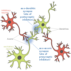
An inhibitory postsynaptic potential (IPSP) is a kind of synaptic potential that makes a postsynaptic neuron less likely to generate an action potential. IPSPs were first investigated in motorneurons by David P. C. Lloyd, John Eccles and Rodolfo Llinás in the 1950s and 1960s. The opposite of an inhibitory postsynaptic potential is an excitatory postsynaptic potential (EPSP), which is a synaptic potential that makes a postsynaptic neuron more likely to generate an action potential. IPSPs can take place at all chemical synapses, which use the secretion of neurotransmitters to create cell to cell signalling. Inhibitory presynaptic neurons release neurotransmitters that then bind to the postsynaptic receptors; this induces a change in the permeability of the postsynaptic neuronal membrane to particular ions. An electric current that changes the postsynaptic membrane potential to create a more negative postsynaptic potential is generated, i.e. the postsynaptic membrane potential becomes more negative than the resting membrane potential, and this is called hyperpolarisation. To generate an action potential, the postsynaptic membrane must depolarize—the membrane potential must reach a voltage threshold more positive than the resting membrane potential. Therefore, hyperpolarisation of the postsynaptic membrane makes it less likely for depolarisation to sufficiently occur to generate an action potential in the postsynaptic neurone.

<span class="mw-page-title-main">Stimulus (physiology)</span> Detectable change in the internal or external surroundings

In physiology, a stimulus is a detectable change in the physical or chemical structure of an organism's internal or external environment. The ability of an organism or organ to detect external stimuli, so that an appropriate reaction can be made, is called sensitivity (excitability). Sensory receptors can receive information from outside the body, as in touch receptors found in the skin or light receptors in the eye, as well as from inside the body, as in chemoreceptors and mechanoreceptors. When a stimulus is detected by a sensory receptor, it can elicit a reflex via stimulus transduction. An internal stimulus is often the first component of a homeostatic control system. External stimuli are capable of producing systemic responses throughout the body, as in the fight-or-flight response. In order for a stimulus to be detected with high probability, its level of strength must exceed the absolute threshold; if a signal does reach threshold, the information is transmitted to the central nervous system (CNS), where it is integrated and a decision on how to react is made. Although stimuli commonly cause the body to respond, it is the CNS that finally determines whether a signal causes a reaction or not.

<span class="mw-page-title-main">Nociceptor</span> Sensory neuron that detects pain

A nociceptor is a sensory neuron that responds to damaging or potentially damaging stimuli by sending "possible threat" signals to the spinal cord and the brain. The brain creates the sensation of pain to direct attention to the body part, so the threat can be mitigated; this process is called nociception.

<span class="mw-page-title-main">End-plate potential</span>

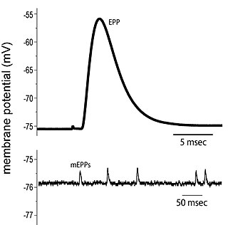
End plate potentials (EPPs) are the voltages which cause depolarization of skeletal muscle fibers caused by neurotransmitters binding to the postsynaptic membrane in the neuromuscular junction. They are called "end plates" because the postsynaptic terminals of muscle fibers have a large, saucer-like appearance. When an action potential reaches the axon terminal of a motor neuron, vesicles carrying neurotransmitters are exocytosed and the contents are released into the neuromuscular junction. These neurotransmitters bind to receptors on the postsynaptic membrane and lead to its depolarization. In the absence of an action potential, acetylcholine vesicles spontaneously leak into the neuromuscular junction and cause very small depolarizations in the postsynaptic membrane. This small response (~0.4mV) is called a miniature end plate potential (MEPP) and is generated by one acetylcholine-containing vesicle. It represents the smallest possible depolarization which can be induced in a muscle.

<span class="mw-page-title-main">Dorsal root ganglion</span> Cluster of neurons in a dorsal root of a spinal nerve

A dorsal root ganglion is a cluster of neurons in a dorsal root of a spinal nerve. The cell bodies of sensory neurons known as first-order neurons are located in the dorsal root ganglia.

<span class="mw-page-title-main">Glycine receptor</span> Widely distributed inhibitory receptor in the central nervous system

The glycine receptor is the receptor of the amino acid neurotransmitter glycine. GlyR is an ionotropic receptor that produces its effects through chloride current. It is one of the most widely distributed inhibitory receptors in the central nervous system and has important roles in a variety of physiological processes, especially in mediating inhibitory neurotransmission in the spinal cord and brainstem.

Molecular neuroscience is a branch of neuroscience that observes concepts in molecular biology applied to the nervous systems of animals. The scope of this subject covers topics such as molecular neuroanatomy, mechanisms of molecular signaling in the nervous system, the effects of genetics and epigenetics on neuronal development, and the molecular basis for neuroplasticity and neurodegenerative diseases. As with molecular biology, molecular neuroscience is a relatively new field that is considerably dynamic.

<span class="mw-page-title-main">Golgi cell</span>

In neuroscience, Golgi cells are inhibitory interneurons found within the granular layer of the cerebellum. They were first identified as inhibitory in 1964. It was also the first example of an inhibitory feedback network, where the inhibitory interneuron was identified anatomically. These cells synapse onto the dendrite of granule cells and unipolar brush cells. They receive excitatory input from mossy fibres, also synapsing on granule cells, and parallel fibers, which are long granule cell axons. Thereby this circuitry allows for feed-forward and feed-back inhibition of granule cells.

Depolarization-induced suppression of inhibition is the classical and original electrophysiological example of endocannabinoid function in the central nervous system. Prior to the demonstration that depolarization-induced suppression of inhibition was dependent on the cannabinoid CB1 receptor function, there was no way of producing an in vitro endocannabinoid mediated effect.

<span class="mw-page-title-main">Substantia gelatinosa of Rolando</span>

The apex of the posterior grey column, one of the three grey columns of the spinal cord, is capped by a V-shaped or crescentic mass of translucent, gelatinous neuroglia, termed the substantia gelatinosa of Rolando, which contains both neuroglia cells, and small nerve cells. The gelatinous appearance is due to a very low concentration of myelinated fibers. It extends the entire length of the spinal cord and into the medulla oblongata where it becomes the spinal nucleus of the trigeminal nerve.

<span class="mw-page-title-main">Group C nerve fiber</span> One of three classes of nerve fiber in the central nervous system and peripheral nervous system

Group C nerve fibers are one of three classes of nerve fiber in the central nervous system (CNS) and peripheral nervous system (PNS). The C group fibers are unmyelinated and have a small diameter and low conduction velocity, whereas Groups A and B are myelinated. Group C fibers include postganglionic fibers in the autonomic nervous system (ANS), and nerve fibers at the dorsal roots. These fibers carry sensory information.

<span class="mw-page-title-main">Summation (neurophysiology)</span>

Summation, which includes both spatial summation and temporal summation, is the process that determines whether or not an action potential will be generated by the combined effects of excitatory and inhibitory signals, both from multiple simultaneous inputs, and from repeated inputs. Depending on the sum total of many individual inputs, summation may or may not reach the threshold voltage to trigger an action potential.

<span class="mw-page-title-main">Spinal interneuron</span> Interneuron relaying signals between sensory and motor neurons in the spinal cord

A spinal interneuron, found in the spinal cord, relays signals between (afferent) sensory neurons, and (efferent) motor neurons. Different classes of spinal interneurons are involved in the process of sensory-motor integration. Most interneurons are found in the grey column, a region of grey matter in the spinal cord.

An axo-axonic synapse is a type of synapse, formed by one neuron projecting its axon terminals onto another neuron's axon.

References

  1. Oleson, Erik B. (2012-01-26). "Endocannabinoids shape accumbal encoding of cue-motivated behavior via CB1 receptor activation in the ventral tegmentum". Neuron. 73 (2): 360–373. doi:10.1016/j.neuron.2011.11.018. PMC   3269037 . PMID   22284189.
  2. McGann JP (July 2013). "Presynaptic inhibition of olfactory sensory neurons: new mechanisms and potential functions". Chemical Senses. 38 (6): 459–474. doi:10.1093/chemse/bjt018. PMC   3685425 . PMID   23761680.
  3. Comitato A, Bardoni R (January 2021). "Presynaptic Inhibition of Pain and Touch in the Spinal Cord: From Receptors to Circuits". International Journal of Molecular Sciences. 22 (1): 414. doi: 10.3390/ijms22010414 . PMC   7795800 . PMID   33401784.
  4. Manske RH (September 1932). "The Alkaloids of Fumaraceous Plants: II. Dicentra Cucullaria (L.) Bernh". Canadian Journal of Research. 7 (3): 265–269. Bibcode:1932CJRes...7..265M. doi:10.1139/cjr32-078. ISSN   1923-4287.
  5. Roberts LA, Beyer C, Komisaruk BR (November 1986). "Nociceptive responses to altered GABAergic activity at the spinal cord". Life Sciences. 39 (18): 1667–74. doi:10.1016/0024-3205(86)90164-5. PMID   3022091.
  6. Price TJ, Cervero F, Gold MS, Hammond DL, Prescott SA (April 2009). "Chloride regulation in the pain pathway". Brain Research Reviews. 60 (1): 149–170. doi:10.1016/j.brainresrev.2008.12.015. PMC   2903433 . PMID   19167425.
  7. Caron, Guillaume; Bilchak, Jadwiga N.; Côté, Marie‐Pascale (2020). "Direct evidence for decreased presynaptic inhibition evoked by PBSt group I muscle afferents after chronic SCI and recovery with step‐training in rats". The Journal of Physiology. 598 (20): 4621–4642. doi:10.1113/JP280070. ISSN   0022-3751. PMC   7719595 . PMID   32721039.
  8. Deidda G, Bozarth IF, Cancedda L (2014). "Modulation of GABAergic transmission in development and neurodevelopmental disorders: investigating physiology and pathology to gain therapeutic perspectives". Frontiers in Cellular Neuroscience. 8: 119. doi: 10.3389/fncel.2014.00119 . PMC   4033255 . PMID   24904277.
  9. Zeilhofer HU, Wildner H, Yévenes GE (January 2012). "Fast synaptic inhibition in spinal sensory processing and pain control". Physiological Reviews. 92 (1): 193–235. doi:10.1152/physrev.00043.2010. PMC   3590010 . PMID   22298656.
  10. Lee E, Lee J, Kim E (May 2017). "Excitation/Inhibition Imbalance in Animal Models of Autism Spectrum Disorders". Biological Psychiatry. 81 (10): 838–847. doi: 10.1016/j.biopsych.2016.05.011 . PMID   27450033.
  11. D'Hulst C, Kooy RF (August 2007). "The GABAA receptor: a novel target for treatment of fragile X?". Trends in Neurosciences. 30 (8): 425–431. doi:10.1016/j.tins.2007.06.003. PMID   17590448. S2CID   7340813.
  12. Benarroch EE (February 2007). "GABAA receptor heterogeneity, function, and implications for epilepsy". Neurology. 68 (8): 612–614. doi:10.1212/01.wnl.0000255669.83468.dd. PMID   17310035. S2CID   11101571.
  13. 1 2 3 4 Guo D, Hu J (December 2014). "Spinal presynaptic inhibition in pain control". Neuroscience. 283: 95–106. doi: 10.1016/j.neuroscience.2014.09.032 . PMID   25255936.
  14. Kahle KT, Staley KJ, Nahed BV, Gamba G, Hebert SC, Lifton RP, Mount DB (September 2008). "Roles of the cation-chloride cotransporters in neurological disease". Nature Clinical Practice. Neurology. 4 (9): 490–503. doi:10.1038/ncpneuro0883. PMID   18769373. S2CID   15424963.
  15. Price TJ, Cervero F, Gold MS, Hammond DL, Prescott SA (April 2009). "Chloride regulation in the pain pathway". Brain Research Reviews. 60 (1): 149–170. doi:10.1016/j.brainresrev.2008.12.015. PMC   2903433 . PMID   19167425.
  16. Willis WD (February 1999). "Dorsal root potentials and dorsal root reflexes: a double-edged sword". Experimental Brain Research. 124 (4): 395–421. doi:10.1007/s002210050637. PMID   10090653. S2CID   40738560.
  17. 1 2 Cattaert D, El Manira A (July 1999). "Shunting versus inactivation: analysis of presynaptic inhibitory mechanisms in primary afferents of the crayfish". The Journal of Neuroscience. 19 (14): 6079–6089. doi:10.1523/JNEUROSCI.19-14-06079.1999. PMC   6783106 . PMID   10407044.
  18. 1 2 3 Willis WD (2006-02-01). "John Eccles' studies of spinal cord presynaptic inhibition". Progress in Neurobiology. 78 (3–5): 189–214. doi:10.1016/j.pneurobio.2006.02.007. PMID   16650518. S2CID   38669996.
  19. 1 2 Cattaert D, Libersat F, El Manira A (February 2001). "Presynaptic inhibition and antidromic spikes in primary afferents of the crayfish: a computational and experimental analysis". The Journal of Neuroscience. 21 (3): 1007–1021. doi:10.1523/JNEUROSCI.21-03-01007.2001. PMC   6762302 . PMID   11157086.
  20. 1 2 Panek I, French AS, Seyfarth EA, Sekizawa S, Torkkeli PH (July 2002). "Peripheral GABAergic inhibition of spider mechanosensory afferents". The European Journal of Neuroscience. 16 (1): 96–104. doi:10.1046/j.1460-9568.2002.02065.x. PMID   12153534. S2CID   20750558.
  21. 1 2 French AS, Panek I, Torkkeli PH (June 2006). "Shunting versus inactivation: simulation of GABAergic inhibition in spider mechanoreceptors suggests that either is sufficient". Neuroscience Research. 55 (2): 189–196. doi:10.1016/j.neures.2006.03.002. PMID   16616790. S2CID   2099107.
  22. Miller RJ (1998). "Presynaptic receptors". Annual Review of Pharmacology and Toxicology. 38: 201–227. doi:10.1146/annurev.pharmtox.38.1.201. PMID   9597154.
  23. Zhang SJ, Jackson MB (March 1995). "Properties of the GABAA receptor of rat posterior pituitary nerve terminals". Journal of Neurophysiology. 73 (3): 1135–1144. doi:10.1152/jn.1995.73.3.1135. PMID   7608760.
  24. Zhang SJ, Jackson MB (March 1995). "GABAA receptor activation and the excitability of nerve terminals in the rat posterior pituitary". The Journal of Physiology. 483 (3): 583–595. doi:10.1113/jphysiol.1995.sp020608. PMC   1157804 . PMID   7776245.
  25. Graham B, Redman S (February 1994). "A simulation of action potentials in synaptic boutons during presynaptic inhibition". Journal of Neurophysiology. 71 (2): 538–549. doi:10.1152/jn.1994.71.2.538. PMID   8176423.
  26. Gasser HS, Graham HT (January 1933). "Potentials produced in the spinal cord by stimulation of dorsal roots". American Journal of Physiology. 103 (2): 303–320. doi:10.1152/ajplegacy.1933.103.2.303.
  27. Barron DH, Matthews BH (April 1938). "The interpretation of potential changes in the spinal cord". The Journal of Physiology. 92 (3): 276–321. doi:10.1113/jphysiol.1938.sp003603. PMC   1395290 . PMID   16994975.
  28. Frank K, Fuortes MG (1957). "Presynaptic and Postsynaptic inhibition of monsynaptic reflexes". Federation Proceedings. 16: 39–40.
  29. Eccles JC, Eccles RM, Magni F (November 1961). "Central inhibitory action attributable to presynaptic depolarization produced by muscle afferent volleys". The Journal of Physiology. 159 (1): 147–166. doi:10.1113/jphysiol.1961.sp006798. PMC   1359583 . PMID   13889050.