Protectin D1

Last updated

Contents

Protectin D1
Protectin D1.svg
Names
Preferred IUPAC name
(4Z,7Z,10R,11E,13E,15Z,17S,19Z)-10,17-Dihydroxydocosa-4,7,11,13,15,19-hexaenoic acid
Other names
10R,17S-Dihydroxy-docosa-4Z,7Z,11E,13E,15Z,19Z-hexaenoate; 10R,17S-Dihydroxy-docosa-4Z,7Z,11E,13E,15Z,19Z-hexaenoic acid; Neuroprotectin D1
Identifiers
3D model (JSmol)
ChemSpider
PubChem CID
UNII
  • InChI=1S/C22H32O4/c1-2-3-10-15-20(23)17-12-8-9-13-18-21(24)16-11-6-4-5-7-14-19-22(25)26/h3,5-13,17-18,20-21,23-24H,2,4,14-16,19H2,1H3,(H,25,26)/b7-5-,9-8+,10-3-,11-6-,17-12-,18-13+/t20-,21+/m0/s1 Yes check.svgY
    Key: CRDZYJSQHCXHEG-SFVBTVKNSA-N Yes check.svgY
  • O=C(O)CC\C=C/C/C=C\C[C@@H](O)\C=C\C=C\C=C/[C@@H](O)C\C=C/CC
Properties
C22H32O4
Molar mass 360.4871 g/mol
Density 1.049 g/cm3
Boiling point 559.379 °C (1,038.882 °F; 832.529 K)
0.0069
log P 4.95
Acidity (pKa)4.82
Except where otherwise noted, data are given for materials in their standard state (at 25 °C [77 °F], 100 kPa).

Protectin D1 also known as neuroprotectin D1 (when it acts in the nervous system) and abbreviated most commonly as PD1 or NPD1 is a member of the class of specialized proresolving mediators. Like other members of this class of polyunsaturated fatty acid metabolites, it possesses strong anti-inflammatory, anti-apoptotic and neuroprotective activity. PD1 is an aliphatic acyclic alkene 22 carbons in length with two hydroxyl groups at the 10 and 17 carbon positions and one carboxylic acid group at the one carbon position. [1]

Specifically, PD1 is an endogenous stereoselective lipid mediator classified as an autocoid protectin. Autacoids are enzymatically derived chemical mediators with distinct biological activities and molecular structures. Protectins are signaling molecules that are produced enzymatically from unsaturated fatty acids. Their molecular structure is characterized by the presence of a conjugated system of double bonds. [1] PD1, like other protectins, is produced by the oxygenation of the ω-3 polyunsaturated fatty acid docosahexaenoic acid (DHA) and it is found in many tissues, such as the retina, the lungs and the nervous system. [2] [3]

PD1 has a significant role as an anti-inflammatory, anti-apoptotic and neuroprotective molecule. Studies in Alzheimer's disease animal models, in stroke patients and in human retina pigment epithelial cells (RPE) have shown that PD1 can potentially reduce inflammation induced by oxidative stress and inhibit the pro-apoptotic signal, thereby preventing cellular degeneration. [2] [3] [4] [5] Finally, recent studies examining the pathogenicity of influenza viruses, including the avian flu (H5N1), have suggested that PD1 can potentially halt the proliferation of the virus, thus protecting respiratory cells from lethal viral infections. [6] [7]

Biosynthesis of PD1

In vivo, PD1 is mainly produced as a response to inflammatory signals and it is found in various tissues, such as the retina pigment epithelial cells, lung epithelial cells, peripheral blood mononuclear cells (PBMC) and neural tissues. Studies in PBMC have shown that endogenous DHA, the main precursor of PD1, is released by the activity of phospholipase A2. [1] [2] [3] According to these studies, PD1 is preferentially synthesized in PBMC cells skewed to the Type 2 T helper cell phenotype (TH2). [1] This suggests that T-cell differentiation plays an important role in the activation of the PD1 biosynthetic pathway. The interaction of PBMC with interleukin 4 (IL-4), a potent inflammatory signal, leads to the differentiation of PBMC to TH2 type lymphocytes. [1] In addition, activated TH2 cells further release IL-4, leading to the up-regulation of the enzyme 15-lipoxygenase-1 (15-LO-1). [1] 15-LO-1 is a non-heme iron-carrying dioxygenase that adds oxygen atoms in a stereospecific manner on free and esterified ω-3 polyunsaturated fatty acids like DHA. [3] Overall, the biosynthesis of PD1 proceeds through three distinct steps throughout which the activity of 15-LO-1 is essential. In the first step of the biosynthetic pathway, the binding of 15-LO-1 to its substrate (DHA) leads to the formation of the (17S)-hydro(peroxy)-DHA intermediate. This intermediate is rapidly processed to form a 16(17)-epoxide-containing molecule, which is the second intermediate. Finally, in the third step of the pathway, enzymatic hydrolysis of the 16(17)-epoxide-containing intermediate leads to the formation of PD1. [1]

Protectin D1 (PD1) Biosynthesis.jpg

Functions of PD1

In general, PD1 in vivo exhibits a potent anti-apoptotic and anti-inflammatory activity in the tissues in which it is localized. DHA, the main PD1 precursor, is mostly found in tissues such as the retinal synapses, photoreceptors, the lungs and the brain, suggesting that these tissues are more likely to be benefited from the protecting activity of PD1. [1] [2] [3] [4] [7] [8]

Activity of PD1 in the retina

RPE are essential in the survival and renewal of the photoreceptors in the retina. These cells exhibit a potent phagocytic activity that ensures the proper function of the retina. Therefore, oxidative stress can potentially damage the RPE cells and cause vision impairment. Studies in human RPE cells have suggested that the presence of oxidative stress triggering molecules, such as H2O2 causes the fragmentation of the DNA that in turn triggers apoptosis. [2] These studies have proposed that PD1 acts as a signaling molecule and through its ligand-receptor interaction down-regulates the expression of genes, such as the transcription factor NF-κB. The inhibition of NF-κB results in the down-regulation of the pro-inflammatory gene COX-2 (cyclooxygenase-2) which is responsible for the release of prostaglandins, a potent pro-inflammatory mediator. [2] In addition, PD1 has an important role in regulating the expression of the Bcl-2 family proteins (Bcl-2, Bcl-xL, Bax and Bad) that precedes the release of the cytochrome c complex from the mitochondria and the formation of the apoptosome. [2] [3] [4] The presence of PD1 up-regulates the expression of the anti-apoptotic proteins Bcl-2 and Bcl-xL, while it inhibits the expression of the pro-apoptotic proteins Bax and Bad. [2] Specifically, PD1 regulates this protein family by promoting the dephosphorylation of Bcl-xL by protein phosphatase 2A (PP2A) at residue Ser-62 which in turn heterodimerizes with the pro-apoptotic protein Bax and inactivates it. [4] Consequently, the activity of the Bcl-2 family proteins results in the inhibition of the caspase 3 enzyme, thus preventing apoptosis and promoting RPE cell survival. [2] [4]

Effects of PD1 in Alzheimer's disease

Among others, Alzheimer's disease is characterized by the reduced concentration of PD1 and by the increased concentration of the amyloid-β peptide (Aβ42) that is responsible for the formation of senile plaques and also induces inflammation and apoptosis in neuronal tissues. [5] [9] Aβ42 is generated by the enzymatic cleavage of the β-amyloid precursor protein (βΑPP) through β- and γ- secretases. Like other pro-inflammatory mediators, Aβ42 induces inflammation through the activation of the pro-inflammatory enzyme COX-2 and the release of prostaglandins. Moreover, the release of Aβ42 down-regulates the anti-apoptotic proteins Bcl-2 and Bcl-xL and up-regulates the pro-apoptotic proteins Bax and Bad that ultimately lead to the formation of the apoptosome. [5] [9] PD1 in human neuronal glial cells (HNG) has been shown to trigger the down-regulation of βΑPP, thus decreasing the Aβ42 content in neuronal tissues and reducing inflammation and apoptosis. [5] Specifically, PD1 in Alzheimer's disease models has been shown to respond to the increased concentration of the pro-inflammatory molecule Aβ42 by binding and activating the peroxisome proliferator-activated receptor gamma (PPARγ) either directly or via other mechanisms. According to some models the activation of PPARγ leads to increased ubiquitination and degradation of βAPP, thus reducing the release of Aβ42. [5] Furthermore, PD1 inhibits the production of Aβ42 peptide by down-regulating β-secretase-1 (BACE1), while up-regulating the α-secretase ADAM10 and the secreted amyloid precursor protein-α (sAPPα). Overall, the above mechanism leads to the cleavage of βAPP protein though a non-amyloidogenic pathway that halts the formation of Aβ42 and prevents the premature neuronal degeneration. [5] [9]

Antiviral activity of PD1

Studies in cultured human lung epithelial cells infected with the influenza virus H1N1 or H5N1 have found that endogenous production of PD1 decreases dramatically during infection due to the inhibition of 15-LO-1. [6] [7] Furthermore, the same studies have shown that in vivo administration of PD1 to H1N1 infected mice can potentially inhibit both the proliferation of the virus and the inflammation caused by the infection, thus increasing survival. PD1 protects against viral infections by disrupting the virus life cycle. Specifically, PD1 inhibits the binding of viral RNA to specific nuclear export factors in the host cells, thus blocking the export of viral RNA from the nucleus to the cytosol. [6] [7] The nuclear RNA export factor 1(NXF1) is of particular interest in the attenuation of viral infections via the activity of PD1. Specifically, the NXF1 transporter through its middle and C-terminal domains binds to the phenylalanine/glycine repeats in the nucleoporins (Nups) that line the nuclear pore. [7] In the absence of PD1, influenza viral RNA binds to the NXF1 transporter that later binds specifically to Nup62 nucleoporin and exports the viral RNA into the cytosol. However, the administration of PD1 has shown that this lipid mediator specifically inhibits the binding of the viral RNA to NXF1, thus disrupting the proliferation of the virus. [7]

Laboratory Synthesis of PD1

The large scale industrial production of PD1 is of great interest for pharmaceutical companies in order to harvest the potent anti-inflammatory and anti-apoptotic activities of this lipid mediator. So far, very few stereoselective laboratory syntheses of PD1 have been reported, but with a relatively low yield. [10] [11]

Convergent Stereoselective Synthesis

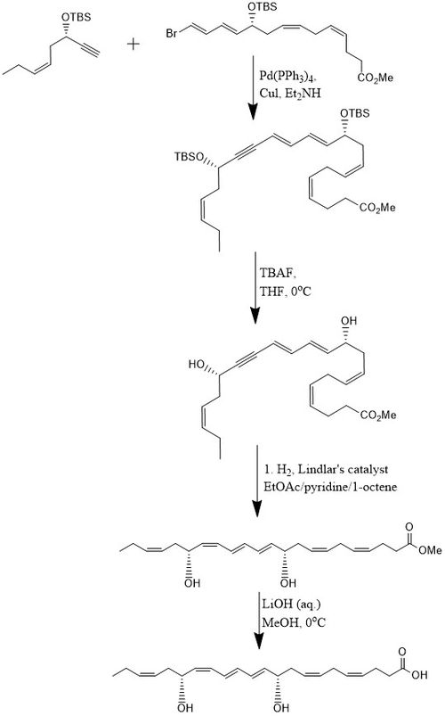
According to one method, PD1 is synthesized in 15% yield through an 8-step convergent stereoselective process. [10] Initially, the alkyne, (Z)-3-tertbutyldimethylsiloxy-oct-5-en-1-yne reacts with bromo-E,E,Z,Z-tetraene ester in a Sonogashira cross-coupling reaction at room temperature in the presence of Pd-(PPh3)4 and CuI using diethylamine as a solvent which produces the bis-hydroxyl-protected methyl ester. Removal of the two tert-butyldimethylsilyl ethers (TBS-protecting groups) is attained with an excess of TBAF in THF at 0 °C which produces a diol containing a conjugated alkyne. The conjugated alkyne is reduced to the methyl ester. In addition, the diol is hydrogenated using the Lindlar catalyst, with 1-octene added as a sacrificial olefin, to produce a highly stereoselective triene, while water is eliminated from the diol through a Boland reduction. Finally, the methyl ester undergoes saponification at 0 °C with dilute LiOH (aq.) in methanol followed by acidic work-up with NaH2PO4 (aq.) in order to produce PD1. [10]

Convergent Stereoselective Synthesis of PD1.jpg

Alternative Stereoselective Synthesis

Alternatively, PD1 laboratory synthesis proceeds through a different stereoselective method. [11] Initially, hydroboration of a TBS-protected acetylene with Sia2BH produces a TBS-protected vinylborane. The TBS-protected vinylborane reacts with vinyliodide in the presence of a Pd-catalyst, sodium hydroxide (NaOH) and THF to produce a TBS-protected alcohol. Later treatment of the TBS-protected alcohol with TBAF removes the protecting group and produces a diol. Finally, the diol is hydrolyzed with LiOH in THF (aq.) to produce PD1. [11]

Laboratory synthesis of Protectin D1 (PD1).jpg

Other PDs

22-hydroxy-NPD1

22-hydroxy-PD1 (22-OH-PD1; i.e. 10R,17S,20-trihydroxy-4Z,7Z,11E,13E,15Z,19Z-docosahexaenoic acid) is an omega oxidation product of PD1 probably formed in cells by the action of an unidentified Cytochrome P450 omega hydroxylase (see specialized proresolving mediators#Protectins/neuroprotectins). While the omega oxidation of many bioactive fatty acid metabolites such as leukotriene B4, 5-HETE, 5-oxo-eicosatetraenoic acid (i.e. 5-oxo-ETE) results in a ~100-fold fall in their activity, the omega oxidized product of PD1 has been shown to possess potent ease exhibits potent anti-inflammatory and proresolving actions by inhibiting PMN chemotaxis in vivo and in vitro and decreased pro-inflammatory mediator levels in inflammatory exudates of an animal model at levels comparable to PD1. [12] [13]

Protectin DX

Protectin DX (PDX; i.e. 10S,17S-dihydroxy-4Z,7Z,11E,13Z,15E,19Z-docosahexaenoic acid) is the 13Z,15E,19Z isomer of NPD1 (which has the 13E,15Z,19Z double bond configuration)(see specialized proresolving mediators#Protectins/neuroprotectins). An early study mistakenly used PDX instead of PD1 in attributing anti-replicative and clinically beneficial effects in viral influenza disease in a mouse model to PD1. [14] PDX also inhibits influx of circulating leukocytes into the peritoneum in a mouse model of inflammation. [15] PDX has other anti-inflammatory actions. It inhibits COX-1 and COX-2 thereby blocking the formation of pro-inflammatory prostaglandins; it also inhibits the platelet-aggregating action of thromboxane A2 thereby blocking the platelet aggregations responses to agents that depend on platelets to release thromboxane A2. [16]

Aspirin-triggered PD1

Aspirin-triggered PD1 (AT-PD1 or 17-epi-PD1: i.e. 10R,17R-dihydroxy-4Z,7Z,11E,13E,15Z,19Z-docosahexaenoic acid) is the 10R-hydroxy isomer of PD1 (which has the 10S hydroxy residue) (see specialized proresolving mediators#Protectins/neuroprotectins). AT-PD1 has been shown to a) reduce the infiltration of neutrophils into the peritoneum in a mouse model of inflammatory disease; b) stimulate the Efferocytosis (i.e. engulfment and removal) of neutrophils; and c) reduce brain infarction and stroke in a rodent model. [17]

10-epi-PD1

10-Epi-PD1 (ent-AT-NPD1: i.e. 10S,17S-Dihydroxy-4Z,7Z,11E,13E,15Z,19Z-docosahexaenoic acid) is the 10S-hydroxy isomer of AT-PD1 (which has a 10R-hydroxy residue) (see specialized proresolving mediators#Protectins/neuroprotectins). 10-Epi-PD1 was detected in only a small amount in human PMN extracts but was more potent than PD1 or PDX in blocking the inflammatory response to zymosan A-induced murine acute peritonitis. [13]

Related Research Articles

<span class="mw-page-title-main">Tumor necrosis factor</span> Protein

Tumor necrosis factor is an adipokine and a cytokine. TNF is a member of the TNF superfamily, which consists of various transmembrane proteins with a homologous TNF domain.

<span class="mw-page-title-main">Lipoxin</span> Acronym for lipoxygenase interaction product

A lipoxin (LX or Lx), an acronym for lipoxygenase interaction product, is a bioactive autacoid metabolite of arachidonic acid made by various cell types. They are categorized as nonclassic eicosanoids and members of the specialized pro-resolving mediators (SPMs) family of polyunsaturated fatty acid (PUFA) metabolites. Like other SPMs, LXs form during, and then act to resolve, inflammatory responses. Initially, two lipoxins were identified, lipoxin A4 (LXA4) and LXB4, but more recent studies have identified epimers of these two LXs: the epi-lipoxins, 15-epi-LXA4 and 15-epi-LXB4 respectively.

<span class="mw-page-title-main">GSK-3</span> Class of enzymes

Glycogen synthase kinase 3 (GSK-3) is a serine/threonine protein kinase that mediates the addition of phosphate molecules onto serine and threonine amino acid residues. First discovered in 1980 as a regulatory kinase for its namesake, glycogen synthase (GS), GSK-3 has since been identified as a protein kinase for over 100 different proteins in a variety of different pathways. In mammals, including humans, GSK-3 exists in two isozymes encoded by two homologous genes GSK-3α (GSK3A) and GSK-3β (GSK3B). GSK-3 has been the subject of much research since it has been implicated in a number of diseases, including type 2 diabetes, Alzheimer's disease, inflammation, cancer, addiction and bipolar disorder.

<span class="mw-page-title-main">Resolvin</span> Class of chemical compounds

Resolvins are specialized pro-resolving mediators (SPMs) derived from omega-3 fatty acids, primarily eicosapentaenoic acid (EPA) and docosahexaenoic acid (DHA), as well as docosapentaenoic acid (DPA) and clupanodonic acid. As autacoids similar to hormones acting on local tissues, resolvins are under preliminary research for their involvement in promoting restoration of normal cellular function following the inflammation that occurs after tissue injury. Resolvins belong to a class of polyunsaturated fatty acid (PUFA) metabolites termed specialized proresolving mediators (SPMs).

Neuroprotection refers to the relative preservation of neuronal structure and/or function. In the case of an ongoing insult the relative preservation of neuronal integrity implies a reduction in the rate of neuronal loss over time, which can be expressed as a differential equation. It is a widely explored treatment option for many central nervous system (CNS) disorders including neurodegenerative diseases, stroke, traumatic brain injury, spinal cord injury, and acute management of neurotoxin consumption. Neuroprotection aims to prevent or slow disease progression and secondary injuries by halting or at least slowing the loss of neurons. Despite differences in symptoms or injuries associated with CNS disorders, many of the mechanisms behind neurodegeneration are the same. Common mechanisms of neuronal injury include decreased delivery of oxygen and glucose to the brain, energy failure, increased levels in oxidative stress, mitochondrial dysfunction, excitotoxicity, inflammatory changes, iron accumulation, and protein aggregation. Of these mechanisms, neuroprotective treatments often target oxidative stress and excitotoxicity—both of which are highly associated with CNS disorders. Not only can oxidative stress and excitotoxicity trigger neuron cell death but when combined they have synergistic effects that cause even more degradation than on their own. Thus limiting excitotoxicity and oxidative stress is a very important aspect of neuroprotection. Common neuroprotective treatments are glutamate antagonists and antioxidants, which aim to limit excitotoxicity and oxidative stress respectively.

Docosapentaenoic acid (DPA) designates any straight open chain polyunsaturated fatty acid (PUFA) which contains 22 carbons and 5 double bonds. DPA is primarily used to designate two isomers, all-cis-4,7,10,13,16-docosapentaenoic acid and all-cis-7,10,13,16,19-docosapentaenoic acid. They are also commonly termed n-6 DPA and n-3 DPA, respectively; these designations describe the position of the double bond being 6 or 3 carbons closest to the (omega) carbon at the methyl end of the molecule and is based on the biologically important difference that n-6 and n-3 PUFA are separate PUFA classes, i.e. the omega-6 fatty acids and omega-3 fatty acids, respectively. Mammals, including humans, can not interconvert these two classes and therefore must obtain dietary essential PUFA fatty acids from both classes in order to maintain normal health.

Inhibitors of apoptosis are a group of proteins that mainly act on the intrinsic pathway that block programmed cell death, which can frequently lead to cancer or other effects for the cell if mutated or improperly regulated. Many of these inhibitors act to block caspases, a family of cysteine proteases that play an integral role in apoptosis. Some of these inhibitors include the Bcl-2 family, viral inhibitor crmA, and IAP's.

Arachidonate 5-lipoxygenase, also known as ALOX5, 5-lipoxygenase, 5-LOX, or 5-LO, is a non-heme iron-containing enzyme that in humans is encoded by the ALOX5 gene. Arachidonate 5-lipoxygenase is a member of the lipoxygenase family of enzymes. It transforms essential fatty acids (EFA) substrates into leukotrienes as well as a wide range of other biologically active products. ALOX5 is a current target for pharmaceutical intervention in a number of diseases.

In biochemistry, docosanoids are signaling molecules made by the metabolism of twenty-two-carbon fatty acids (EFAs), especially the omega-3 fatty acid, Docosahexaenoic acid (DHA) by lipoxygenase, cyclooxygenase, and cytochrome P450 enzymes. Other docosanoids are metabolites of n-3 docosapentaenoic acid, n-6 DHA (i.e. 4Z,7Z,10Z,13Z,16Z-docosahexaenoic acid, and docosatetraenoic acid. Prominent docosanoid metabolites of DHA and n-3 DHA are members of the specialized proresolving mediator class of polyunsaturated fatty acid metabolites that possess potent anti-inflammation, tissue healing, and other activities.

<span class="mw-page-title-main">ALOX15</span> Lipoxygenase found in humans

ALOX15 is, like other lipoxygenases, a seminal enzyme in the metabolism of polyunsaturated fatty acids to a wide range of physiologically and pathologically important products. ▼ Gene Function

<span class="mw-page-title-main">Formyl peptide receptor 2</span> Protein-coding gene in the species Homo sapiens

N-formyl peptide receptor 2 (FPR2) is a G-protein coupled receptor (GPCR) located on the surface of many cell types of various animal species. The human receptor protein is encoded by the FPR2 gene and is activated to regulate cell function by binding any one of a wide variety of ligands including not only certain N-Formylmethionine-containing oligopeptides such as N-Formylmethionine-leucyl-phenylalanine (FMLP) but also the polyunsaturated fatty acid metabolite of arachidonic acid, lipoxin A4 (LXA4). Because of its interaction with lipoxin A4, FPR2 is also commonly named the ALX/FPR2 or just ALX receptor.

<span class="mw-page-title-main">NAGly receptor</span> Protein-coding gene in the species Homo sapiens

N-Arachidonyl glycine receptor, also known as G protein-coupled receptor 18 (GPR18), is a protein that in humans is encoded by the GPR18 gene. Along with the other previously "orphan" receptors GPR55 and GPR119, GPR18 has been found to be a receptor for endogenous lipid neurotransmitters, several of which also bind to cannabinoid receptors. It has been found to be involved in the regulation of intraocular pressure.

<span class="mw-page-title-main">GPR32</span> Human biochemical receptor

G protein-coupled receptor 32, also known as GPR32 or the RvD1 receptor, is a human receptor (biochemistry) belonging to the rhodopsin-like subfamily of G protein-coupled receptors.

<span class="mw-page-title-main">DNA damage-inducible transcript 3</span> Protein-coding gene in the species Homo sapiens

DNA damage-inducible transcript 3, also known as C/EBP homologous protein (CHOP), is a pro-apoptotic transcription factor that is encoded by the DDIT3 gene. It is a member of the CCAAT/enhancer-binding protein (C/EBP) family of DNA-binding transcription factors. The protein functions as a dominant-negative inhibitor by forming heterodimers with other C/EBP members, preventing their DNA binding activity. The protein is implicated in adipogenesis and erythropoiesis and has an important role in the cell's stress response.

In cell biology, efferocytosis is the process by which apoptotic cells are removed by phagocytic cells. It can be regarded as the 'burying of dead cells'.

<span class="mw-page-title-main">Bcl-2 family</span>

The Bcl-2 family consists of a number of evolutionarily-conserved proteins that share Bcl-2 homology (BH) domains. The Bcl-2 family is most notable for their regulation of apoptosis, a form of programmed cell death, at the mitochondrion. The Bcl-2 family proteins consists of members that either promote or inhibit apoptosis, and control apoptosis by governing mitochondrial outer membrane permeabilization (MOMP), which is a key step in the intrinsic pathway of apoptosis. A total of 25 genes in the Bcl-2 family were identified by 2008.

<span class="mw-page-title-main">Maresin</span> Chemical compound

Maresin 1 (MaR1 or 7R,14S-dihydroxy-4Z,8E,10E,12Z,16Z,19Z-docosahexaenoic acid) is a macrophage-derived mediator of inflammation resolution coined from macrophage mediator in resolving inflammation. Maresin 1, and more recently defined maresins, are 12-lipoxygenase-derived metabolites of the omega-3 fatty acid, docosahexaenoic acid (DHA), that possess potent anti-inflammatory, pro-resolving, protective, and pro-healing properties similar to a variety of other members of the specialized proresolving mediators (SPM) class of polyunsaturated fatty acid (PUFA) metabolites. SPM are dihydroxy, trihydroxy, and epoxy-hydroxy metabolites of long chain PUFA made by certain dioxygenase enzymes viz., cyclooxygenases and lipoxygenases. In addition to the maresins, this class of mediators includes: the 15-lipoxygenase (i.e. ALOX15 and/or possibly ALOX15B)-derived Lipoxin A4 and B4 metabolites of the omega 6 fatty acid, arachidonic acid; the cyclooxygenase 2-derived Resolvin E series metabolites of the omega 3 fatty acid, eicosapentaenoic acid; certain 15-lipoxygenase-derived Resolvin D series metabolites of DHA; certain other 15-lipoxygenase-derived protectin D1 and related metabolites of DHA; and the more recently defined and therefore less fully studied 15-lipoxygenase-derived Resolvin Dn-3DPA metabolites of the omega-3 fatty acid n-3 docosapentaenoic acid (n-3 DPA or clupanodonic acid), the cyclooxygenase 2-derived Resolvin T metabolites of this clupanodonic acid, and the 15-lipoxygenase-derived products of the N-acetylated fatty acid amide of the DHA metabolite, docosahexaenoyl ethanolamide (see resolvins).

<span class="mw-page-title-main">15-Hydroxyeicosatetraenoic acid</span> Chemical compound

15-Hydroxyeicosatetraenoic acid (also termed 15-HETE, 15(S)-HETE, and 15S-HETE) is an eicosanoid, i.e. a metabolite of arachidonic acid. Various cell types metabolize arachidonic acid to 15(S)-hydroperoxyeicosatetraenoic acid (15(S)-HpETE). This initial hydroperoxide product is extremely short-lived in cells: if not otherwise metabolized, it is rapidly reduced to 15(S)-HETE. Both of these metabolites, depending on the cell type which forms them, can be further metabolized to 15-oxo-eicosatetraenoic acid (15-oxo-ETE), 5S,15S-dihydroxy-eicosatetraenoic acid (5(S),15(S)-diHETE), 5-oxo-15(S)-hydroxyeicosatetraenoic acid (5-oxo-15(S)-HETE, a subset of specialized pro-resolving mediators viz., the lipoxins, a class of pro-inflammatory mediators, the eoxins, and other products that have less well-defined activities and functions. Thus, 15(S)-HETE and 15(S)-HpETE, in addition to having intrinsic biological activities, are key precursors to numerous biologically active derivatives.

Specialized pro-resolving mediators are a large and growing class of cell signaling molecules formed in cells by the metabolism of polyunsaturated fatty acids (PUFA) by one or a combination of lipoxygenase, cyclooxygenase, and cytochrome P450 monooxygenase enzymes. Pre-clinical studies, primarily in animal models and human tissues, implicate SPM in orchestrating the resolution of inflammation. Prominent members include the resolvins and protectins.

Dihydroxy-E,Z,E-PUFA are metabolites of polyunsaturated fatty acids (PUFA) that possess two hydroxyl residues and three in series conjugated double bonds having the E,Z,E cis-trans configuration. These recently classified metabolites are distinguished from the many other dihydroxy-PUFA with three conjugated double bonds that do not have this critical E,Z,E configuration: they inhibit the function of platelets and therefore may be involved in controlling and prove useful for inhibiting human diseases which involve the pathological activation of these blood-borne elements.

References

  1. 1 2 3 4 5 6 7 8 Ariel A, Li PL, Wang W, Tang WX, Fredman G, Hong S, Serhan CN (October 2010). "The docosatriene protectin D1 is produced by TH2 skewing and promotes human T cell apoptosis via lipid raft clustering". J. Biol. Chem. 280 (52): 43079–43086. doi: 10.1074/jbc.M509796200 . PMID   16216871.
  2. 1 2 3 4 5 6 7 8 9 Mukherjee PK, Marcheselli VL, Serhan CN, Bazan NG (June 2004). "Neuroprotectin D1: a docosahexaenoic acid-derived docosatriene protects human retinal pigment epithelial cells from oxidative stress". Proc. Natl. Acad. Sci. USA. 101 (22): 8491–8496. doi: 10.1073/pnas.0402531101 . PMC   420421 . PMID   15152078.
  3. 1 2 3 4 5 6 Calandria JM, Marcheselli VL, Mukherjee PK, Uddin J, Winkler JW, Petasis NA, Bazan NG (April 2009). "Selective survival rescue in 15-lipoxygenase-1-deficient retinal pigment epithelial cells by the novel docosahexaenoic acid-derived mediator, neuroprotectin D1". J. Biol. Chem. 284 (26): 17877–17882. doi: 10.1074/jbc.M109.003988 . PMC   2719426 . PMID   19403949.
  4. 1 2 3 4 5 Antony R, Lukiw WJ, Bazan NG (April 2010). "Neuroprotectin D1 induces dephosphorylation of Bcl-xL in a PP2A-dependent manner during oxidative stress and promotes retinal pigment epithelial cell survival". J. Biol. Chem. 285 (24): 18301–18308. doi: 10.1074/jbc.M109.095232 . PMC   2881755 . PMID   20363734.
  5. 1 2 3 4 5 6 Zhao Y, Calon F, Julien C, Winkler JW, Petasis NA, Lukiw WJ, Bazan NG (January 2011). "Docosahexaenoic acid-derived neuroprotectin D1 induces neuronal survival via secretase-and PPARγ-mediated mechanisms in Alzheimer's disease models". PLOS ONE. 6 (1): e15816. Bibcode:2011PLoSO...615816Z. doi: 10.1371/journal.pone.0015816 . PMC   3016440 . PMID   21246057.
  6. 1 2 3 García-Sastre A (July 2013). "Lessons from Lipids in the Fight against Influenza". Cell. 154 (1): 22–23. doi: 10.1016/j.cell.2013.06.024 . PMID   23827671.
  7. 1 2 3 4 5 6 Morita M, Kuba K, Ichikawa A, Nakayama M, Katahira J, Iwamoto R, Imai Y (March 2013). "The lipid mediator protectin D1 inhibits influenza virus replication and improves severe influenza". Cell. 153 (1): 112–125. doi: 10.1016/j.cell.2013.02.027 . PMID   23477864.
  8. Serhan CN, Gotlinger K, Hong S, Lu Y, Siegelman J, Baer T, Petasis NA (February 2006). "Anti-inflammatory actions of neuroprotectin D1/protectin D1 and its natural stereoisomers: assignments of dihydroxy-containing docosatrienes". J. Immunol. 176 (3): 1848–59. doi: 10.4049/jimmunol.176.3.1848 . PMID   16424216.
  9. 1 2 3 Clementi ME, Pezzotti M, Orsini F, Sampaolese B, Mezzogori D, Grassi C, Misiti F (March 2006). "Alzheimer's amyloid β-peptide (1–42) induces cell death in human neuroblastoma via bax/bcl-2 ratio increase: An intriguing role for methionine 35". Biochem Biophys Res Commun. 342 (1): 206–213. doi:10.1016/j.bbrc.2006.01.137. PMID   16472763.
  10. 1 2 3 Aursnes M, Tungen JE, Vik A, Dalli J, Hansen TV (November 2013). "Stereoselective synthesis of protectin D1: a potent anti-inflammatory and proresolving lipid mediator". Org Biomol Chem. 12 (3): 432–437. doi:10.1039/c3ob41902a. PMC   3904955 . PMID   24253202.
  11. 1 2 3 Ogawa N, Kobayashi Y (June 2011). "Total synthesis of the antiinflammatory and proresolving protectin D1". Tetrahedron Lett. 52 (23): 3001–3004. doi:10.1016/j.tetlet.2011.03.152.
  12. Tungen JE, Aursnes M, Vik A, Ramon S, Colas RA, Dalli J, Serhan CN, Hansen TV (2014). "Synthesis and anti-inflammatory and pro-resolving activities of 22-OH-PD1, a monohydroxylated metabolite of protectin D1". Journal of Natural Products. 77 (10): 2241–7. doi:10.1021/np500498j. PMC   4208671 . PMID   25247845.
  13. 1 2 Balas L, Durand T (2016). "Dihydroxylated E,E,Z-docosatrienes. An overview of their synthesis and biological significance". Progress in Lipid Research. 61: 1–18. doi:10.1016/j.plipres.2015.10.002. PMID   26545300.
  14. Balas L, Guichardant M, Durand T, Lagarde M (2014). "Confusion between protectin D1 (PD1) and its isomer protectin DX (PDX). An overview on the dihydroxy-docosatrienes described to date". Biochimie. 99: 1–7. doi:10.1016/j.biochi.2013.11.006. PMID   24262603.
  15. Stein K, Stoffels M, Lysson M, Schneiker B, Dewald O, Krönke G, Kalff JC, Wehner S (2016). "A role for 12/15-lipoxygenase-derived proresolving mediators in postoperative ileus: protectin DX-regulated neutrophil extravasation". Journal of Leukocyte Biology. 99 (2): 231–9. doi: 10.1189/jlb.3HI0515-189R . PMID   26292977. S2CID   28347830.
  16. Lagarde M, Véricel E, Liu M, Chen P, Guichardant M (2014). "Structure-function relationships of non-cyclic dioxygenase products from polyunsaturated fatty acids: poxytrins as a class of bioactive derivatives". Biochimie. 107 Pt A: 91–4. doi:10.1016/j.biochi.2014.09.008. PMID   25223888.
  17. Serhan CN, Dalli J, Colas RA, Winkler JW, Chiang N (2015). "Protectins and maresins: New pro-resolving families of mediators in acute inflammation and resolution bioactive metabolome". Biochimica et Biophysica Acta (BBA) - Molecular and Cell Biology of Lipids. 1851 (4): 397–413. doi:10.1016/j.bbalip.2014.08.006. PMC   4324013 . PMID   25139562.