
The stomach is a muscular, hollow organ in the gastrointestinal tract of humans and many other animals, including several invertebrates. The stomach has a dilated structure and functions as a vital digestive organ. In the digestive system the stomach is involved in the second phase of digestion, following chewing. It performs a chemical breakdown by means of enzymes and hydrochloric acid.

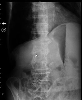
Peptic ulcer disease (PUD) is a break in the inner lining of the stomach, the first part of the small intestine, or sometimes the lower esophagus. An ulcer in the stomach is called a gastric ulcer, while one in the first part of the intestines is a duodenal ulcer. The most common symptoms of a duodenal ulcer are waking at night with upper abdominal pain and upper abdominal pain that improves with eating. With a gastric ulcer, the pain may worsen with eating. The pain is often described as a burning or dull ache. Other symptoms include belching, vomiting, weight loss, or poor appetite. About a third of older people have no symptoms. Complications may include bleeding, perforation, and blockage of the stomach. Bleeding occurs in as many as 15% of cases.

The pylorus, or pyloric part, connects the stomach to the duodenum. The pylorus is considered as having two parts, the pyloric antrum and the pyloric canal. The pyloric canal ends as the pyloric orifice, which marks the junction between the stomach and the duodenum. The orifice is surrounded by a sphincter, a band of muscle, called the pyloric sphincter. The word pylorus comes from Greek πυλωρός, via Latin. The word pylorus in Greek means "gatekeeper", related to "gate" and is thus linguistically related to the word "pylon".

A feeding tube is a medical device used to provide nutrition to people who cannot obtain nutrition by mouth, are unable to swallow safely, or need nutritional supplementation. The state of being fed by a feeding tube is called gavage, enteral feeding or tube feeding. Placement may be temporary for the treatment of acute conditions or lifelong in the case of chronic disabilities. A variety of feeding tubes are used in medical practice. They are usually made of polyurethane or silicone. The diameter of a feeding tube is measured in French units. They are classified by the site of insertion and intended use.

Esophagogastroduodenoscopy, also called by various other names, is a diagnostic endoscopic procedure that visualizes the upper part of the gastrointestinal tract down to the duodenum. It is considered a minimally invasive procedure since it does not require an incision into one of the major body cavities and does not require any significant recovery after the procedure. However, a sore throat is common.

Pyloric stenosis is a narrowing of the opening from the stomach to the first part of the small intestine. Symptoms include projectile vomiting without the presence of bile. This most often occurs after the baby is fed. The typical age that symptoms become obvious is two to twelve weeks old.

A gastrectomy is a partial or total surgical removal of the stomach.

Gastrointestinal diseases refer to diseases involving the gastrointestinal tract, namely the oesophagus, stomach, small intestine, large intestine and rectum, and the accessory organs of digestion, the liver, gallbladder, and pancreas.
Dumping syndrome occurs when food, especially sugar, moves too quickly from the stomach to the duodenum—the first part of the small intestine—in the upper gastrointestinal (GI) tract. This condition is also called rapid gastric emptying. It is mostly associated with conditions following gastric or esophageal surgery, though it can also arise secondary to diabetes or to the use of certain medications; it is caused by an absent or insufficiently functioning pyloric sphincter, the valve between the stomach and the duodenum.

A gastroenterostomy is the surgical creation of a connection between the stomach and the jejunum. The operation can sometimes be performed at the same time as a partial gastrectomy. Gastroenterostomy was in the past typically performed to treat peptic ulcers, but today it is usually carried out to enable food to pass directly to the middle section of the small intestine when it is necessary to bypass the first section because of duodenal damage. The procedure is still being used to treat gastroparesis that is refractory to other treatments, but it is now rarely used to treat peptic ulcers because most cases thereof are bacterial in nature and there are many new drugs available to treat the gastric reflux often experienced with peptic ulcer disease. Reported cure rates for H. pylori infection range from 70% to 90% after antibiotic treatment.

In anatomy, the gastroduodenal artery is a small blood vessel in the abdomen. It supplies blood directly to the pylorus and proximal part of the duodenum. It also indirectly supplies the pancreatic head.
In medicine, Valentino's syndrome is pain presenting in the right lower quadrant of the abdomen caused by a duodenal ulcer with perforation through the retroperitoneum.

A vagotomy is a surgical procedure that involves removing part of the vagus nerve.
Gastric outlet obstruction (GOO) is a medical condition where there is an obstruction at the level of the pylorus, which is the outlet of the stomach. Individuals with gastric outlet obstruction will often have recurrent vomiting of food that has accumulated in the stomach, but which cannot pass into the small intestine due to the obstruction. The stomach often dilates to accommodate food intake and secretions. Causes of gastric outlet obstruction include both benign causes, as well as malignant causes, such as gastric cancer.
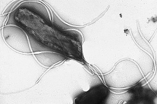
This is a timeline of the events relating to the discovery that peptic ulcer disease and some cancers are caused by H. pylori. In 2005, Barry Marshall and Robin Warren were awarded the Nobel Prize in Physiology or Medicine for their discovery that peptic ulcer disease (PUD) was primarily caused by Helicobacter pylori, a bacterium with affinity for acidic environments, such as the stomach. As a result, PUD that is associated with H. pylori is currently treated with antibiotics used to eradicate the infection. For decades prior to their discovery, it was widely believed that PUD was caused by excess acid in the stomach. During this time, acid control was the primary method of treatment for PUD, to only partial success. Among other effects, it is now known that acid suppression alters the stomach milieu to make it less amenable to H. pylori infection.

A perforated ulcer is a condition in which an untreated ulcer has burned through the mucosal wall in a segment of the gastrointestinal tract allowing gastric contents to leak into the abdominal cavity.

The abdominopelvic cavity is a body cavity that consists of the abdominal cavity and the pelvic cavity. It contains the stomach, liver, pancreas, spleen, gallbladder, kidneys, and most of the small and large intestines. It also contains the urinary bladder and internal reproductive organs. The abdominal pelvic cavity is a little pocket sac that lies way low in the base of the abdominal pelvis cavity. There's no membrane that separates out the abdominal cavity from the pelvic cavity so it is sometimes referred to as the abdominal pelvis or the peritoneal cavity. There are many diseases and disorders associated with the organs of the abdominopelvic cavity.
Biliary reflux, bile reflux (gastritis), duodenogastroesophageal reflux (DGER) or duodenogastric reflux is a condition that occurs when bile and/or other contents like bicarbonate, and pancreatic enzymes flow upward (refluxes) from the duodenum into the stomach and esophagus.
The Pyloromyotomy is a surgical procedure in which a portion of the muscle fibers of the pyloric muscle are cut. This is typically done in cases where the contents from the stomach are inappropriately stopped by the pyloric muscle, causing the stomach contents to build up in the stomach and unable to be appropriately digested. The procedure is typically performed in cases of “hypertrophic pyloric stenosis” in young children. In most cases, the procedure can be performed with either an open approach or a laparoscopic approach and the patients typically have good outcomes with minimal complications.

The gastrointestinal wall of the gastrointestinal tract is made up of four layers of specialised tissue. From the inner cavity of the gut outwards, these are:
