Related Research Articles

The parietal lobe is one of the four major lobes of the cerebral cortex in the brain of mammals. The parietal lobe is positioned above the temporal lobe and behind the frontal lobe and central sulcus.
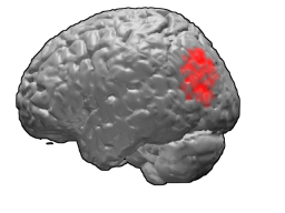
The occipital lobe is one of the four major lobes of the cerebral cortex in the brain of mammals. The name derives from its position at the back of the head, from the Latin ob, 'behind', and caput, 'head'.
Brodmann area 39, or BA39, is part of the parietal cortex in the human brain. BA39 encompasses the angular gyrus, lying near to the junction of temporal, occipital and parietal lobes.

The fusiform gyrus, also known as the lateral occipitotemporal gyrus,is part of the temporal lobe and occipital lobe in Brodmann area 37. The fusiform gyrus is located between the lingual gyrus and parahippocampal gyrus above, and the inferior temporal gyrus below. Though the functionality of the fusiform gyrus is not fully understood, it has been linked with various neural pathways related to recognition. Additionally, it has been linked to various neurological phenomena such as synesthesia, dyslexia, and prosopagnosia.
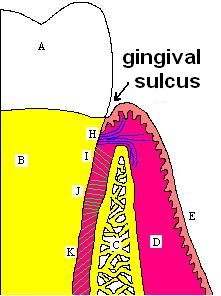
In biological morphology and anatomy, a sulcus is a furrow or fissure. It may be a groove, natural division, deep furrow, elongated cleft, or tear in the surface of a limb or an organ, most notably on the surface of the brain, but also in the lungs, certain muscles, as well as in bones, and elsewhere. Many sulci are the product of a surface fold or junction, such as in the gums, where they fold around the neck of the tooth.

The lobes of the brain are the major identifiable zones of the cerebral cortex, and they comprise the surface of each hemisphere of the cerebrum. The two hemispheres are roughly symmetrical in structure, and are connected by the corpus callosum. They traditionally have been divided into four lobes, but are today considered as having six lobes each. The lobes are large areas that are anatomically distinguishable, and are also functionally distinct to some degree. Each lobe of the brain has numerous ridges, or gyri, and furrows, the sulci that constitute further subzones of the cortex. The expression "lobes of the brain" usually refers only to those of the cerebrum, not to the distinct areas of the cerebellum.

The cuneus is a smaller lobe in the occipital lobe of the brain. The cuneus is bounded anteriorly by the parieto-occipital sulcus and inferiorly by the calcarine sulcus.

In human brain anatomy, an operculum, may refer to the frontal, temporal, or parietal operculum, which together cover the insula as the opercula of insula. It can also refer to the occipital operculum, part of the occipital lobe.

The inferior temporal gyrus is one of three gyri of the temporal lobe and is located below the middle temporal gyrus, connected behind with the inferior occipital gyrus; it also extends around the infero-lateral border on to the inferior surface of the temporal lobe, where it is limited by the inferior sulcus. This region is one of the higher levels of the ventral stream of visual processing, associated with the representation of objects, places, faces, and colors. It may also be involved in face perception, and in the recognition of numbers and words.

In neuroanatomy, the parieto-occipital sulcus is a deep sulcus in the cerebral cortex that marks the boundary between the cuneus and precuneus, and also between the parietal and occipital lobes. Only a small part can be seen on the lateral surface of the hemisphere, its chief part being on the medial surface.

In neuroanatomy, a sulcus is a depression or groove in the cerebral cortex. It surrounds a gyrus, creating the characteristic folded appearance of the brain in humans and other mammals. The larger sulci are usually called fissures.

The superior longitudinal fasciculus (SLF) is an association tract in the brain that is composed of three separate components. It is present in both hemispheres and can be found lateral to the centrum semiovale and connects the frontal, occipital, parietal, and temporal lobes. This bundle of tracts (fasciculus) passes from the frontal lobe through the operculum to the posterior end of the lateral sulcus where they either radiate to and synapse on neurons in the occipital lobe, or turn downward and forward around the putamen and then radiate to and synapse on neurons in anterior portions of the temporal lobe.

In the occipital lobe, the lateral occipital sulcus, where present, divides the lateral, or middle occipital gyrus into a superior and an inferior part, which are then continuous in front with the parietal and temporal lobes. The anterior portion is often incomplete, but in some individuals it may encounter the superior temporal sulcus whilst the posterior portion originates from the middle of the curved lunate sulcus, or from a curved portion of the transverse occipital sulcus if absent.
The neuroanatomy of memory encompasses a wide variety of anatomical structures in the brain.
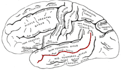
The superior temporal sulcus (STS) is the sulcus separating the superior temporal gyrus from the middle temporal gyrus in the temporal lobe of the brain. A sulcus is a deep groove that curves into the largest part of the brain, the cerebrum, and a gyrus is a ridge that curves outward of the cerebrum.

In brain anatomy, the lunate sulcus or simian sulcus, also known as the sulcus lunatus, is a fissure in the occipital lobe variably found in humans and more often larger when present in apes and monkeys. The lunate sulcus marks the transition between V1 and V2.

The inferior surface of the temporal lobe is concave, and is continuous posteriorly with the tentorial surface of the occipital lobe. It is traversed by the inferior temporal sulcus, which extends from near the occipital pole behind, to within a short distance of the temporal pole in front, but is frequently subdivided by bridging gyri.
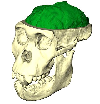
Paleoneurobiology is the study of brain evolution by analysis of brain endocasts to determine endocranial traits and volumes. Considered a subdivision of neuroscience, paleoneurobiology combines techniques from other fields of study including paleontology and archaeology. It reveals specific insight concerning human evolution. The cranium is unique in that it grows in response to the growth of brain tissue rather than genetic guidance, as is the case with bones that support movement. Fossil skulls and their endocasts can be compared to each other, to the skulls and fossils of recently deceased individuals, and even compared to those of other species to make inferences about functional anatomy, physiology and phylogeny. Paleoneurobiology is in large part influenced by developments in neuroscience as a whole; without substantial knowledge about current functionality, it would be impossible to make inferences about the functionality of ancient brains.

Yakovlevian torque is the tendency of the right side of the human brain to be warped slightly forward relative to the left and the left side of the human brain to be warped slightly backward relative to the right. This is responsible for certain asymmetries, such as how the lateral sulcus of the human brain is often longer and less curved on the left side of the brain relative to the right. Stated in another way, Yakovlevian torque can be defined by the existence of right-frontal and left-occipital petalias, which are protrusions of the surface of one hemisphere relative to the other. It is named for Paul Ivan Yakovlev (1894–1983), a Russian-American neuroanatomist from Harvard Medical School.

The occipital gyri (OcG) are three gyri in parallel, along the lateral portion of the occipital lobe, also referred to as a composite structure in the brain. The gyri are the superior occipital gyrus, the middle occipital gyrus, and the inferior occipital gyrus, and these are also known as the occipital face area. The superior and inferior occipital sulci separates the three occipital gyri.
References
- ↑ Balter, Michael (2007-11-27). "In Study of Brain Evolution, Zeal and Bitter Debate". The New York Times. Retrieved 2010-05-22.