
The lymphatic system, or lymphoid system, is an organ system in vertebrates that is part of the immune system, and complementary to the circulatory system. It consists of a large network of lymphatic vessels, lymph nodes, lymphoid organs, lymphatic tissue and lymph. Lymph is a clear fluid carried by the lymphatic vessels back to the heart for re-circulation. The Latin word for lymph, lympha, refers to the deity of fresh water, "Lympha".

In human anatomy, the subclavian arteries are paired major arteries of the upper thorax, below the clavicle. They receive blood from the aortic arch. The left subclavian artery supplies blood to the left arm and the right subclavian artery supplies blood to the right arm, with some branches supplying the head and thorax. On the left side of the body, the subclavian comes directly off the aortic arch, while on the right side it arises from the relatively short brachiocephalic artery when it bifurcates into the subclavian and the right common carotid artery.

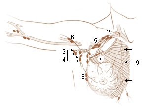
In human anatomy, the thoracic duct is the larger of the two lymph ducts of the lymphatic system. The thoracic duct usually begins from the upper aspect of the cisterna chyli, passing out of the abdomen through the aortic hiatus into first the posterior mediastinum and then the superior mediastinum, extending as high up as the root of the neck before descending to drain into the systemic (blood) circulation at the venous angle.

The subclavian vein is a paired large vein, one on either side of the body, that is responsible for draining blood from the upper extremities, allowing this blood to return to the heart. The left subclavian vein plays a key role in the absorption of lipids, by allowing products that have been carried by lymph in the thoracic duct to enter the bloodstream. The diameter of the subclavian veins is approximately 1–2 cm, depending on the individual.
The cisterna chyli or receptaculum chyli is a dilated sac at the lower end of the thoracic duct in most mammals into which lymph from the intestinal trunk and two lumbar lymphatic trunks flow. It receives fatty chyle from the intestines and thus acts as a conduit for the lipid products of digestion. It is the most common drainage trunk of most of the body's lymphatics. The cisterna chyli is a retroperitoneal structure.

In human anatomy, the superior epigastric artery is a terminal branch of the internal thoracic artery that provides arterial supply to the abdominal wall, and upper rectus abdominis muscle. It enters the rectus sheath to descend upon the inner surface of the rectus abdominis muscle. It ends by anastomosing with the inferior epigastric artery.

The hemiazygos vein is a vein running superiorly in the lower thoracic region, just to the left side of the vertebral column.

The anterior jugular vein is a vein in the neck.

The retromandibular vein is a major vein of the face. It is formed within the parotid gland by the confluence of the maxillary vein, and superficial temporal vein. It descends in the gland and splits into two branches upon emerging from the gland. Its anterior branch then joins the (anterior) facial vein forming the common facial vein, while its posterior branch joins the posterior auricular vein forming the external jugular vein.

The deep cervical lymph nodes are a group of cervical lymph nodes in the neck that form a chain along the internal jugular vein within the carotid sheath.

The jugular trunk is a lymphatic vessel in the neck. It is formed by vessels that emerge from the superior deep cervical lymph nodes and unite to efferents of the inferior deep cervical lymph nodes.
The efferent vessels of the tracheobronchial lymph nodes ascend upon the trachea and unite with efferents of the internal mammary and anterior mediastinal glands to form the right and left bronchomediastinal trunks.
The parasternal lymph nodes are placed at the anterior ends of the intercostal spaces, by the side of the internal thoracic artery.
The efferent vessels of the subclavicular group unite to form the subclavian trunk, which opens either directly into the junction of the internal jugular and subclavian veins or into the jugular lymphatic trunk; on the left side it may end in the thoracic duct.
The superficial lateral cervical lymph nodes are found along the course of the external jugular vein, between the inferior aspect of the parotid gland and the supraclavicular nodes. The nodes are intercalated along the course of the vessels draining the parotid nodes and the infraauricular nodes. These nodes drain into the supraclavicular nodes, and on to the jugular trunk, followed by the thoracic duct on the left or the right lymphatic duct.
The pretracheal lymph nodes are lymph nodes located anterior to the trachea in the neck.
Lymph trunk is a collection of lymph vessels that carries lymph, and is formed by confluence of many efferent lymph vessels. It in turn drains into one of the two lymph ducts.

A lymph duct is a great lymphatic vessel that empties lymph into one of the subclavian veins. There are two lymph ducts in the human body—the right lymphatic duct and the thoracic duct. The right lymphatic duct drains lymph from the right upper limb, right side of thorax and right halves of head and neck. The thoracic duct drains lymph into the circulatory system at the left brachiocephalic vein between the left subclavian and left internal jugular veins.
Lymph sacs are a part of the development of the lymphatic system, known as lymphangiogenesis. The lymph sacs are precursors of the lymph vessels. These sacs develop through the processes of vasculogenesis and angiogenesis. However, there is evidence of both of these processes in different organisms. In mice, it is thought that the lymphatic components form through an angiogenic process. But, there is evidence from bird embryos that gives rise to the idea that lymphatic vessels arise in the embryos through a vasculogenesis-like process from the lymphangioblastic endothelial precursor cells.


