Righting reflex

Last updated

The righting reflex, also known as the labyrinthine righting reflex, is a reflex that corrects the orientation of the body when it is taken out of its normal upright position. It is initiated by the vestibular system, which detects that the body is not erect and causes the head to move back into position as the rest of the body follows. The perception of head movement involves the body sensing linear acceleration or the force of gravity through the otoliths, and angular acceleration through the semicircular canals. The reflex uses a combination of visual system inputs, vestibular inputs, and somatosensory inputs to make postural adjustments when the body becomes displaced from its normal vertical position. These inputs are used to create what is called an efference copy. This means that the brain makes comparisons in the cerebellum between expected posture and perceived posture, and corrects for the difference. The reflex takes 6 or 7 weeks to perfect, but can be affected by various types of balance disorders. [1]

Contents

The righting reflex has also been studied in cats and other non-human mammals.

Overview

Vestibular system

The vestibular system is composed of inner ear organs forming the "labyrinth": the semicircular canals, the otoliths, and the cochlea. The section below is an overview of the vestibular system, as it is crucial to the understanding of the righting reflex. Sensory information from the vestibular system allows the head to move back into position when disturbed as the rest of the body follows. The semicircular canals (brown, see figure) are arranged at angles to the horizontal plane of the head when it is in its normal vertical posture. Each canal has a widened base, called an ampulla, that houses specialized sensory hair cells. [2] Fluid in these canals surrounds the hair cells, and moves across them as the head moves to gather information about the movement and position of the body. [2] The hair cells are covered in tiny sensory hairs called stereocilia, which are sensitive to displacement forces as the body is moved in different positions. When the head is moved, the force moves the hair cells forward, which sends signals to afferent fibers and on to the brain. [2] The brain can then decide which muscles in the body need to become active in order to right itself.

The semicircular canals have a superior, posterior, and horizontal component. Studies have shown that the horizontal canal is most correlated with agility, as shown with several mammals. [3] Curvature and size of these canals seems to affect agility, and may be due to the environments in which animals navigate, such as a mostly two-dimensional landscape as compared to three-dimensional spaces (i.e. in the air, the trees, or the water). [4]

The otoliths have two components: the utricle and the saccule. Both are made of the same sensory tissue containing hair cells, which is covered by a gelatinous layer and the otolithic membrane on top. Embedded in this membrane are calcium carbonate crystals, called otoconia, or "ear rocks." As the head is tilted forward or backward, the otoconia move the hair cells in a similar fashion to the semicircular canal fluid movement and cause depolarization of the hair cells. Signals from these cells are also transmitted along afferent fibers and on to the brain. [2]

Signal transduction

Vestibular afferent signals are carried by type I or type II hair cells, which are distinguished by a larger amount of stereocilia per cell in type I cells than in type II cells. [5] Nerve fibers attached to these hair cells carry signals to the vestibular nuclei in the brain, which are then used to gain information about the body's position. Larger diameter afferent fibers carry information from both type I and type II hair cells, and regular afferent fibers carry signals from type II hair cells. [6] The semicircular canals encode head velocity signals, or angular acceleration, while the otoconia encode linear acceleration signals and gravitational signals. Regular afferent signals and irregular afferent signals travel to the vestibular nuclei in the brain, although irregular signals are at least two times more sensitive. Because of this, it has been questioned why humans have regular afferent signals. Studies have shown that regular afferent signals give information about how long the motion of the head or body lasts, and irregular afferent signals occur when the head is moved more violently, such as in falling. [6]

Function

The righting reflex involves complex muscular movements in response to a stimulus. When startled, the brain can evoke anticipatory postural adjustments, or a series of muscle movements, which involves the function of the midbrain. [7] However, the mechanisms of such an origin are yet to be elucidated. Data support the generation of these movements from circuits in the spine connected to the supplementary motor area, the basal ganglia, and the reticular formation.

Reference frames

Visual input for proper righting reflex function is perceived in the form of reference frames, which create a representation of space for comparison to expected orientation. Three types of reference frames are used to perceive vertical orientation; they are consistently updated and quickly adapting to process changes in vestibular input. [8]

Allocentric reference frame

The allocentric reference frame describes a visual reference frame based on the arrangement of objects in an organism's environment. To test for the use of an allocentric reference frame, a "rod-and-frame" test, in which a subject's perception of virtual objects in an environment are altered, can be used to cause a body tilt as the subject believes to be correcting for the shift. [8]

Egocentric reference frame

The egocentric reference frame refers to a proprioceptive reference frame using the position of an organism's body in a space. This reference frame relies heavily on somatosensory information, or feedback from the body's sensory system. Muscle vibrations can be used to alter a subject's perception of the location of their bodies by creating an abnormal somatosensory signal. [8]

Geocentric reference frame

The geocentric reference frame involves visual inputs to help detect the verticality of an environment through gravitational pull. The sole of the foot contains receptors in the skin to detect the force of gravity, and plays a large role in standing or walking balance. The abdominal organs also contain receptors that provide geocentric information. "Roll-tilt" tests in which a subject's body is mechanically moved can be used to test for geocentric reference frame function. [8]

Pathways

The righting reflex can be described as a three-neuron arc system composed of primary vestibular neurons, vestibular nuclei neurons, and target motorneurons. [6] Input from the vestibular system is received by sensory receptors in the hair cells of the semicircular canals and the otoliths, which are processed in the vestibular nuclei. The cerebellum is also active at this time for processing of what is called an efference copy, which compares expectations of the body's posture with how it is oriented at the time. The difference between expected posture and actual posture is corrected for via motorneurons in the spinal cord, which control muscle movements for righting the body. [9]

These automatic postural adjustments can be explained in terms of two reflexes similar to the righting reflex: the vestibulo-ocular reflex (VOR) and the vestibulocollic reflex (VCR). [10] The VOR involves movement of the eyes while the head turns to remain fixated on a stationary image, and the VCR involves control of neck muscles for correction of the head's orientation. [11] During the VOR, the semicircular canals send information to the brain and correct eye movements in the direction opposite head movement by sending excitatory signals to motor neurons on the side opposite to the head rotation. [11] Neurons in the otoliths control not only these signals for control of eye movements, but also signals for head movement correction through the neck muscles. [11] The righting reflex utilizes the VOR and VCR as it brings the body back into position. Visual information under the control of these reflexes creates greater stability for more accurate postural correction. [12]

Tests for righting reflex function

Vestibular function can be tested through a series of visual acuity tests. The static visual acuity test investigates a patient's ability to see an object from a distance by placing a subject at a certain distance from a letter fixed on a screen. The dynamic visual acuity test involves a patient's ability to control eye movements by following letters that appear on a screen. The difference between these two test results is the patient's fixation ability and vestibuloocular reflex (VOR) efficiency. [13]

Vestibular reflexes can also be examined using body tilt experiments. Patients with vestibular disorders may go through the Dix-Hallpike maneuver, in which the patient is seated with legs extended and rotates the head 45 degrees. The patient is then asked to lie down on the table and checked for nystagmus, or uncontrollable eye movements. Nystagmus in patients indicates dysfunction of the vestibular system, which can lead to dizziness and inability to complete a righting reflex. [1]

Proprioceptive ability tests are important in testing for righting reflex function. A therapist may ask a patient whether they know where a certain limb or joint is located without looking at it. These tests are often conducted on uneven surfaces, including sand and grass. [1]

Recently, vestibular reflexes have been investigated using leg rotation experiments. A leg and foot rotation test can be used to investigate changes in neuron activity within the labyrinth, or the inner ear. When the head is rotated while the leg and foot are rotated 90 degrees, the vestibular signals cause the brain to inhibit movement in the direction of the rotation. At the same time, it activates the muscles on the opposite side in an attempt to correct for the displacement. [14]

Plasticity

Because visual input is so critical in proper righting reflex function, impairment of vision can be detrimental. [15] Blind patients can rely on vestibular input where visual input is not available, and the visual cortex can become rewired to accommodate other senses taking control. Developmentally blind patients have a larger portion of the brain dedicated to vestibular and somatosensory input than patients with normal visual function. Recently blind patients must form new connections where visual inputs once were, and vestibular therapy may enhance this ability. [15] This principle, called neuroplasticity, is of growing interest to researchers today.

Disorders

Many inner ear disorders can cause dizziness, which leads to dysfunctional righting reflex action. Common inner ear disorders can cause vertigo in patients, which can be acute or chronic symptoms. [1] Labyrinthitis, or inflammation of the inner ear, can cause imbalances that must be overcome through therapeutic exercises. Labyrinthectomy, or removal of inner ear organs, is an operation conducted for patients with severe inner ear disorders whose vertigo is debilitating. Imbalances result from the procedure, but therapy can help overcome the symptoms. [16]

Benign paroxysmal positional vertigo

Benign paroxysmal positional vertigo, or BPPV, is a disorder caused by the breaking off of a piece of otoconia from the otoliths. The otoconia floats freely in the inner ear fluid, causing disorientation and vertigo. [1] The disorder can be tested for using a nystagmus test, such as the Dix-Hallpike maneuver. This disorder can disrupt the function of the righting reflex as the symptoms of vertigo and disorientation prevent proper postural control. Treatment for the disorder includes antihistamines and anticholinergics, and the disorder often goes away without surgical removal of the free otoconia. [1]

Ménière's disease

Ménière's disease is thought to be a balance disorder involving fluid buildup in the inner ear. This can result from a number of factors, including head injury, ear infection, genetic predisposition, chemical toxicity, allergies, or syphilis. Syphilis can cause some patients to develop the disease later in life. [1] The disease is characterized by pressure in the ears, ringing in the ears, and vertigo. It also causes nystagmus, or uncontrollable eye movements. There is no known treatment for the disorder, although symptoms can be treated. These include water pills to thin out ear fluid, eating a low-salt diet, and taking anti-nausea medication. [1]

Other causes of righting reflex disorders

Vestibular and balance disorders can have a number of contributing factors. Dietary factors such as a high-salt diet, high caffeine intake, high sugar intake, monosodium glutamate (MSG) intake, dehydration, or food allergies can contribute to symptoms of vertigo and should be avoided in balance disorder patients. Other disorders can have symptoms of vertigo associated with them, such as epilepsy, migraine, stroke, or multiple sclerosis. Infectious diseases such as Lyme disease and meningitis can also cause vertigo. [1]

Righting reflex in animals

As a cat falls, it turns its head, rotates its spine, and aligns its hindquarters to land on its feet. This motion, coupled with free fall, creates a net zero angular momentum. Cat fall 150x300 6fps.gif
As a cat falls, it turns its head, rotates its spine, and aligns its hindquarters to land on its feet. This motion, coupled with free fall, creates a net zero angular momentum.

The righting reflex is not exclusive to humans. A well-known righting reflex in cats allows them to land on their feet after a fall. As a cat falls, it turns its head, rotates its spine, aligns its hindquarters, and arches its back to minimize injury. [18] The cat reaches free fall to accomplish this, which is much lower than that of humans, and they are able to hit the ground in a relaxed body form to prevent serious injury.

Bats, however, have a unique vestibular system anatomy. Their balance system, at an orientation 180 degrees opposite to that of humans, allows them to perform powerful feats of flight while hunting in the dark. This ability couples vestibular function with sensory echolocation to hunt prey. [19] However, they lack a righting reflex similar to most mammals. When exposed to zero-G, bats do not undergo the series of righting reflexes that most mammals do to correct orientation because they are accustomed to resting upside-down. [20]

Related Research Articles

<span class="mw-page-title-main">Inner ear</span> Innermost part of the vertebrate ear

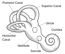
The inner ear is the innermost part of the vertebrate ear. In vertebrates, the inner ear is mainly responsible for sound detection and balance. In mammals, it consists of the bony labyrinth, a hollow cavity in the temporal bone of the skull with a system of passages comprising two main functional parts:

<span class="mw-page-title-main">Sense of balance</span> Physiological sense regarding posture

The sense of balance or equilibrioception is the perception of balance and spatial orientation. It helps prevent humans and nonhuman animals from falling over when standing or moving. Equilibrioception is the result of a number of sensory systems working together; the eyes, the inner ears, and the body's sense of where it is in space (proprioception) ideally need to be intact.

A balance disorder is a disturbance that causes an individual to feel unsteady, for example when standing or walking. It may be accompanied by feelings of giddiness, or wooziness, or having a sensation of movement, spinning, or floating. Balance is the result of several body systems working together: the visual system (eyes), vestibular system (ears) and proprioception. Degeneration or loss of function in any of these systems can lead to balance deficits.

<span class="mw-page-title-main">Vestibulocochlear nerve</span> Cranial nerve VIII, for hearing and balance

The vestibulocochlear nerve or auditory vestibular nerve, also known as the eighth cranial nerve, cranial nerve VIII, or simply CN VIII, is a cranial nerve that transmits sound and equilibrium (balance) information from the inner ear to the brain. Through olivocochlear fibers, it also transmits motor and modulatory information from the superior olivary complex in the brainstem to the cochlea.

<span class="mw-page-title-main">Vestibulo–ocular reflex</span> Reflex where rotation of the head causes eye movement to stabilize vision

The vestibulo-ocular reflex (VOR) is a reflex that acts to stabilize gaze during head movement, with eye movement due to activation of the vestibular system. The reflex acts to stabilize images on the retinas of the eye during head movement. Gaze is held steadily on a location by producing eye movements in the direction opposite that of head movement. For example, when the head moves to the right, the eyes move to the left, meaning the image a person sees stays the same even though the head has turned. Since slight head movement is present all the time, VOR is necessary for stabilizing vision: people with an impaired reflex find it difficult to read using print, because the eyes do not stabilise during small head tremors, and also because damage to reflex can cause nystagmus.

<span class="mw-page-title-main">Utricle (ear)</span> Membranous labyrinth in the vestibule of ear

The utricle and saccule are the two otolith organs in the vertebrate inner ear. They are part of the balancing system in the vestibule of the bony labyrinth. They use small stones and a viscous fluid to stimulate hair cells to detect motion and orientation. The utricle detects linear accelerations and head-tilts in the horizontal plane. The word utricle comes from Latin uter 'leather bag'.

<span class="mw-page-title-main">Vestibular system</span> Sensory system that facilitates body balance

The vestibular system, in vertebrates, is a sensory system that creates the sense of balance and spatial orientation for the purpose of coordinating movement with balance. Together with the cochlea, a part of the auditory system, it constitutes the labyrinth of the inner ear in most mammals.

Spatial disorientation is the inability to determine position or relative motion, commonly occurring during periods of challenging visibility, since vision is the dominant sense for orientation. The auditory system, vestibular system, and proprioceptive system collectively work to coordinate movement with balance, and can also create illusory nonvisual sensations, resulting in spatial disorientation in the absence of strong visual cues.

<span class="mw-page-title-main">Labyrinthitis</span> Medical condition

Labyrinthitis is inflammation of the labyrinth, a maze of fluid-filled channels in the inner ear. Vestibular neuritis is inflammation of the vestibular nerve. Both conditions involve inflammation of the inner ear. Labyrinths that house the vestibular system sense changes in the head's position or the head's motion. Inflammation of these inner ear parts results in a sensation of the world spinning and also possible hearing loss or ringing in the ears. It can occur as a single attack, a series of attacks, or a persistent condition that diminishes over three to six weeks. It may be associated with nausea, vomiting, and eye nystagmus.

<span class="mw-page-title-main">Electronystagmography</span>

Electronystagmography (ENG) is a diagnostic test to record involuntary movements of the eye caused by a condition known as nystagmus. It can also be used to diagnose the cause of vertigo, dizziness or balance dysfunction by testing the vestibular system. Electronystagmography is used to assess voluntary and involuntary eye movements. It evaluates the cochlear nerve and the oculomotor nerve. The ENG can be used to determine the origin of various eye and ear disorders.

<span class="mw-page-title-main">Vertigo</span> Type of dizziness where a person has the sensation of moving or surrounding objects moving

Vertigo is a condition in which a person has the sensation of movement or of surrounding objects moving when they are not. Often it feels like a spinning or swaying movement. It may be associated with nausea, vomiting, perspiration, or difficulties walking. It is typically worse when the head is moved. Vertigo is the most common type of dizziness.

<span class="mw-page-title-main">Vestibular nerve</span> Branch of the vestibulocochlear nerve

The vestibular nerve is one of the two branches of the vestibulocochlear nerve. In humans the vestibular nerve transmits sensory information transmitted by vestibular hair cells located in the two otolith organs and the three semicircular canals via the vestibular ganglion of Scarpa. Information from the otolith organs reflects gravity and linear accelerations of the head. Information from the semicircular canals reflects rotational movement of the head. Both are necessary for the sensation of body position and gaze stability in relation to a moving environment.

<span class="mw-page-title-main">Caloric reflex test</span> Test of the vestibulo-ocular reflex

In medicine, the caloric reflex test is a test of the vestibulo-ocular reflex that involves irrigating cold or warm water or air into the external auditory canal. This method was developed by Robert Bárány, who won a Nobel prize in 1914 for this discovery.

<span class="mw-page-title-main">Vestibulospinal tract</span> Neural tract in the central nervous system

The vestibulospinal tract is a neural tract in the central nervous system. Specifically, it is a component of the extrapyramidal system and is classified as a component of the medial pathway. Like other descending motor pathways, the vestibulospinal fibers of the tract relay information from nuclei to motor neurons. The vestibular nuclei receive information through the vestibulocochlear nerve about changes in the orientation of the head. The nuclei relay motor commands through the vestibulospinal tract. The function of these motor commands is to alter muscle tone, extend, and change the position of the limbs and head with the goal of supporting posture and maintaining balance of the body and head.

<span class="mw-page-title-main">Ampullary cupula</span>

The ampullary cupula, or cupula, is a structure in the vestibular system, providing the sense of spatial orientation.

<span class="mw-page-title-main">Otolithic membrane</span>

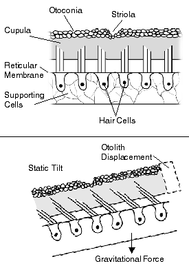
The otolithic membrane is a fibrous structure located in the vestibular system of the inner ear. It plays a critical role in the brain's interpretation of equilibrium. The membrane serves to determine if the body or the head is tilted, in addition to the linear acceleration of the body. The linear acceleration could be in the horizontal direction as in a moving car or vertical acceleration such as that felt when an elevator moves up or down.

The Epley maneuver or repositioning maneuver is a maneuver used by medical professionals to treat one common cause of vertigo, benign paroxysmal positional vertigo (BPPV) of the posterior or anterior canals of the ear. The maneuver works by allowing free-floating particles, displaced otoconia, from the affected semicircular canal to be relocated by using gravity, back into the utricle, where they can no longer stimulate the cupula, therefore relieving the patient of bothersome vertigo. The maneuver was developed by the physician, John M. Epley, and was first described in 1980.

The vestibular evoked myogenic potential is a neurophysiological assessment technique used to determine the function of the otolithic organs of the inner ear. It complements the information provided by caloric testing and other forms of inner ear testing. There are two different types of VEMPs. One is the oVEMP and another is the cVEMP. The oVEMP measures integrity of the utricule and superior vestibular nerve and the cVemp measures the saccule and the inferior vestibular nerve.

Space neuroscience or astroneuroscience is the scientific study of the central nervous system (CNS) functions during spaceflight. Living systems can integrate the inputs from the senses to navigate in their environment and to coordinate posture, locomotion, and eye movements. Gravity has a fundamental role in controlling these functions. In weightlessness during spaceflight, integrating the sensory inputs and coordinating motor responses is harder to do because gravity is no longer sensed during free-fall. For example, the otolith organs of the vestibular system no longer signal head tilt relative to gravity when standing. However, they can still sense head translation during body motion. Ambiguities and changes in how the gravitational input is processed can lead to potential errors in perception, which affects spatial orientation and mental representation. Dysfunctions of the vestibular system are common during and immediately after spaceflight, such as space motion sickness in orbit and balance disorders after return to Earth.

<span class="mw-page-title-main">Vestibular rehabilitation</span> Form of physical therapy for vestibular disorders

Vestibular rehabilitation (VR), also known as vestibular rehabilitation therapy (VRT), is a specialized form of physical therapy used to treat vestibular disorders or symptoms, characterized by dizziness, vertigo, and trouble with balance, posture, and vision. These primary symptoms can result in secondary symptoms such as nausea, fatigue, and lack of concentration. All symptoms of vestibular dysfunction can significantly decrease quality of life, introducing mental-emotional issues such as anxiety and depression, and greatly impair an individual, causing them to become more sedentary. Decreased mobility results in weaker muscles, less flexible joints, and worsened stamina, as well as decreased social and occupational activity. Vestibular rehabilitation therapy can be used in conjunction with cognitive behavioral therapy in order to reduce anxiety and depression resulting from an individual's change in lifestyle. However, there is often confusion about whether vestibular rehabilitation falls under physical therapy (PT) or occupational therapy (OT).

References

  1. 1 2 3 4 5 6 7 8 9 Wazen, Jack J.; Mitchell, Deborah R. (2004). Dizzy: what you need to know about managing and treating balance disorder . New York: Simon Schuster. ISBN   978-0-7432-3622-5. OCLC   52858223.
  2. 1 2 3 4 Purves, Dale. (2008). Neuroscience. Sunderland, Mass.: Sinauer. ISBN   978-0-87893-697-7. OCLC   144771764.
  3. Cox, PG.; Jeffery, N. (Jan 2010). "Semicircular canals and agility: the influence of size and shape measures". J Anat. 216 (1): 37–47. doi:10.1111/j.1469-7580.2009.01172.x. PMC   2807974 . PMID   20002227.
  4. Jusufi, A.; Zeng, Y.; Full, RJ.; Dudley, R. (Dec 2011). "Aerial righting reflexes in flightless animals". Integr Comp Biol. 51 (6): 937–43. doi: 10.1093/icb/icr114 . PMID   21930662.
  5. Moravec, WJ.; Peterson, EH. (Nov 2004). "Differences between stereocilia numbers on type I and type II vestibular hair cells". J Neurophysiol. 92 (5): 3153–60. doi:10.1152/jn.00428.2004. PMID   15201311.
  6. 1 2 3 Cullen, KE. (Mar 2012). "The vestibular system: multimodal integration and encoding of self-motion for motor control". Trends Neurosci. 35 (3): 185–96. doi:10.1016/j.tins.2011.12.001. PMC   4000483 . PMID   22245372.
  7. Delval, A.; Dujardin, K.; Tard, C.; Devanne, H.; Willart, S.; Bourriez, JL.; Derambure, P.; Defebvre, L. (Sep 2012). "Anticipatory postural adjustments during step initiation: elicitation by auditory stimulation of differing intensities". Neuroscience. 219: 166–74. doi:10.1016/j.neuroscience.2012.05.032. PMID   22626643. S2CID   23629664.
  8. 1 2 3 4 Borel, L.; Lopez, C.; Péruch, P.; Lacour, M. (Dec 2008). "Vestibular syndrome: a change in internal spatial representation". Neurophysiol Clin. 38 (6): 375–89. doi:10.1016/j.neucli.2008.09.002. PMID   19026958. S2CID   2704908.
  9. Mohapatra, S.; Krishnan, V.; Aruin, AS. (Mar 2012). "Postural control in response to an external perturbation: effect of altered proprioceptive information". Exp Brain Res. 217 (2): 197–208. doi:10.1007/s00221-011-2986-3. PMC   3325787 . PMID   22198575.
  10. Wilson, VJ; Boyle, R; Fukushima, K; Rose, PK; Shinoda, Y; Sugiuchi, Y; Uchino, Y (1994). "The vestibulocollic reflex". Journal of Vestibular Research: Equilibrium & Orientation. 5 (3): 147–70. doi:10.1016/0957-4271(94)00035-Z. PMID   7627376.
  11. 1 2 3 Uchino, Y.; Kushiro, K. (Dec 2011). "Differences between otolith- and semicircular canal-activated neural circuitry in the vestibular system". Neurosci Res. 71 (4): 315–27. doi:10.1016/j.neures.2011.09.001. PMID   21968226. S2CID   1545618.
  12. Sozzi, S.; Do, MC.; Monti, A.; Schieppati, M. (Jun 2012). "Sensorimotor integration during stance: processing time of active or passive addition or withdrawal of visual or haptic information". Neuroscience. 212: 59–76. doi:10.1016/j.neuroscience.2012.03.044. PMID   22516013. S2CID   338434.
  13. Badaracco C, Labini FS, Meli A, Tufarelli D: Oscillopsia in labyrinthine defective patients: comparison of objective and subjective measures. Am J Otolaryngol 2010, 31(6):399-403.
  14. Grasso C, Barresi M, Scattina E, Orsini P, Vignali E, Bruschini L, Manzoni D: Tuning of human vestibulospinal reflexes by leg rotation. Hum Mov Sci 2011, 30(2):296-313.
  15. 1 2 Seemungal, BM.; Glasauer, S.; Gresty, MA.; Bronstein, AM. (Jun 2007). "Vestibular perception and navigation in the congenitally blind". J Neurophysiol. 97 (6): 4341–56. CiteSeerX   10.1.1.326.5602 . doi:10.1152/jn.01321.2006. PMID   17392406.
  16. Lao Z, Sha Y, Chen B, Dai C-F, Huang W-H, Cheng Y-S: Labyrinthine sequestrum: four case studies. Otolaryngology–Head and Neck Surgery 2012, 147(3):535-537.
  17. Nguyen, H: "How does a cat always land on its feet?" Georgia Institute of Technology, Department of Biomedical Engineering. "My Third Page". Archived from the original on 2001-04-10. Retrieved 2014-08-23.
  18. "Cats: A Cat's Nine Lives." Video. National Geographic 2012
  19. Forman S: "Bats with frikkin' lasers." The Brown Daily Herald. 10 March 2010. http://www.browndailyherald.com/bats-with-frikkin-lasers-1.2186630#.UKqC2rvK3B8
  20. Fejtek M, Delorme M, Wassersug R: Behavioral reactions of the bat Carollia perspicillata to abrupt changes in gravity. Uchu Seibutsu Kagaku. 1995, 9(2):77-81.