Related Research Articles

Chronobiology is a field of biology that examines timing processes, including periodic (cyclic) phenomena in living organisms, such as their adaptation to solar- and lunar-related rhythms. These cycles are known as biological rhythms. Chronobiology comes from the ancient Greek χρόνος, and biology, which pertains to the study, or science, of life. The related terms chronomics and chronome have been used in some cases to describe either the molecular mechanisms involved in chronobiological phenomena or the more quantitative aspects of chronobiology, particularly where comparison of cycles between organisms is required.

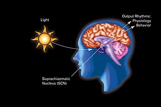
The suprachiasmatic nucleus or nuclei (SCN) is a small region of the brain in the hypothalamus, situated directly above the optic chiasm. The SCN is the principal circadian pacemaker in mammals, responsible for generating circadian rhythms. Reception of light inputs from photosensitive retinal ganglion cells allow the SCN to coordinate the subordinate cellular clocks of the body and entrain to the environment. The neuronal and hormonal activities it generates regulate many different body functions in an approximately 24-hour cycle.

CREB-TF is a cellular transcription factor. It binds to certain DNA sequences called cAMP response elements (CRE), thereby increasing or decreasing the transcription of the genes. CREB was first described in 1987 as a cAMP-responsive transcription factor regulating the somatostatin gene.
Melatonin receptors are G protein-coupled receptors (GPCR) which bind melatonin. Three types of melatonin receptors have been cloned. The MT1 (or Mel1A or MTNR1A) and MT2 (or Mel1B or MTNR1B) receptor subtypes are present in humans and other mammals, while an additional melatonin receptor subtype MT3 (or Mel1C or MTNR1C) has been identified in amphibia and birds. The receptors are crucial in the signal cascade of melatonin. In the field of chronobiology, melatonin has been found to be a key player in the synchrony of biological clocks. Melatonin secretion by the pineal gland has circadian rhythmicity regulated by the suprachiasmatic nucleus (SCN) found in the brain. The SCN functions as the timing regulator for melatonin; melatonin then follows a feedback loop to decrease SCN neuronal firing. The receptors MT1 and MT2 control this process. Melatonin receptors are found throughout the body in places such as the brain, the retina of the eye, the cardiovascular system, the liver and gallbladder, the colon, the skin, the kidneys, and many others. In 2019, X-ray crystal and cryo-EM structures of MT1 and MT2 were reported.
In neuroanatomy, the retinohypothalamic tract (RHT) is a photic neural input pathway involved in the circadian rhythms of mammals. The origin of the retinohypothalamic tract is the intrinsically photosensitive retinal ganglion cells (ipRGC), which contain the photopigment melanopsin. The axons of the ipRGCs belonging to the retinohypothalamic tract project directly, monosynaptically, to the suprachiasmatic nuclei (SCN) via the optic nerve and the optic chiasm. The suprachiasmatic nuclei receive and interpret information on environmental light, dark and day length, important in the entrainment of the "body clock". They can coordinate peripheral "clocks" and direct the pineal gland to secrete the hormone melatonin.
Period circadian protein homolog 1 is a protein in humans that is encoded by the PER1 gene.
Joseph S. Takahashi is a Japanese American neurobiologist and geneticist. Takahashi is a professor at University of Texas Southwestern Medical Center as well as an investigator at the Howard Hughes Medical Institute. Takahashi's research group discovered the genetic basis for the mammalian circadian clock in 1994 and identified the Clock gene in 1997. Takahashi was elected to the National Academy of Sciences in 2003.
Russell Grant Foster, CBE, FRS FMedSci is a British professor of circadian neuroscience, the Director of the Nuffield Laboratory of Ophthalmology and the Head of the Sleep and Circadian Neuroscience Institute (SCNi). He is also a Nicholas Kurti Senior Fellow at Brasenose College at the University of Oxford. Foster and his group are credited with key contributions to the discovery of the non-rod, non-cone, photosensitive retinal ganglion cells (pRGCs) in the mammalian retina which provide input to the circadian rhythm system. He has written and co-authored over a hundred scientific publications.
Steven M. Reppert is an American neuroscientist known for his contributions to the fields of chronobiology and neuroethology. His research has focused primarily on the physiological, cellular, and molecular basis of circadian rhythms in mammals and more recently on the navigational mechanisms of migratory monarch butterflies. He was the Higgins Family Professor of Neuroscience at the University of Massachusetts Medical School from 2001 to 2017, and from 2001 to 2013 was the founding chair of the Department of Neurobiology. Reppert stepped down as chair in 2014. He is currently distinguished professor emeritus of neurobiology.
Michael Menaker, was an American chronobiology researcher, and was Commonwealth Professor of Biology at University of Virginia. His research focused on circadian rhythmicity of vertebrates, including contributing to an understanding of light input pathways on extra-retinal photoreceptors of non-mammalian vertebrates, discovering a mammalian mutation for circadian rhythmicity, and locating a circadian oscillator in the pineal gland of bird. He wrote almost 200 scientific publications.

Douglas G. McMahon is a professor of Biological Sciences and Pharmacology at Vanderbilt University. McMahon has contributed several important discoveries to the field of chronobiology and vision. His research focuses on connecting the anatomical location in the brain to specific behaviors. As a graduate student under Gene Block, McMahon identified that the basal retinal neurons (BRNs) of the molluscan eye exhibited circadian rhythms in spike frequency and membrane potential, indicating they are the clock neurons. He became the 1986 winner of the Society for Neuroscience's Donald B. Lindsley Prize in Behavioral Neuroscience for his work. Later, he moved on to investigate visual, circadian, and serotonergic mechanisms of neuroplasticity. In addition, he helped find that constant light can desynchronize the circadian cells in the suprachiasmatic nucleus (SCN). He has always been interested in the underlying causes of behavior and examining the long term changes in behavior and physiology in the neurological modular system. McMahon helped identifying a retrograde neurotransmission system in the retina involving the melanopsin containing ganglion cells and the retinal dopaminergic amacrine neurons.
Hitoshi Okamura is a Japanese scientist who specializes in chronobiology. He is currently a professor of Systems Biology at Kyoto University Graduate School of Pharmaceutical Sciences and the Research Director of the Japan Science Technology Institute, CREST. Okamura's research group cloned mammalian Period genes, visualized clock oscillation at the single cell level in the central clock of the SCN, and proposed a time-signal neuronal pathway to the adrenal gland. He received a Medal of Honor with Purple Ribbon in 2007 for his research and was awarded Aschoff's Ruler for his work on circadian rhythms in rodents. His lab recently revealed the effects of m6A mRNA methylation on the circadian clock, neuronal communications in jet lag, and the role of dysregulated clocks in salt-induced hypertension.

William Joseph Schwartz is an American neurologist and scientist who serves as Professor and Associate Chair for Research and Education in the neurology department at the University of Texas Dell Medical School. His work on the neurobiology of circadian timekeeping has focused on the mammalian suprachiasmatic nucleus. Schwartz demonstrated that the suprachiasmatic nucleus is rhythmic in vivo using a 2-deoxyglucose radioactive marker for functional brain imaging. As of 2014, he is editor of the Journal of Biological Rhythms.
Hajime Tei is a Japanese neuroscientist specializing in the study of chronobiology. He currently serves as a professor at the Kanazawa University Graduate School of Natural Science & Technology. He is most notable for his contributions to the discovery of the mammalian period genes, which he discovered alongside Yoshiyuki Sakaki and Hitoshi Okamura.
Sato Honma is a Japanese chronobiologist who researches the biological mechanisms of circadian rhythms. She mainly collaborates with Ken-Ichi Honma on publications, and both of their primary research focuses are the human circadian clock under temporal isolation and the mammalian suprachiasmatic nucleus (SCN), its components, and associates. Honma is a retired professor at the Hokkaido University School of Medicine in Sapporo, Japan. She received her Ph.D. in physiology from Hokkaido University. She taught physiology at the School of Medicine and then at the Research and Education Center for Brain Science at Hokkaido University. She is currently the director at the Center for Sleep and Circadian Rhythm Disorders at Sapporo Hanazono Hospital and works as a somnologist.
Johanna H. Meijer is a Dutch scientist who has contributed significantly to the field of chronobiology. Meijer has made notable contributions to the understanding of the neural and molecular mechanisms of circadian pacemakers. She is known for her extensive studies of photic and non-photic effects on the mammalian circadian clocks. Notably, Meijer is the 2016 recipient of the Aschoff and Honma Prize, one of the most prestigious international prizes in the circadian research field. In addition to still unraveling neuronal mechanisms of circadian clocks and their applications to health, Meijer's lab now studies the effects of modern lifestyles on our circadian rhythm and bodily functions.
The food-entrainable oscillator (FEO) is a circadian clock that can be entrained by varying the time of food presentation. It was discovered when a rhythm was found in rat activity. This was called food anticipatory activity (FAA), and this is when the wheel-running activity of mice decreases after feeding, and then rapidly increases in the hours leading up to feeding. FAA appears to be present in non-mammals (pigeons/fish), but research heavily focuses on its presence in mammals. This rhythmic activity does not require the suprachiasmatic nucleus (SCN), the central circadian oscillator in mammals, implying the existence of an oscillator, the FEO, outside of the SCN, but the mechanism and location of the FEO is not yet known. There is ongoing research to investigate if the FEO is the only non-light entrainable oscillator in the body.
Elizabeth Maywood is an English researcher who studies circadian rhythms and sleep in mice. Her studies are focused on the suprachiasmatic nucleus (SCN), a small region of the brain that controls circadian rhythms.
Ken-Ichi Honma is a Japanese chronobiologist who researches the biological mechanisms underlying circadian rhythms. After graduating from Hokkaido University School of Medicine, he practiced clinical psychiatry before beginning his research. His recent research efforts are centered around photic and non-photic entrainment, the structure of circadian clocks, and the ontogeny of circadian clocks. He often collaborates with his wife, Sato Honma, in work involving the mammalian suprachiasmatic nucleus (SCN), its components, and associated topics.
Martin R. Ralph is a circadian biologist who serves as a professor in the Psychology Department at the University of Toronto. His research primarily focuses on circadian rhythmicity in the fields of neuroscience, psychology, and endocrinology. His most notable work was has been on the suprachiasmatic nucleus, now recognized as the central circadian pacemaker in mammals, but has also investigated circadian rhythms in the context of time, memory, and light.
References
- 1 2 3 4 Moore, Robert Y. (2013-01-01). "The Suprachiasmatic Nucleus and the Circadian Timing System". In Gillette, Martha U. (ed.). Chronobiology: Biological Timing in Health and Disease. pp. 1–28. doi:10.1016/B978-0-12-396971-2.00001-4. ISBN 9780123969712. PMID 23899592.
{{cite book}}:|journal=ignored (help) - ↑ "Robert Y. Moore, MD, PhD". www.upmc.com. Retrieved 2017-04-13.
- 1 2 3 4 5 6 7 8 9 10 11 12 13 14 15 16 Squire, Larry (2011-09-09). History of Neuroscience in Autobiography, Volume 7. Oxford University Press, Inc. pp. 530–561. ISBN 978-0-19-539613-3.
- ↑ Cassone, V. M.; Speh, J. C.; Card, J. P.; Moore, R. Y. (1988-01-01). "Comparative anatomy of the mammalian hypothalamic suprachiasmatic nucleus". Journal of Biological Rhythms. 3 (1): 71–91. doi: 10.1177/074873048800300106 . ISSN 0748-7304. PMID 2979633. S2CID 29581353.
- ↑ Moore, Robert Y. (1996). "Chapter 8 Entrainment pathways and the functional organization of the circadian system". Progress in Brain Research. Elsevier B.V. pp. 103–119.
- 1 2 Moore, Robert Y.; Speh, Joan C.; Leak, Rehana K. (2002-07-01). "Suprachiasmatic nucleus organization". Cell and Tissue Research. 309 (1): 89–98. doi:10.1007/s00441-002-0575-2. ISSN 0302-766X. PMID 12111539. S2CID 25249691.
- ↑ Moore, Robert Y. (2007-12-01). "Suprachiasmatic nucleus in sleep–wake regulation". Sleep Medicine. The roles of melatonin and the suprachiasmatic nucleus in sleep regulation. Proceedings from a Roundtable Discussion. 8, Supplement 3: 27–33. doi:10.1016/j.sleep.2007.10.003. PMID 18032104.
- ↑ "Parkinson's symptoms shown on PET scans - ScienceBlog.com". ScienceBlog.com. 2003-03-28. Retrieved 2017-04-27.