Schamberg disease

Last updated
Schamberg's disease
Schambergdisease-26male.png
Rust-colored spots typical of Schamberg disease on the lower left leg and left foot of a 26-year-old Caucasian male.
Specialty Dermatology   OOjs UI icon edit-ltr-progressive.svg

Schamberg's disease, (also known as "progressive pigmentary dermatosis of Schamberg", [1] "purpura pigmentosa progressiva" (PPP), [1] and "Schamberg's purpura" [1] ) is a chronic discoloration of the skin found in people of all ages, usually only affecting the feet, legs or thighs or a combination. It may occur as a single event or subsequent bouts may cause further spread. It is most common in males. [2] :829 It is named after Jay Frank Schamberg, who described it in 1901. There is no known cure for this disease but it is not a life-threatening condition and is mainly of cosmetic concern, although, because it can appear so suddenly, so extensively and because it usually leaves permanent discoloration of the skin, it can cause understandable psychological concern. The skin lesions sometimes cause itching, which can be treated by applying cortisone cream. The cortisone cream will only help with the itching and does not improve the discoloration of the skin. Schamberg's disease causes no other symptoms beside skin discoloration and itching. The condition is caused by inflammation of capillaries near the surface of skin and subsequent leaking of red blood cells into surrounding tissues. As the red blood cells break down and get mostly resorbed, some of the iron released by the red blood cells remains in the skin and causes the characteristic rust-colored appearance. The cause of the capillary inflammation is usually unknown.

Contents

Symptoms

The lesions are most frequent on the lower limbs, but may occur anywhere on the body, including the hands, arms, torso and even the neck. They may vary in number and erupt in mass numbers. They consist of irregular patches of orange or brown pigmentation with characteristic "cayenne pepper" spots appearing within and at the edge of old lesions. There are usually no symptoms, although there may be some slight itching, but there is no pain. The eruption may persist for many years. The pattern of the eruption changes, with slow extension and often some clearing of the original lesions.

Schamberg's disease, or progressive pigmented purpuric dermatosis, is a chronic discoloration of the skin which usually affects the legs and often spreads slowly. This disease is more common in males and may occur at any age from childhood onward. This condition is observed worldwide and has nothing to do with race or ethnic background. [3]

Causes

Schamberg's disease is caused by leaky blood vessels near the surface of the skin, capillaries, which allow red blood cells to slip through into the skin. [3] The red blood cells in the skin then fall apart and release their iron, which is released from hemoglobin. [3] The iron causes a rust color and this accounts for the orange tint of the rash. [4] Although the underlying cause for the leaky blood vessels is almost always unknown, researchers suggest some potential triggers. [5] These include the body's inflammatory reaction to some agent, such as a viral infection or a prescription or over the counter medication or supplement, such as thiamine and aspirin. [5] Even though there is no correlation with genetics, there have been a few cases where few people in a family had this condition. [5]

Although a definite cause for capillary inflammation is almost always unknown, certain preventive measures can be taken. [6] Doctors may prescribe medications that enhance the circulation of blood, which can keep blood vessels strong and healthy. [6]

Mechanism

Schamberg's disease is a skin disorder that causes a discoloration of the lower extremities. [4] It usually occurs in the lower extremities and rarely elsewhere. [4] This condition is caused by leaky blood vessels near the surface of the skin. [7] The cause of the leaky capillaries is usually not known. [7] When the red blood cells escape the blood vessels, they end up close under the skin surface, where they break apart, releasing hemoglobin, which in turn breaks apart, releasing Iron. [7] (Iron is the part of hemoglobin that enables it to transfer oxygen from the lungs to the cells and carbon dioxide from the cells to the lungs.) The iron released into the skin gets bound up into a complex called hemosiderin, which causes the discoloration of the skin. [7]

Diagnosis

With a complete history, the results from visual examination, and the aid of appropriate laboratory testing, a dermatologist can usually determine whether the skin lesions are in fact due Schamberg's disease.

Schamberg's disease can only be properly diagnosed by a healthcare provider. For a trained skin specialist such as a dermatologist, the condition is often readily diagnosed, because the visual appearance of the lesions on the skin itself usually suggests the possibility that the cause may be Schamberg's disease. While reviewing medical history is important to diagnose this condition, it is essential that the skin be physically examined.

To ensure that the skin lesions are not caused by other skin conditions or infections, a doctor will often order a complete blood count (CBC) and other blood tests. [3] Blood test results are usually normal. They are performed primarily to rule out other bleeding disorders that cause purpura. Since Schamberg's disease is usually asymptomatic beyond the visible lesions themselves, few other tests are usually indicated.

Micrograph of Schamberg disease, suggesting the spongiotic stage of a purpuric dermatitis. Histopathology of Schamberg disease.jpg
Micrograph of Schamberg disease, suggesting the spongiotic stage of a purpuric dermatitis.

Additional testing may aid diagnosis. A skin biopsy may be taken to determine capillaritis of dermal vessels. [8] Capillaritis or pigmented purpura is skin condition that has brown-reddish patches on the skin, which is caused by leaky capillaries. [9] Such skin biopsies are sent to a laboratory for a pathological examination, where each biopsy is observed under a microscope. [3] A dermatologist may also perform a dermatoscopy. [3]

Treatment

There is no cure for Schamberg's disease, however, this condition is not life-threatening or a major health concern. The most usual problems that patients will encounter is discoloration of the skin and, occasionally, itching. [10] Itching may be improved by applying a cortisone cream. Rarely, in very severe or concerning cases, Colchicine treatment has been used to prevent recurrence. [10] Some recommend that patients take a vitamin C supplement to promote collagen production, but this is not proven to be helpful. [8] In cases where there is a known trigger, people should avoid re-exposure to that trigger, e.g., people suspected to be sensitive to food with artificial colors or preservatives should avoid foods containing those items. [7] This is because some people have been observed to be sensitive to these agents, and the body initiates an inflammatory reaction if exposed to them again, which causes further capillary inflammation and red blood cell leakage. [7] Several research studies have indicated that Schamberg's disease can be controlled and the number of lesions can be reduced with the use of a drug called aminaphtone. [11] This drug helps reduce capillary fragility and red blood cell leakage. [11]

A study published in 2014 on the Journal of the German Society of Dermatology (Deutsche Dermatologische Gesellschaft) concludes that oral rutoside and ascorbic acid may be an efficient and well tolerated treatment for PPPD, with a recommendation for early treatment for best clinical outcome. [12]

Many topical and systemic therapies for Schamberg disease have been tried without consistent results. A case series published in 2012 describes the treatment of five patients with Schamberg's disease of the lower extremities using Advanced Fluorescence Technology (AFT) pulsed light with favorable results. [13]

Prognosis

A patient with Schamberg's disease can live a normal and healthy life. Since there is no proven cure for this condition, the patient will have to endure the lesions on his or her skin. With appropriate treatments, the condition may get better. [3] Although the skin lesions are not life-threatening, it may cause a cosmetic concern for some individuals. [3] Skin lesions may cause psychological discomfort, where patients may require reassurance to help with stress and anxiety. [3] There are a few rare cases of T-cell lymphoma that have developed from Schamberg's disease. [3] This is not a cause for concern, since the risk factors associated with Schamberg's disease are relatively low.

Recent research

A few very small non-blinded studies of treatment with narrow-band ultraviolet light have been reported as promising. [14]

Related Research Articles

<span class="mw-page-title-main">Chilblains</span> Medical condition

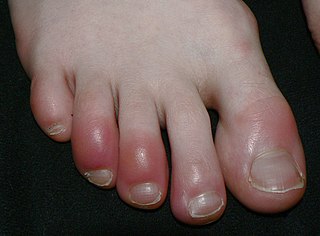
Chilblains, also known as pernio, is a medical condition in which damage occurs to capillary beds in the skin, most often in the hands or feet, when blood perfuses into the nearby tissue, resulting in redness, itching, inflammation, and possibly blisters.
<span class="mw-page-title-main">Bruise</span> Type of localized bleeding in tissues outside blood vessels

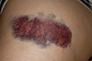
A bruise, also known as a contusion, is a type of hematoma of tissue, the most common cause being capillaries damaged by trauma, causing localized bleeding that extravasates into the surrounding interstitial tissues. Most bruises occur close enough to the epidermis such that the bleeding causes a visible discoloration. The bruise then remains visible until the blood is either absorbed by tissues or cleared by immune system action. Bruises which do not blanch under pressure can involve capillaries at the level of skin, subcutaneous tissue, muscle, or bone.

<span class="mw-page-title-main">Vasculitis</span> Medical disorders that destroy blood vessels by inflammation

Vasculitis is a group of disorders that destroy blood vessels by inflammation. Both arteries and veins are affected. Lymphangitis is sometimes considered a type of vasculitis. Vasculitis is primarily caused by leukocyte migration and resultant damage. Although both occur in vasculitis, inflammation of veins (phlebitis) or arteries (arteritis) on their own are separate entities.

<span class="mw-page-title-main">Rosacea</span> Skin condition resulting in redness, pimples and swelling, usually on the face

Rosacea is a long-term skin condition that typically affects the face. It results in redness, pimples, swelling, and small and superficial dilated blood vessels. Often, the nose, cheeks, forehead, and chin are most involved. A red, enlarged nose may occur in severe disease, a condition known as rhinophyma.

<span class="mw-page-title-main">Henoch–Schönlein purpura</span> Medical condition

Henoch–Schönlein purpura (HSP), also known as IgA vasculitis, is a disease of the skin, mucous membranes, and sometimes other organs that most commonly affects children. In the skin, the disease causes palpable purpura, often with joint pain and abdominal pain. With kidney involvement, there may be a loss of small amounts of blood and protein in the urine, but this usually goes unnoticed; in a small proportion of cases, the kidney involvement proceeds to chronic kidney disease. HSP is often preceded by an infection, such as a throat infection.

<span class="mw-page-title-main">Purpura</span> Hemorrhagic lesion of the skin caused by bleeding underneath the skin

Purpura is a condition of red or purple discolored spots on the skin that do not blanch on applying pressure. The spots are caused by bleeding underneath the skin secondary to platelet disorders, vascular disorders, coagulation disorders, or other causes. They measure 3–10 mm, whereas petechiae measure less than 3 mm, and ecchymoses greater than 1 cm.

Vulvitis is inflammation of the vulva, the external female mammalian genitalia that include the labia majora, labia minora, clitoris, and introitus. It may co-occur as vulvovaginitis with vaginitis, inflammation of the vagina, and may have infectious or non-infectious causes. The warm and moist conditions of the vulva make it easily affected. Vulvitis is prone to occur in any female especially those who have certain sensitivities, infections, allergies, or diseases that make them likely to have vulvitis. Postmenopausal women and prepubescent girls are more prone to be affected by it, as compared to women in their menstruation period. It is so because they have low estrogen levels which makes their vulvar tissue thin and dry. Women having diabetes are also prone to be affected by vulvitis due to the high sugar content in their cells, increasing their vulnerability. Vulvitis is not a disease, it is just an inflammation caused by an infection, allergy or injury. Vulvitis may also be symptom of any sexually transmitted infection or a fungal infection.

<span class="mw-page-title-main">Febrile neutrophilic dermatosis</span> Medical condition

Sweet syndrome (SS), or acute febrile neutrophilic dermatosis, is a skin disease characterized by the sudden onset of fever, an elevated white blood cell count, and tender, red, well-demarcated papules and plaques that show dense infiltrates by neutrophil granulocytes on histologic examination.
<span class="mw-page-title-main">Pyogenic granuloma</span> Vascular tumor on both mucosa and skin

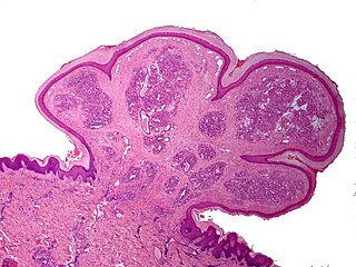
A pyogenic granuloma or lobular capillary hemangioma is a vascular tumor that occurs on both mucosa and skin, and appears as an overgrowth of tissue due to irritation, physical trauma, or hormonal factors. It is often found to involve the gums, skin, or nasal septum, and has also been found far from the head, such as in the thigh.

<span class="mw-page-title-main">Angiokeratoma</span> Medical condition

Angiokeratoma is a benign cutaneous lesion of capillaries, resulting in small marks of red to blue color and characterized by hyperkeratosis. Angiokeratoma corporis diffusum refers to Fabry's disease, but this is usually considered a distinct condition.

<span class="mw-page-title-main">Pruritic urticarial papules and plaques of pregnancy</span> Chronic rash that occurs during pregnancy

Pruritic urticarial papules and plaques of pregnancy (PUPPP), known in United Kingdom as polymorphic eruption of pregnancy (PEP), is a chronic hives-like rash that strikes some women during pregnancy. Some skin changes are known to occur in people who are pregnant while other skin conditions, or dermatoses, that people have prior to getting pregnant will become altered or symptoms will increase. Pruritic urticarial papules and plaques of pregnancy (PUPPP) is one of many skin conditions that is specific to pregnancy and occurs in about 1 in every 160 (0.625%) of pregnancies.

<span class="mw-page-title-main">Granuloma annulare</span> Medical condition

Granuloma annulare (GA) is a common, sometimes chronic skin condition which presents as reddish bumps on the skin arranged in a circle or ring. It can initially occur at any age, though two-thirds of patients are under 30 years old, and it is seen most often in children and young adults. Females are two times as likely to have it as males.

<span class="mw-page-title-main">Transient acantholytic dermatosis</span> Medical condition

Grover's disease (GD) is a polymorphic, pruritic, papulovesicular dermatosis characterized histologically by acantholysis with or without dyskeratosis. Once confirmed, most cases of Grover's disease last six to twelve months, which is why it was originally called "transient". However it may last much longer. Nevertheless, it is not to be confused with relapsing linear acantholytic dermatosis.

Cerebral vasculitis is vasculitis involving the brain and occasionally the spinal cord. It affects all of the vessels: very small blood vessels (capillaries), medium-size blood vessels, or large blood vessels. If blood flow in a vessel with vasculitis is reduced or stopped, the parts of the body that receive blood from that vessel begins to die. It may produce a wide range of neurological symptoms, such as headache, skin rashes, feeling very tired, joint pains, difficulty moving or coordinating part of the body, changes in sensation, and alterations in perception, thought or behavior, as well as the phenomena of a mass lesion in the brain leading to coma and herniation. Some of its signs and symptoms may resemble multiple sclerosis. 10% have associated bleeding in the brain.

<span class="mw-page-title-main">Dermatitis herpetiformis</span> Chronic autoimmune disorder leading to blistering skin

Dermatitis herpetiformis (DH) is a chronic autoimmune blistering skin condition, characterised by intensely itchy blisters filled with a watery fluid. DH is a cutaneous manifestation of coeliac disease, although the exact causal mechanism is not known. DH is neither related to nor caused by herpes virus; the name means that it is a skin inflammation having an appearance similar to herpes.

Necrotizing vasculitis, also called systemic necrotizing vasculitus, is a category of vasculitis, comprising vasculitides that present with necrosis. Examples include giant cell arteritis, microscopic polyangiitis, and granulomatosis with polyangiitis. ICD-10 uses the variant "necrotizing vasculopathy". ICD-9, while classifying these conditions together, does not use a dedicated phrase, instead calling them "polyarteritis nodosa and allied conditions".
<span class="mw-page-title-main">Pigmented purpuric dermatosis</span> Medical condition

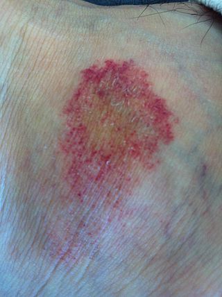
Pigmented purpuric dermatosis refers to one of the three major classes of skin conditions characterized by purpuric skin eruptions.

<span class="mw-page-title-main">Cutaneous small-vessel vasculitis</span> Medical condition

Cutaneous small-vessel vasculitis (CSVV), is inflammation of small blood vessels, usually accompanied by small lumps beneath the skin. The condition is also known as hypersensitivity vasculitis, cutaneous leukocytoclastic vasculitis, hypersensitivity angiitis, cutaneous leukocytoclastic angiitis, cutaneous necrotizing vasculitis and cutaneous necrotizing venulitis,

Cutaneous manifestations of COVID-19 are characteristic signs or symptoms of the Coronavirus disease 2019 that occur in the skin. The American Academy of Dermatology reports that skin lesions such as morbilliform, pernio, urticaria, macular erythema, vesicular purpura, papulosquamous purpura and retiform purpura are seen in people with COVID-19. Pernio-like lesions were more common in mild disease while retiform purpura was seen only in critically ill patients. The major dermatologic patterns identified in individuals with COVID-19 are urticarial rash, confluent erythematous/morbilliform rash, papulovesicular exanthem, chilbain-like acral pattern, livedo reticularis and purpuric "vasculitic" pattern. Chilblains and Multisystem inflammatory syndrome in children are also cutaneous manifestations of COVID-19.

Retiform purpura is the result of total vascular blockage and damage to the skin's blood vessels. These lesions can appear due to intravascular issues where clots, proteins, or emboli block skin vessels. They can also result from direct harm to the vessel walls, as seen in conditions like vasculitis, calciphylaxis, and certain severe opportunistic infections.

References

  1. 1 2 3 Rapini, Ronald P.; Bolognia, Jean L.; Jorizzo, Joseph L. (2007). Dermatology: 2-Volume Set. St. Louis: Mosby. ISBN   978-1-4160-2999-1.
  2. James, William D.; Berger, Timothy G.; et al. (2006). Andrews' Diseases of the Skin: clinical Dermatology. Saunders Elsevier. ISBN   0-7216-2921-0.
  3. 1 2 3 4 5 6 7 8 9 10 "Schamberg Disease". DoveMed. Retrieved 2017-11-05.
  4. 1 2 3 "Causes Of Schamberg's Disease: Symptoms, Diagnosis & Treatment". www.tandurust.com. Retrieved 2017-11-05.
  5. 1 2 3 "What is Schamberg's Disease? - Skin Site". Skin Site. Retrieved 2017-11-05.
  6. 1 2 "Schamberg's Disease Treatment - Home Remedies for Schamberg's Disease". www.best-home-remedies.com. Retrieved 2017-11-06.
  7. 1 2 3 4 5 6 "Progressive Pigmentary Purpura - American Osteopathic College of Dermatology (AOCD)". www.aocd.org. Retrieved 2017-11-07.
  8. 1 2 "Schamberg's Disease. Read About Schamberg's Disease". patient.info. Retrieved 2017-11-06.
  9. "Capillaritis | DermNet New Zealand". www.dermnetnz.org. Retrieved 2017-11-07.
  10. 1 2 "How to get rid of Schamberg's Disease | Chronic Lesions on Legs". www.depression-guide.com. Retrieved 2017-11-06.
  11. 1 2 de Godoy, José Maria Pereira; Batigália, Fernando (2009-06-11). "Aminaphtone in the control of Schamberg's disease". Thrombosis Journal. 7: 8. doi: 10.1186/1477-9560-7-8 . ISSN   1477-9560. PMC   2703626 . PMID   19515261.
  12. Schober, S. M.; Peitsch, W. K.; Bonsmann, G.; Metze, D.; Thomas, K.; Goerge, T.; Luger, T. A.; Schneider, S. W. (2014). "Early treatment with rutoside and ascorbic acid is highly effective for progressive pigmented purpuric dermatosis". Journal of the German Society of Dermatology. 12 (12): 1112–9. doi:10.1111/ddg.12520. PMID   25482694. S2CID   13538334.
  13. Manolakos, Danielle; Weiss, Jonathan; Glick, Brad (2012). "Treatment of Schamberg's Disease with Advanced Fluorescence Technology". Journal of Drugs in Dermatology . 11 (4): 528–29. PMID   22453593 . Retrieved 8 September 2020. Lock-green.svg
  14. Summarized in 2015 by Dhali TK, Chahar M, Haroon MA (2015). "Phototherapy as an effective treatment for Majocchi's disease--case report". An Bras Dermatol. 90 (1): 96–9. doi:10.1590/abd1806-4841.20153067. PMC   4323703 . PMID   25672304.{{cite journal}}: CS1 maint: multiple names: authors list (link)