
A cell wall is a structural layer that surrounds some cell types, found immediately outside the cell membrane. It can be tough, flexible, and sometimes rigid. Primarily, it provides the cell with structural support, shape, protection, and functions as a selective barrier. Another vital role of the cell wall is to help the cell withstand osmotic pressure and mechanical stress. While absent in many eukaryotes, including animals, cell walls are prevalent in other organisms such as fungi, algae and plants, and are commonly found in most prokaryotes, with the exception of mollicute bacteria.

Cellulose is an organic compound with the formula (C
6H
10O
5)
n, a polysaccharide consisting of a linear chain of several hundred to many thousands of β(1→4) linked D-glucose units. Cellulose is an important structural component of the primary cell wall of green plants, many forms of algae and the oomycetes. Some species of bacteria secrete it to form biofilms. Cellulose is the most abundant organic polymer on Earth. The cellulose content of cotton fiber is 90%, that of wood is 40–50%, and that of dried hemp is approximately 57%.
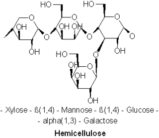
A hemicellulose is one of a number of heteropolymers, such as arabinoxylans, present along with cellulose in almost all terrestrial plant cell walls. Cellulose is crystalline, strong, and resistant to hydrolysis. Hemicelluloses are branched, shorter in length than cellulose, and also show a propensity to crystallize. They can be hydrolyzed by dilute acid or base as well as a myriad of hemicellulase enzymes.

Plant cells are the cells present in green plants, photosynthetic eukaryotes of the kingdom Plantae. Their distinctive features include primary cell walls containing cellulose, hemicelluloses and pectin, the presence of plastids with the capability to perform photosynthesis and store starch, a large vacuole that regulates turgor pressure, the absence of flagella or centrioles, except in the gametes, and a unique method of cell division involving the formation of a cell plate or phragmoplast that separates the new daughter cells.

Polysaccharides, or polycarbohydrates, are the most abundant carbohydrates found in food. They are long-chain polymeric carbohydrates composed of monosaccharide units bound together by glycosidic linkages. This carbohydrate can react with water (hydrolysis) using amylase enzymes as catalyst, which produces constituent sugars. They range in structure from linear to highly branched. Examples include storage polysaccharides such as starch, glycogen and galactogen and structural polysaccharides such as cellulose and chitin.

Phloem is the living tissue in vascular plants that transports the soluble organic compounds made during photosynthesis and known as photosynthates, in particular the sugar sucrose, to the rest of the plant. This transport process is called translocation. In trees, the phloem is the innermost layer of the bark, hence the name, derived from the Ancient Greek word φλοιός (phloiós), meaning "bark". The term was introduced by Carl Nägeli in 1858. Different types of phloem can be distinguished. The early phloem formed in the growth apices is called protophloem. Protophloem eventually becomes obliterated once it connects to the durable phloem in mature organs, the metaphloem. Further, secondary phloem is formed during the thickening of stem structures.

In biology, tissue is an assembly of similar cells and their extracellular matrix from the same embryonic origin that together carry out a specific function. Tissues occupy a biological organizational level between cells and a complete organ. Accordingly, organs are formed by the functional grouping together of multiple tissues.

Bark is the outermost layer of stems and roots of woody plants. Plants with bark include trees, woody vines, and shrubs. Bark refers to all the tissues outside the vascular cambium and is a nontechnical term. It overlays the wood and consists of the inner bark and the outer bark. The inner bark, which in older stems is living tissue, includes the innermost layer of the periderm. The outer bark on older stems includes the dead tissue on the surface of the stems, along with parts of the outermost periderm and all the tissues on the outer side of the periderm. The outer bark on trees which lies external to the living periderm is also called the rhytidome.

Lignin is a class of complex organic polymers that form key structural materials in the support tissues of most plants. Lignins are particularly important in the formation of cell walls, especially in wood and bark, because they lend rigidity and do not rot easily. Chemically, lignins are polymers made by cross-linking phenolic precursors.

The Casparian strip is a band-like thickening in the center of the root endodermis of vascular plants. The composition of the region is mainly suberin, lignin and some structural proteins, which are capable of reducing the diffusive apoplastic flow of water and solutes into the stele and its width varies between species. The Casparian strip is impervious to water so can control the transportation of water and inorganic salts between the cortex and the vascular bundle, preventing water and inorganic salts from being transported to the stele through the apoplast, so that it must enter the cell membrane and move to the stele through the symplastic pathway, blocking the internal and external objects of the cell. The function of mass transportation are similar to that of animal tissues.. The development of the Casparian strip is regulated by transcription factors such as SHORT-ROOT (SHR), SCARECROW (SCR) and MYB36, as well as polypeptide hormone synthesised by midcolumn cells.

The ground tissue of plants includes all tissues that are neither dermal nor vascular. It can be divided into three types based on the nature of the cell walls. This tissue system is present between the dermal tissue and forms the main bulk of the plant body.
- Parenchyma cells have thin primary walls and usually remain alive after they become mature. Parenchyma forms the "filler" tissue in the soft parts of plants, and is usually present in cortex, pericycle, pith, and medullary rays in primary stem and root.
- Collenchyma cells have thin primary walls with some areas of secondary thickening. Collenchyma provides extra mechanical and structural support, particularly in regions of new growth.
- Sclerenchyma cells have thick lignified secondary walls and often die when mature. Sclerenchyma provides the main structural support to the plant.

Fibrils are structural biological materials found in nearly all living organisms. Not to be confused with fibers or filaments, fibrils tend to have diameters ranging from 10–100 nanometers. Fibrils are not usually found alone but rather are parts of greater hierarchical structures commonly found in biological systems. Due to the prevalence of fibrils in biological systems, their study is of great importance in the fields of microbiology, biomechanics, and materials science.
A microfibril is a very fine fibril, or fiber-like strand, consisting of glycoproteins and cellulose. It is usually, but not always, used as a general term in describing the structure of protein fiber, e.g. hair and sperm tail. Its most frequently observed structural pattern is the 9+2 pattern in which two central protofibrils are surrounded by nine other pairs. Cellulose inside plants is one of the examples of non-protein compounds that are using this term with the same purpose. Cellulose microfibrils are laid down in the inner surface of the primary cell wall. As the cell absorbs water, its volume increases and the existing microfibrils separate and new ones are formed to help increase cell strength.

Xylan is a type of hemicellulose, a polysaccharide consisting mainly of xylose residues. It is found in plants, in the secondary cell walls of dicots and all cell walls of grasses. Xylan is the third most abundant biopolymer on Earth, after cellulose and chitin.
Xyloglucan is a hemicellulose that occurs in the primary cell wall of all vascular plants; however, all enzymes responsible for xyloglucan metabolism are found in Charophyceae algae. In many dicotyledonous plants, it is the most abundant hemicellulose in the primary cell wall. Xyloglucan binds to the surface of cellulose microfibrils and may link them together. It is the substrate of xyloglucan endotransglycosylase, which cuts and ligates xyloglucans, as a means of integrating new xyloglucans into the cell wall. It is also thought to be the substrate of alpha-expansin, which promotes cell wall enlargement.

Cinnamoyl-CoA reductase (EC 1.2.1.44), systematically named cinnamaldehyde:NADP+ oxidoreductase (CoA-cinnamoylating) but commonly referred to by the acronym CCR, is an enzyme that catalyzes the reduction of a substituted cinnamoyl-CoA to its corresponding cinnamaldehyde, utilizing NADPH and H+ and releasing free CoA and NADP+ in the process. Common biologically relevant cinnamoyl-CoA substrates for CCR include p-coumaroyl-CoA and feruloyl-CoA, which are converted into p-coumaraldehyde and coniferaldehyde, respectively, though most CCRs show activity toward a variety of other substituted cinnamoyl-CoA's as well. Catalyzing the first committed step in monolignol biosynthesis, this enzyme plays a critical role in lignin formation, a process important in plants both for structural development and defense response.

The UDP-forming form of cellulose synthase is the main enzyme that produces cellulose. Systematically, it is known as UDP-glucose:(1→4)-β-D-glucan 4-β-D-glucosyltransferase in enzymology. It catalyzes the chemical reaction:
Arabinoxylan is a form of the hemicellulose xylan found in both the primary and secondary cell walls of plants which in addition to xylose contains substantial amounts of another pentose sugar, arabinose. The term arabinoxylan usually refers to feruloyl-arabinoxylan from grasses and other commelinids containing moieties of the phenolic ferulic acid that can undergo oxidative coupling forming crosslinks between arabinoxylan chains and with lignin. Whilst arabinose has been found linked to xylan in non-commelinid plants, ferulic acid has not been reported on these and unlike feruloyl-arabinoxylan these arabinoxylans are not monophyletic. The remainder of this article refers to feruloyl-arabinoxylan from cell walls of grasses and other commelinid species.

Bacterial cellulose is an organic compound with the formula (C
6H
10O
5)
n produced by certain types of bacteria. While cellulose is a basic structural material of most plants, it is also produced by bacteria, principally of the genera Komagataeibacter, Acetobacter, Sarcina ventriculi and Agrobacterium. Bacterial, or microbial, cellulose has different properties from plant cellulose and is characterized by high purity, strength, moldability and increased water holding ability. In natural habitats, the majority of bacteria synthesize extracellular polysaccharides, such as cellulose, which form protective envelopes around the cells. While bacterial cellulose is produced in nature, many methods are currently being investigated to enhance cellulose growth from cultures in laboratories as a large-scale process. By controlling synthesis methods, the resulting microbial cellulose can be tailored to have specific desirable properties. For example, attention has been given to the bacteria Komagataeibacter xylinus due to its cellulose's unique mechanical properties and applications to biotechnology, microbiology, and materials science.

The exodermis is a physiological barrier that has a role in root function and protection. The exodermis is a membrane of variable permeability responsible for the radial flow of water, ions, and nutrients. It is the outer layer of a plant's cortex. The exodermis serves a double function as it can protect the root from invasion by foreign pathogens and ensures that the plant does not lose too much water through diffusion through the root system and can properly replenish its stores at an appropriate rate.
