Shoulder reduction

Last updated
Shoulder reduction
GreekReduction.jpg
Reduction of a dislocated shoulder with a Hippocratic device.

Shoulder reduction is the process of returning the shoulder to its normal position following a shoulder dislocation. Normally, closed reduction, in which the relationship of bone and joint is manipulated externally without surgical intervention, is used. A variety of techniques exist, but some are preferred due to fewer complications or easier execution. [1] In cases where closed reduction is not successful, open (surgical) reduction may be needed. [2] X-rays are often used to confirm success and absence of associated fractures. The arm should be kept in a sling or immobilizer for several days, prior to supervised recovery of motion and strength.

Contents

Various non-operative reduction techniques are employed. They have certain principles in common, including gentle in-line traction, reduction or abolition of muscle spasm, and gentle external rotation. They all strive to avoid inadvertent injury. Two of them, the Milch and Stimson techniques, have been compared in a randomized trial. [3] Pain can be managed during the procedures either by procedural sedation and analgesia or by injecting lidocaine into the shoulder joint. [4]

External rotation

The person's arm is brought against their side. [2] The elbow is then bent to 90 degrees and the forearm is slowly and gently externally rotated. [2] Any discomfort or spasm interrupts the process until the person is able to relax. Reduction usually takes place by the time full external rotation has been achieved. [5]

Milch

This is an extension of the external rotation technique. The externally rotated arm is gently abducted (brought away from the body into an overhead position) while external rotation is maintained. Gentle in-line traction is applied to the arm while some pressure is applied to the humeral head via the operator's thumb in the armpit to keep the head from moving inferiorly. [2] [6]

Stimson

An example of Stimson maneuver. The person is lying on their stomach with a 4 kg weight tied to their wrist. Stimson2017.jpg
An example of Stimson maneuver. The person is lying on their stomach with a 4 kg weight tied to their wrist.

In the Stimson technique, also known as prone technique, the person lies on their stomach on a bed or bench and the arm hangs off the side, being allowed to drop toward the ground. A 5–10 kg weight is suspended from the wrist to overcome spasm and to permit reduction by the force of gravity; [3]

Spaso

The person is on their back and gentle upward traction is applied to the extremity coupled with external rotation. [2]

Cunningham technique

Cunningham technique Cunningham technique demonstration.jpg
Cunningham technique

The Cunningham technique was originally published in 2003 and is an anatomically based method of shoulder reduction that utilizes positioning (analgesic position), voluntary scapular retraction, and bicipital massage. [7] If performed correctly most patients do not require analgesia for the performance of this technique. Inappropriate use of traction will result in pain for the patient with subsequent spasm and failure to reduce. If the patient is unable to adduct the humerus or unable to cooperate with positioning the technique should not be attempted. The patient may require analgesia or sedation if they are in pain or unable to relax spasming muscles.

FARES method

FARES stands for fast, reliable, and safe. With the person lying on their back, the operator holds the person's hand on the affected side while the arm is at the side and the elbow is fully extended. The forearm is in neutral position. Next, the operator gently applies longitudinal traction and slowly the arm is abducted. At the same time, continuous vertical oscillating movement at a rate of 2–3 "cycles" per second is applied throughout the whole reduction process. [8] [9]

Traction countertraction

Traction countertraction involves pulling the dislocated arm down and outwards while an assistant pulls the body upwards. [2]

Kocher's method

Kocher's method was described in 1870 and has been incorrectly associated with neuromuscular complications and humeral fractures due to the inappropriate addition of traction by later users. It was designed for subcoracoid dislocations. "This method: Pressing the arm bent at the elbow towards the body, turning outward until resistance is felt, lifting of the outwardly rotated upper arm in the sagittal plane as far as possible, and finally slowly turning it inward." [10]

Related Research Articles

<span class="mw-page-title-main">Rotator cuff</span> Group of muscles

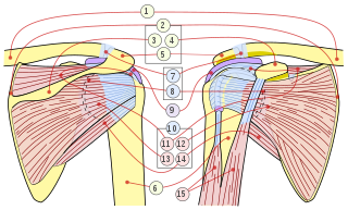
The rotator cuff is a group of muscles and their tendons that act to stabilize the human shoulder and allow for its extensive range of motion. Of the seven scapulohumeral muscles, four make up the rotator cuff. The four muscles are:

<span class="mw-page-title-main">Shoulder problem</span> Medical condition

Shoulder problems including pain, are one of the more common reasons for physician visits for musculoskeletal symptoms. The shoulder is the most movable joint in the body. However, it is an unstable joint because of the range of motion allowed. This instability increases the likelihood of joint injury, often leading to a degenerative process in which tissues break down and no longer function well.

<span class="mw-page-title-main">Deltoid muscle</span> Shoulder muscle

The deltoid muscle is the muscle forming the rounded contour of the human shoulder. It is also known as the 'common shoulder muscle', particularly in other animals such as the domestic cat. Anatomically, the deltoid muscle appears to be made up of three distinct sets of muscle fibers, namely the

  1. anterior or clavicular part
  2. posterior or scapular part
  3. intermediate or acromial part
<span class="mw-page-title-main">Joint dislocation</span> Medical injury

A joint dislocation, also called luxation, occurs when there is an abnormal separation in the joint, where two or more bones meet. A partial dislocation is referred to as a subluxation. Dislocations are often caused by sudden trauma on the joint like an impact or fall. A joint dislocation can cause damage to the surrounding ligaments, tendons, muscles, and nerves. Dislocations can occur in any major joint or minor joint. The most common joint dislocation is a shoulder dislocation.

<span class="mw-page-title-main">Distal radius fracture</span> Fracture of the radius bone near the wrist

A distal radius fracture, also known as wrist fracture, is a break of the part of the radius bone which is close to the wrist. Symptoms include pain, bruising, and rapid-onset swelling. The ulna bone may also be broken.

<span class="mw-page-title-main">Teres minor muscle</span> Muscle of the rotator cuff

The teres minor is a narrow, elongated muscle of the rotator cuff. The muscle originates from the lateral border and adjacent posterior surface of the corresponding right or left scapula and inserts at both the greater tubercle of the humerus and the posterior surface of the joint capsule.

<span class="mw-page-title-main">Nerve block</span> Deliberate inhibition of nerve impulses

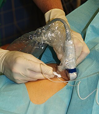
Nerve block or regional nerve blockade is any deliberate interruption of signals traveling along a nerve, often for the purpose of pain relief. Local anesthetic nerve block is a short-term block, usually lasting hours or days, involving the injection of an anesthetic, a corticosteroid, and other agents onto or near a nerve. Neurolytic block, the deliberate temporary degeneration of nerve fibers through the application of chemicals, heat, or freezing, produces a block that may persist for weeks, months, or indefinitely. Neurectomy, the cutting through or removal of a nerve or a section of a nerve, usually produces a permanent block. Because neurectomy of a sensory nerve is often followed, months later, by the emergence of new, more intense pain, sensory nerve neurectomy is rarely performed.

<span class="mw-page-title-main">Shoulder joint</span> Synovial ball and socket joint in the shoulder

The shoulder joint is structurally classified as a synovial ball-and-socket joint and functionally as a diarthrosis and multiaxial joint. It involves an articulation between the glenoid fossa of the scapula and the head of the humerus. Due to the very loose joint capsule that gives a limited interface of the humerus and scapula, it is the most mobile joint of the human body.

<span class="mw-page-title-main">Hip dislocation</span> Orthopedic injury

A hip dislocation is when the thighbone (femur) separates from the hip bone (pelvis). Specifically it is when the ball–shaped head of the femur separates from its cup–shaped socket in the hip bone, known as the acetabulum. The joint of the femur and pelvis is very stable, secured by both bony and soft-tissue constraints. With that, dislocation would require significant force which typically results from significant trauma such as from a motor vehicle collision or from a fall from elevation. Hip dislocations can also occur following a hip replacement or from a developmental abnormality known as hip dysplasia.

<span class="mw-page-title-main">Separated shoulder</span> Medical condition

A separated shoulder, also known as acromioclavicular joint injury, is a common injury to the acromioclavicular joint. The AC joint is located at the outer end of the clavicle where it attaches to the acromion of the scapula. Symptoms include non-radiating pain which may make it difficult to move the shoulder. The presence of swelling or bruising and a deformity in the shoulder is also common depending on how severe the dislocation is.

<span class="mw-page-title-main">Dislocated shoulder</span> Injury

A dislocated shoulder is a condition in which the head of the humerus is detached from the glenoid fossa. Symptoms include shoulder pain and instability. Complications may include a Bankart lesion, Hill-Sachs lesion, rotator cuff tear, or injury to the axillary nerve.

<span class="mw-page-title-main">Bankart lesion</span> Medical condition

A Bankart lesion is a type of shoulder injury that occurs following a dislocated shoulder. It is an injury of the anterior (inferior) glenoid labrum of the shoulder. When this happens, a pocket at the front of the glenoid forms that allows the humeral head to dislocate into it. It is an indication for surgery and often accompanied by a Hill-Sachs lesion, damage to the posterior humeral head.

<span class="mw-page-title-main">Hill–Sachs lesion</span> Cortical depression in the posterolateral head of the humerus

A Hill–Sachs lesion, or Hill–Sachs fracture, is a cortical depression in the posterolateral head of the humerus. It results from forceful impaction of the humeral head against the anteroinferior glenoid rim when the shoulder is dislocated anteriorly.

<span class="mw-page-title-main">Elbow</span> Joint between the upper and lower parts of the arm

The elbow is the region between the upper arm and the forearm that surrounds the elbow joint. The elbow includes prominent landmarks such as the olecranon, the cubital fossa, and the lateral and the medial epicondyles of the humerus. The elbow joint is a hinge joint between the arm and the forearm; more specifically between the humerus in the upper arm and the radius and ulna in the forearm which allows the forearm and hand to be moved towards and away from the body. The term elbow is specifically used for humans and other primates, and in other vertebrates it is not used. In those cases, forelimb plus joint is used.

Procedural sedation and analgesia (PSA) is a technique in which a sedating/dissociative medication is given, usually along with an analgesic medication, in order to perform non-surgical procedures on a patient. The overall goal is to induce a decreased level of consciousness while maintaining the patient's ability to breathe on their own. Airway protective reflexes are not compromised by this process and therefore endotracheal intubation is not required. PSA is commonly used in the emergency department, in addition to the operating room.

<span class="mw-page-title-main">Dislocation of jaw</span> Medical condition

Dislocations occur when two bones that originally met at the joint detach. Dislocations should not be confused with subluxation. Subluxation is when the joint is still partially attached to the bone.

<span class="mw-page-title-main">Cunningham shoulder reduction</span>

Cunningham shoulder reduction was originally published in 2003 and is an anatomically based method of shoulder reduction that utilizes positioning, voluntary scapular retraction, and bicipital massage. It is designed for true anterior/subcoracoid glenohumeral dislocations in patients who can fully adduct their humerus. This is distinct from anteroinferior/subglenoid glenohumeral dislocations for which alternative techniques should be used. The method is one of several techniques used for shoulder reduction.

<span class="mw-page-title-main">Watsu</span> Form of aquatic bodywork

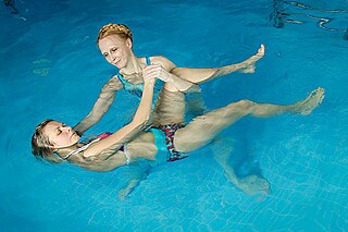
Watsu is a form of aquatic bodywork used for deep relaxation and passive aquatic therapy. Watsu is characterized by one-on-one sessions in which a practitioner or therapist gently cradles, moves, stretches, and massages a receiver in chest-deep warm water.

The Latarjet operation, also known as the Latarjet-Bristow procedure, is a surgical procedure used to treat recurrent shoulder dislocations, typically caused by bone loss or a fracture of the glenoid. The procedure was first described by French surgeon Dr. Michel Latarjet in 1954.

<span class="mw-page-title-main">Knee dislocation</span> Medical condition

A knee dislocation is an injury in which there is disruption of the knee joint between the tibia and the femur. Symptoms include pain and instability of the knee. Complications may include injury to an artery, most commonly the popliteal artery behind the knee, or compartment syndrome.

References

  1. Dislocations, Shoulder~workup at eMedicine
  2. 1 2 3 4 5 6 Bonz, J; Tinloy, B (May 2015). "Emergency department evaluation and treatment of the shoulder and humerus". Emergency Medicine Clinics of North America. 33 (2): 297–310. doi:10.1016/j.emc.2014.12.004. PMID   25892723.
  3. 1 2 Amar, E; Maman, E; Khashan, M; Kauffman, E; et al. (Nov 2012). "Milch versus Stimson technique for nonsedated reduction of anterior shoulder dislocation: a prospective randomized trial and analysis of factors affecting success". J Shoulder Elbow Surg. 21 (11): 1443–9. doi:10.1016/j.jse.2012.01.004. PMID   22516569.
  4. Fitch, RW; Kuhn, JE (August 2008). "Intraarticular lidocaine versus intravenous procedural sedation with narcotics and benzodiazepines for reduction of the dislocated shoulder: a systematic review". Academic Emergency Medicine. 15 (8): 703–8. doi:10.1111/j.1553-2712.2008.00164.x. PMID   18783486.
  5. Harnroongroj, T; Wangphanich, J; Harnroongroj, T (Dec 2011). "Efficacy of gentle traction, abduction and external rotation maneuver under sedative-free for reduction of acute anterior shoulder dislocation: retrospective comparative study". J Med Assoc Thai. 94 (12): 1482–6. PMID   22295736.
  6. Singh, S; Yong, CK; Mariapan, S (Dec 2012). "Closed reduction techniques in acute anterior shoulder dislocation: modified Milch technique compared with traction-countertraction technique". J Shoulder Elbow Surg. 21 (12): 1706–11. doi:10.1016/j.jse.2012.04.004. PMID   22819577.
  7. "Cunningham | DISLOCATION.COM.AU". dislocation.com.au. Archived from the original on 2020-02-13.
  8. Sayegh, Fares E.; Kenanidis, Eustathios I.; Papavasiliou, Kyriakos A.; Potoupnis, Michael E.; Kirkos, John M.; Kapetanos, George A. (2009-12-01). "Reduction of acute anterior dislocations: a prospective randomized study comparing a new technique with the Hippocratic and Kocher methods". The Journal of Bone and Joint Surgery. American Volume. 91 (12): 2775–2782. doi:10.2106/JBJS.H.01434. ISSN   1535-1386. PMID   19952238.
  9. "FARES method to reduce acute anterior shoulder dislocation: a case series and an efficacy analysis". Hong Kong Journal of Emergency Medicine. January 2012.
  10. "Welcome to DISLOCATION.COM.AU". DISLOCATION.COM.AU. Retrieved 14 October 2021.