
The lungs are the primary organs of the respiratory system in humans and many other animals including a few fish and some snails. In mammals and most other vertebrates, two lungs are located near the backbone on either side of the heart. Their function in the respiratory system is to extract oxygen from the atmosphere and transfer it into the bloodstream, and to release carbon dioxide from the bloodstream into the atmosphere, in a process of gas exchange. Respiration is driven by different muscular systems in different species. Mammals, reptiles and birds use their different muscles to support and foster breathing. In early tetrapods, air was driven into the lungs by the pharyngeal muscles via buccal pumping, a mechanism still seen in amphibians. In humans, the main muscle of respiration that drives breathing is the diaphragm. The lungs also provide airflow that makes vocal sounds including human speech possible.

The thoracic diaphragm, or simply the diaphragm, is a sheet of internal skeletal muscle in humans and other mammals that extends across the bottom of the thoracic cavity. The diaphragm separates the thoracic cavity, containing the heart and lungs, from the abdominal cavity and performs an important function in respiration: as the diaphragm contracts, the volume of the thoracic cavity increases, creating a negative pressure there, which draws air into the lungs.

A pulmonary artery is an artery in the pulmonary circulation that carries deoxygenated blood from the right side of the heart to the lungs. The largest pulmonary artery is the main pulmonary artery or pulmonary trunk from the heart, and the smallest ones are the arterioles, which lead to the capillaries that surround the pulmonary alveoli.

Patent ductus arteriosus (PDA) is a medical condition in which the ductus arteriosus fails to close after birth: this allows a portion of oxygenated blood from the left heart to flow back to the lungs by flowing from the aorta, which has a higher pressure, to the pulmonary artery. Symptoms are uncommon at birth and shortly thereafter, but later in the first year of life there is often the onset of an increased work of breathing and failure to gain weight at a normal rate. With time, an uncorrected PDA usually leads to pulmonary hypertension followed by right-sided heart failure.

Situs inversus is a congenital condition in which the major visceral organs are reversed or mirrored from their normal positions. The normal arrangement of internal organs is known as situs solitus while situs inversus is generally the mirror image of situs solitus. Although cardiac problems are more common than in the general population, most people with situs inversus have no medical symptoms or complications resulting from the condition, and until the advent of modern medicine it was usually undiagnosed.
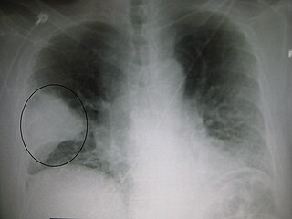
Radiology (X-rays) is used in the diagnosis of tuberculosis. Abnormalities on chest radiographs may be suggestive of, but are never diagnostic of, TB but can be used to rule out pulmonary TB.
Transient tachypnea of the newborn is a respiratory problem that can be seen in the newborn shortly after delivery. It is caused by retained fetal lung fluid due to impaired clearance mechanisms. it is the most common cause of respiratory distress in term neonates. It consists of a period of rapid breathing. Usually, this condition resolves over 24-72 hours. Treatment is supportive and may include supplemental oxygen and antibiotics. The chest x-ray shows hyperinflation of the lungs including prominent pulmonary vascular markings, flattening of the diaphragm, and fluid in the horizontal fissure of the right lung.
Atelectasis is the collapse or closure of a lung resulting in reduced or absent gas exchange. It is usually unilateral, affecting part or all of one lung. It is a condition where the alveoli are deflated down to little or no volume, as distinct from pulmonary consolidation, in which they are filled with liquid. It is often called a collapsed lung, although that term may also refer to pneumothorax.

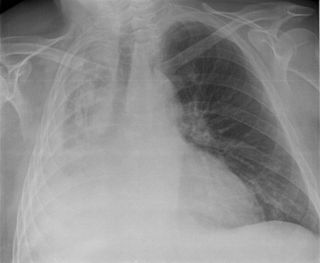
A chest radiograph, colloquially called a chest X-ray (CXR), or chest film, is a projection radiograph of the chest used to diagnose conditions affecting the chest, its contents, and nearby structures. Chest radiographs are the most common film taken in medicine.

A pulmonary sequestration is a medical condition wherein a piece of tissue that ultimately develops into lung tissue is not attached to the pulmonary arterial blood supply, as is the case in normally developing lung. This sequestered tissue is therefore not connected to the normal bronchial airway architecture, and fails to function in, and contribute to, respiration of the organism.

The fossa ovalis is a depression in the right atrium of the heart, at the level of the interatrial septum, the wall between right and left atrium. The fossa ovalis is the remnant of a thin fibrous sheet that covered the foramen ovale during fetal development.

In animals that give live birth, the fetal circulation is the circulatory system of a fetus. The term usually encompasses the entire fetoplacental circulation, which includes the umbilical cord and the blood vessels within the placenta that carry fetal blood.

A respiratory examination, or lung examination, is performed as part of a physical examination, in response to respiratory symptoms such as shortness of breath, cough, or chest pain, and is often carried out with a cardiac examination.

Pericardial effusion is an abnormal accumulation of fluid in the pericardial cavity. Because of the limited amount of space in the pericardial cavity, fluid accumulation leads to an increased intrapericardial pressure which can negatively affect heart function. A pericardial effusion with enough pressure to adversely affect heart function is called cardiac tamponade. Pericardial effusion usually results from a disturbed equilibrium between the production and re-absorption of pericardial fluid, or from a structural abnormality that allows fluid to enter the pericardial cavity.
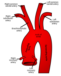
The ascending aorta (AAo) is a portion of the aorta commencing at the upper part of the base of the left ventricle, on a level with the lower border of the third costal cartilage behind the left half of the sternum.
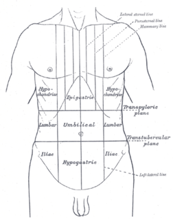
Traube's (semilunar) space is an anatomic space of some clinical importance. It is a crescent-shaped space, encompassed by the lower edge of the left lung, the anterior border of the spleen, the left costal margin and the inferior margin of the left lobe of the liver. Thus, its surface markings are respectively the left sixth rib superiorly, the left mid axillary line laterally, and the left costal margin inferiorly.

Projectional radiography is a form of radiography and medical imaging that produces two-dimensional images by x-ray radiation. The image acquisition is generally performed by radiographers, and the images are often examined by radiologists. Both the procedure and any resultant images are often simply called "X-ray". Plain radiography generally refers to projectional radiography. Plain radiography can also refer to radiography without a radiocontrast agent or radiography that generates single static images, as contrasted to fluoroscopy, which are technically also projectional.

In human anatomy, an azygos lobe is a normal anatomical variation of the upper lobe of the right lung. It is seen in 1% of the population. Embryologically, it arises from an anomalous lateral course of the azygos vein in a pleural septum within the apical segment of the right upper lobe or in other words an azygos lobe is formed when the right posterior cardinal vein, one of the precursors of the azygos vein, fails to migrate over the apex of the lung and penetrates it instead, carrying along two pleural layers that invaginates into the upper portion of the right upper lobe. As it has no bronchi, veins and arteries of its own or corresponding alteration in the segmental architecture of the lung, so it is not a true (misnomer), or even accessory, pulmonary lobe, but rather an anatomically separated part of the upper lobe. It is usually an incidental finding on chest x-ray or computed tomography and is as such not associated with any morbidity but can cause technical problems in thoracoscopic procedures.
In medicine, the Golden S sign is a sign seen on imaging of the chest that suggests a central lung mass or lung collapse. It was first described by Dr. Ross Golden (1889-1975) in 1925 in association with bronchial carcinoma, but it is also seen in metastatic cancer, enlarged lymph nodes, and collapse of the right upper lobe of the lung.
Pneumonia can be classified in several ways, most commonly by where it was acquired, but may also by the area of lung affected or by the causative organism. There is also a combined clinical classification, which combines factors such as age, risk factors for certain microorganisms, the presence of underlying lung disease or systemic disease, and whether the person has recently been hospitalized.


