Chest pain in children

Last updated

Chest pain in children
Doctor uses a stethoscope to examine a young patient.JPEG
A doctor listens to a young girl's heart and lungs with a stethoscope
Specialty Pediatrics

Chest pain in children is the pain felt in the chest by infants, children and adolescents. In most cases the pain is not associated with the heart. It is primarily identified by the observance or report of pain by the infant, child or adolescent by reports of distress by parents or caregivers. Chest pain is not uncommon in children. Many children are seen in ambulatory clinics, emergency departments and hospitals and cardiology clinics. Most often there is a benign cause for the pain for most children. Some have conditions that are serious and possibly life-threatening. Chest pain in pediatric patients requires careful physical examination and a detailed history that would indicate the possibility of a serious cause. Studies of pediatric chest pain are sparse. It has been difficult to create evidence-based guidelines for evaluation. [1]

Contents

Differential diagnosis

Chest pain in children is usually evaluated in the emergency departments. It can be distressing for parents and children. Pediatric chest pain differs from chest pain in adults because it is most often unrelated to the heart. [2] The causes of pediatric chest pain vary according to the organ or tissue in the child. that generates the pain. Generally, muscular skeletal pain, which includes costochondritis, is the reason for the emergency department visit. Pain that is felt in the chest but is due to muscular skeletal inflammation or an unknown cause and accounts for 7% and 69% visits. Muscular skeletal pain is described and defined differently as a diagnosis of exclusion or is documented as being associated with idiopathic causes. Asthma and other respiratory symptoms are the second most common presentation. Respiratory associated causes compose 13% to 24% of pediatric chest pain symptoms. Gastrointestinal and psychogenic symptoms reported by parents and patients occur less than 10% of the time. Cardiac causes of pediatric chest pain are found infrequently and are not identified more than 5% of the time. Unknown causes, were estimated to account for 20% to 61% of the final diagnosis given. Patients who receive a diagnosis of cardiac disease are more apt to have acute pain. This pain often awakes them from sleep or presents with fever or abnormal observations found during the physical examination. Trauma can also be a cause for chest pain and has been found to be associated with the pain in 5% of the patients. [1]

Children can present with chest pain can have a sudden onset related to vigorous physical activity and coughing. These symptoms seem to be closely associated with asthma. [1] Infection with Haemophilus influenzae can cause chest pain. [3]

Preliminary

Since most causes of pediatric and adolescent chest pain are not considered life-threatening, parents and their children are often reassured that in the majority of cases, the cause of the pain can be determined. If the child or adolescent appears to have some dehydration, and intravenous line along with administration of saline is done. The clinician may or may not decide to perform diagnostic testing . This is especially true if the child or adolescent has symptoms of chronic pain. If an obvious cause of the chest pain is not readily apparent, testing may begin with an x-ray and an electrocardiogram . This helps the clinician to determine whether or not the cause of pain is related to pulmonary or cardiac causes. [4]

Some preliminary diagnosis include:[ citation needed ]

Diagnosis

Brugada syndrome Brugada syndrome type1 example2 (CardioNetworks ECGpedia).png
Brugada syndrome

Preliminary "red flags" are identified that are more likely to suggest chest pain associated with heart. These are:

Testing

If the diagnosis is unclear, the clinician may order additional testing. These tests may be an electrocardiogram, stress test, pulmonary function test, drug screen and ultrasound. [4] [6]

Muscular skeletal causes

Fractured ribs marked Fracturedribsmarked.jpg
Fractured ribs marked

Respiratory causes

pneumothorax 05-Spontanpneumothorax.jpg
pneumothorax

Cardiovascular causes

Gastrointestinal causes

Non-organic causes

Epidemiology

Percentages of the causes of chest pain in children and adolescents Cause of chest pain in children and adolescents.PNG
Percentages of the causes of chest pain in children and adolescents

Chest pain is relatively common in children and adolescents. Unlike adults, the cause is rarely cardiac. [2] Approximately 0.3% to 0.6% of emergency department visits by pediatric patients are for chest pain. The emergency department visits are consistent throughout the seasons and times of year. The median age pediatric patients that complain of pain is from 12 to 13 years old both males and females display the symptoms and signs at approximately the same ratio. [2] Those that do complain of the chest pain usually present with acute pain that they have experienced for less than one day. This is not universal though. A small study in Turkey evaluated patients and found that 59% complained of pain that they had had for more than one month in duration. Estimates of 13% to 24% of patients seen in the emergency department are found to have a pulmonary association with their pain. [1]

Treatment

Children who have chest pain that is related to asthma and exercise stress can be treated with bronchodilator use. [1] If there is obviously distress and difficulty breathing, treatment to provide the support of airway and breathing is provided. In addition, treatment can include measures to support circulation. [4]

Part of effective treatment includes obtaining a detailed medical history, [2] an important step that is frequently absent in emergency department visits and connected with hospitalization prevention. [12]

Related Research Articles

<span class="mw-page-title-main">Pneumonia</span> Inflammation of the alveoli of the lungs

Pneumonia is an inflammatory condition of the lung primarily affecting the small air sacs known as alveoli. Symptoms typically include some combination of productive or dry cough, chest pain, fever, and difficulty breathing. The severity of the condition is variable.

<span class="mw-page-title-main">Shortness of breath</span> Feeling of difficulty breathing

Shortness of breath (SOB), also medically known as dyspnea or dyspnoea, is an uncomfortable feeling of not being able to breathe well enough. The American Thoracic Society defines it as "a subjective experience of breathing discomfort that consists of qualitatively distinct sensations that vary in intensity", and recommends evaluating dyspnea by assessing the intensity of its distinct sensations, the degree of distress and discomfort involved, and its burden or impact on the patient's activities of daily living. Distinct sensations include effort/work to breathe, chest tightness or pain, and "air hunger". The tripod position is often assumed to be a sign.

<span class="mw-page-title-main">Pleural effusion</span> Accumulation of excess fluid in the pleural cavity

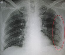
A pleural effusion is accumulation of excessive fluid in the pleural space, the potential space that surrounds each lung. Under normal conditions, pleural fluid is secreted by the parietal pleural capillaries at a rate of 0.6 millilitre per kilogram weight per hour, and is cleared by lymphatic absorption leaving behind only 5–15 millilitres of fluid, which helps to maintain a functional vacuum between the parietal and visceral pleurae. Excess fluid within the pleural space can impair inspiration by upsetting the functional vacuum and hydrostatically increasing the resistance against lung expansion, resulting in a fully or partially collapsed lung.

<span class="mw-page-title-main">Chest pain</span> Discomfort or pain in the chest as a medical symptom

Chest pain is pain or discomfort in the chest, typically the front of the chest. It may be described as sharp, dull, pressure, heaviness or squeezing. Associated symptoms may include pain in the shoulder, arm, upper abdomen, or jaw, along with nausea, sweating, or shortness of breath. It can be divided into heart-related and non-heart-related pain. Pain due to insufficient blood flow to the heart is also called angina pectoris. Those with diabetes or the elderly may have less clear symptoms.

<span class="mw-page-title-main">Pericardiocentesis</span> Procedure where fluid is aspirated from the pericardium

Pericardiocentesis (PCC), also called pericardial tap, is a medical procedure where fluid is aspirated from the pericardium.

<span class="mw-page-title-main">Costochondritis</span> Human disease

Costochondritis, also known as chest wall pain syndrome or costosternal syndrome, is a benign inflammation of the upper costochondral and sternocostal joints. 90% of patients are affected in multiple ribs on a single side, typically at the 2nd to 5th ribs. Chest pain, the primary symptom of costochondritis, is considered a symptom of a medical emergency, making costochondritis a common presentation in the emergency department. One study found costochondritis was responsible for 30% of patients with chest pain in an emergency department setting.

Cardiac asthma is the medical condition of intermittent wheezing, coughing, and shortness of breath that is associated with underlying congestive heart failure (CHF). Symptoms of cardiac asthma are related to the heart's inability to effectively and efficiently pump blood in a CHF patient. This can lead to accumulation of fluid in and around the lungs, disrupting the lung's ability to oxygenate blood.

<span class="mw-page-title-main">Respiratory disease</span> Disease of the respiratory system

Respiratory diseases, or lung diseases, are pathological conditions affecting the organs and tissues that make gas exchange difficult in air-breathing animals. They include conditions of the respiratory tract including the trachea, bronchi, bronchioles, alveoli, pleurae, pleural cavity, the nerves and muscles of respiration. Respiratory diseases range from mild and self-limiting, such as the common cold, influenza, and pharyngitis to life-threatening diseases such as bacterial pneumonia, pulmonary embolism, tuberculosis, acute asthma, lung cancer, and severe acute respiratory syndromes, such as COVID-19. Respiratory diseases can be classified in many different ways, including by the organ or tissue involved, by the type and pattern of associated signs and symptoms, or by the cause of the disease.

Reactive airway disease (RAD) is an informal label that physicians apply to patients with symptoms similar to those of asthma. An exact definition of the condition does not exist. Individuals who are typically labeled as having RAD generally have a history of wheezing, coughing, dyspnea, and production of sputum that may or may not be caused by asthma. Symptoms may also include, but are not limited to, coughing, shortness of breath, excess mucus in the bronchial tube, swollen mucous membrane in the bronchial tube, and/or hypersensitive bronchial tubes. Physicians most commonly label patients with RAD when they are hesitant about formally diagnosing a patient with asthma, which is most prevalent in the pediatric setting. While some physicians may use RAD and asthma synonymously, there is controversy over this usage.

Mixed connective tissue disease, commonly abbreviated as MCTD, is an autoimmune disease characterized by the presence of elevated blood levels of a specific autoantibody, now called anti-U1 ribonucleoprotein (RNP) together with a mix of symptoms of systemic lupus erythematosus (SLE), scleroderma, and polymyositis. The idea behind the "mixed" disease is that this specific autoantibody is also present in other autoimmune diseases such as systemic lupus erythematosus, polymyositis, scleroderma, etc. MCTD was characterized as an individual disease in 1972 by Sharp et al., and the term was introduced by Leroy in 1980.

<span class="mw-page-title-main">Lymphangiomatosis</span> Medical condition

Lymphangiomatosis is a condition where a lymphangioma is not present in a single localised mass, but in a widespread or multifocal manner. It is a rare type of tumor which results from an abnormal development of the lymphatic system.

<span class="mw-page-title-main">Lupus erythematosus</span> Human disease

Lupus erythematosus is a collection of autoimmune diseases in which the human immune system becomes hyperactive and attacks healthy tissues. Symptoms of these diseases can affect many different body systems, including joints, skin, kidneys, blood cells, heart, and lungs. The most common and most severe form is systemic lupus erythematosus.

Vocal cord dysfunction (VCD) is a pathology affecting the vocal folds characterized by full or partial vocal fold closure causing difficulty and distress during respiration, especially during inhalation.

Obstructive shock is one of the four types of shock, caused by a physical obstruction in the flow of blood. Obstruction can occur at the level of the great vessels or the heart itself. Causes include pulmonary embolism, cardiac tamponade, and tension pneumothorax. These are all life-threatening. Symptoms may include shortness of breath, weakness, or altered mental status. Low blood pressure and tachycardia are often seen in shock. Other symptoms depend on the underlying cause.

<span class="mw-page-title-main">Fibrothorax</span> Medical condition involving fibrosis of the pleural space

Fibrothorax is a medical condition characterised by severe scarring (fibrosis) and fusion of the layers of the pleural space surrounding the lungs resulting in decreased movement of the lung and ribcage. The main symptom of fibrothorax is shortness of breath. There also may be recurrent fluid collections surrounding the lungs. Fibrothorax may occur as a complication of many diseases, including infection of the pleural space known as an empyema or bleeding into the pleural space known as a haemothorax.

<span class="mw-page-title-main">Lupus</span> Human autoimmune disease

Lupus, technically known as systemic lupus erythematosus (SLE), is an autoimmune disease in which the body's immune system mistakenly attacks healthy tissue in many parts of the body. Symptoms vary among people and may be mild to severe. Common symptoms include painful and swollen joints, fever, chest pain, hair loss, mouth ulcers, swollen lymph nodes, feeling tired, and a red rash which is most commonly on the face. Often there are periods of illness, called flares, and periods of remission during which there are few symptoms.

<span class="mw-page-title-main">Asbestos-related diseases</span> Medical condition

Asbestos-related diseases are disorders of the lung and pleura caused by the inhalation of asbestos fibres. Asbestos-related diseases include non-malignant disorders such as asbestosis, diffuse pleural thickening, pleural plaques, pleural effusion, rounded atelectasis and malignancies such as lung cancer and malignant mesothelioma.

<span class="mw-page-title-main">Foreign body aspiration</span> Medical condition

Foreign body aspiration occurs when a foreign body enters the airway which can cause difficulty breathing or choking. Objects may reach the respiratory tract and the digestive tract from the mouth and nose, but when an object enters the respiratory tract it is termed aspiration. The foreign body can then become lodged in the trachea or further down the respiratory tract such as in a bronchus. Regardless of the type of object, any aspiration can be a life-threatening situation and requires timely recognition and action to minimize risk of complications. While advances have been made in management of this condition leading to significantly improved clinical outcomes, there were still 2,700 deaths resulting from foreign body aspiration in 2018. Approximately one child dies every five days due to choking on food in the United States, highlighting the need for improvements in education and prevention.

<span class="mw-page-title-main">Urinothorax</span> Presence of urine in the fluid-filled cavity that surrounds the lungs

Urinothorax is defined as urine in the fluid-filled cavity that surrounds the lungs. It is usually caused by obstructive uropathy. It is mainly diagnosed by analyzing the pleural fluid. Treatment involves treating the underlying condition, which typically results in resolution of the urinothorax. It is an extremely rare cause of pleural effusion.

<span class="mw-page-title-main">Lupus vasculitis</span> Medical condition

Lupus vasculitis is one of the secondary vasculitides that occurs in approximately 50% of patients with systemic lupus erythematosus (SLE).

References

  1. 1 2 3 4 5 6 7 8 9 10 Thull-Freedman, Jennifer (2010). "Evaluation of Chest Pain in the Pediatric Patient". Medical Clinics of North America. 94 (2): 327–347. doi:10.1016/j.mcna.2010.01.004. ISSN   0025-7125. PMID   20380959: Access provided by the University of Pittsburgh{{cite journal}}: CS1 maint: postscript (link)
  2. 1 2 3 4 5 6 Collins, S. A.; Griksaitis, M. J.; Legg, J. P. (2013). "15-minute consultation: A structured approach to the assessment of chest pain in a child". Archives of Disease in Childhood: Education and Practice Edition. 99 (4): 122–126. doi: 10.1136/archdischild-2013-303919 . ISSN   1743-0585. PMID   24301714.
  3. Yeh, Y-H; Chu, P-H; Yeh, C-H; Jan Wu, Y-J; Lee, M-H.; Jung, S-M; Kuo, C-T (2004). "Haemophilus influenzae pericarditis with tamponade as the initial presentation of systemic lupus erythematosus". International Journal of Clinical Practice. 58 (11): 1045–1047. doi:10.1111/j.1742-1241.2004.00041.x. ISSN   1368-5031. PMID   15605669. S2CID   21831622.
  4. 1 2 3 4 5 6 7 8 9 10 11 12 McMahon, Maureen (2011). Pediatrics a competency-based companion. Philadelphia, PA: Saunders/Elsevier. ISBN   978-1-4160-5350-7: Access provided by the University of Pittsburgh{{cite book}}: CS1 maint: postscript (link)
  5. Maharaj, Satish S; Chang, Simone M (2015). "Cardiac tamponade as the initial presentation of systemic lupus erythematosus: a case report and review of the literature". Pediatric Rheumatology. 13 (1): 9. doi: 10.1186/s12969-015-0005-0 . ISSN   1546-0096. PMC   4369869 . PMID   25802493.
  6. Brass, Patrick; Hellmic, Martin; Kolodziej, Laurentius; Schick, Guido; Smith, Andrew F (2015). "Ultrasound guidance versus anatomical landmarks for subclavian or femoral vein catheterization". Cochrane Database of Systematic Reviews. 1 (12): CD011447. doi: 10.1002/14651858.CD011447 . PMC   6516998 . PMID   25575245.
  7. Keech, Brian M. (2015). "Thoracic epidural analgesia in a child with multiple traumatic rib fractures". Journal of Clinical Anesthesia. 27 (8): 685–691. doi:10.1016/j.jclinane.2015.05.008. ISSN   0952-8180. PMID   26118312.
  8. Bounakis, Nikolaos; Karampalis, Christos; Sharp, Hilary; Tsirikos, Athanasios I (2015). "Surgical treatment of scoliosis in Rubinstein-Taybi syndrome type 2: a case report". Journal of Medical Case Reports. 9 (1): 10. doi: 10.1186/1752-1947-9-10 . ISSN   1752-1947. PMC   4334754 . PMID   25596810.
  9. Stroud, Andrea M.; Tulanont, Darena D.; Coates, Thomasena E.; Goodney, Philip P.; Croitoru, Daniel P. (2014). "Epidural analgesia versus intravenous patient-controlled analgesia following minimally invasive pectus excavatum repair: a systematic review and meta-analysis". Journal of Pediatric Surgery. 49 (5): 798–806. doi:10.1016/j.jpedsurg.2014.02.072. ISSN   0022-3468. PMC   5315444 . PMID   24851774.
  10. Punja, Mohan; Mark, Dustin G.; McCoy, Jonathan V.; Javan, Ramin; Pines, Jesse M.; Brady, William (2010). "Electrocardiographic manifestations of cardiac infectious-inflammatory disorders". The American Journal of Emergency Medicine. 28 (3): 364–377. doi:10.1016/j.ajem.2008.12.017. ISSN   0735-6757. PMID   20223398:Access provided by the University of Pittsburgh{{cite journal}}: CS1 maint: postscript (link)
  11. Legg, Julian P.; Griksaitis, Michael J.; Collins, Samuel A. (1 August 2014). "15-minute consultation: A structured approach to the assessment of chest pain in a child". Archives of Disease in Childhood: Education and Practice Edition. 99 (4): 122–126. doi: 10.1136/archdischild-2013-303919 . ISSN   1743-0585. PMID   24301714.
  12. Martin, Molly A.; Press, Valerie G.; Nyenhuis, Sharmilee M.; Krishnan, Jerry A.; Erwin, Kim; Mosnaim, Giselle; Margellos-Anast, Helen; Paik, S. Margaret; Ignoffo, Stacy; McDermott, Michael (2016). "Care transition interventions for children with asthma in the emergency department". Journal of Allergy and Clinical Immunology. 138 (6): 1518–1525. doi:10.1016/j.jaci.2016.10.012. PMC   5327498 . PMID   27931533.