Fourth heart sound

Last updated
Fourth heart sound
Other namesAtrial gallop, presystolic gallop
Diagram of the human heart (cropped).svg
Diagram of the heart.
Specialty Cardiology

The fourth heart sound or S4 is an extra heart sound that occurs during late diastole, immediately before the normal two "lub-dub" heart sounds (S1 and S2). It occurs just after atrial contraction and immediately before the systolic S1 and is caused by the atria contracting forcefully in an effort to overcome an abnormally stiff or hypertrophic ventricle.

Contents

This produces a rhythm classically compared to the cadence of the word "Tennessee." [1] [ full citation needed ] [2] One can also use the phrase "A-stiff-wall" to help with the cadence (a S4, stiff S1, wall S2), as well as the pathology of the S4 sound. [3]

Physiology

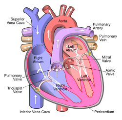
The normal heart sounds, S1 and S2, are produced during the closing of the atrioventricular valves and semilunar valves, respectively. The closing of these valves produces a brief period of turbulent flow, which produces sound. [4]

The S4 sound occurs, by definition, immediately before S1, while the atria of the heart are vigorously contracting. [5] It is manifest as a vibration of 20 to 30 Hz within the ventricle. [5] While the mechanism is not absolutely certain, it is generally accepted that S4 is caused by stiffening of the walls of the ventricles (usually the left), which produces abnormally turbulent flow as the atria contract to force blood into the ventricle. [5] [4] This for example occurs in conditions that decrease ventricular compliance like left ventricular hypertrophy. [4]

S4 is sometimes audible in the elderly due to a more rigid ventricle. When loud, it is a sign of a pathologic state, [6] usually a failing left ventricle. If the problem lies with the left ventricle, the gallop rhythm will be heard best at the cardiac apex. It will become more apparent with exercise, with the patient lying on the left-hand side, or with the patient holding expiration. If the problem is in the right ventricle, the abnormal sound will be most evident on the lower left hand side of the sternum and will get louder with exercise and quick, deep inspiration. [7]

S4 has also been termed an atrial gallop or a presystolic gallop because of its occurrence late in the heart cycle. It is a type of gallop rhythm by virtue of having an extra sound; the other gallop rhythm is called S3. The two are quite different, but they may sometimes occur together forming a quadruple gallop. If the heart rate is also very fast (tachycardia), it can become difficult to distinguish between S3 and S4 thus producing a single sound called a summation gallop.

Treatment

The S4 heart sound itself does not require treatment; rather plans should be laid to stop the progression of whatever causes the underlying ventricular dysfunction. The S4 heart sound is a secondary manifestation of a primary disease process and treatment should be focused on treating the underlying, primary disease.

Related Research Articles

<span class="mw-page-title-main">Heart</span> Organ found inside most animals

The heart is a muscular organ found in most animals. This organ pumps blood through the blood vessels of the circulatory system. The pumped blood carries oxygen and nutrients to the body, while carrying metabolic waste such as carbon dioxide to the lungs. In humans, the heart is approximately the size of a closed fist and is located between the lungs, in the middle compartment of the chest, called the mediastinum.

<span class="mw-page-title-main">Heart sounds</span> Noise generated by the beating heart

Heart sounds are the noises generated by the beating heart and the resultant flow of blood through it. Specifically, the sounds reflect the turbulence created when the heart valves snap shut. In cardiac auscultation, an examiner may use a stethoscope to listen for these unique and distinct sounds that provide important auditory data regarding the condition of the heart.

<span class="mw-page-title-main">Mitral valve</span> Valve in the heart connecting the left atrium and left ventricle

The mitral valve, also known as the bicuspid valve or left atrioventricular valve, is one of the four heart valves. It has two cusps or flaps and lies between the left atrium and the left ventricle of the heart. The heart valves are all one-way valves allowing blood flow in just one direction. The mitral valve and the tricuspid valve are known as the atrioventricular valves because they lie between the atria and the ventricles.

<span class="mw-page-title-main">Heart murmur</span> Medical condition

Heart murmurs are unique heart sounds produced when blood flows across a heart valve or blood vessel. This occurs when turbulent blood flow creates a sound loud enough to hear with a stethoscope. Turbulent blood flow is not smooth. The sound differs from normal heart sounds by their characteristics. For example, heart murmurs may have a distinct pitch, duration and timing. The major way health care providers examine the heart on physical exam is heart auscultation; another clinical technique is palpation, which can detect by touch when such turbulence causes the vibrations called cardiac thrill. A murmur is a sign found during the cardiac exam. Murmurs are of various types and are important in the detection of cardiac and valvular pathologies.

<span class="mw-page-title-main">Systole</span> Part of the cardiac cycle when a heart chamber contracts

Systole is the part of the cardiac cycle during which some chambers of the heart contract after refilling with blood.

<span class="mw-page-title-main">Diastole</span> Part of the cardiac cycle

Diastole is the relaxed phase of the cardiac cycle when the chambers of the heart are refilling with blood. The contrasting phase is systole when the heart chambers are contracting. Atrial diastole is the relaxing of the atria, and ventricular diastole the relaxing of the ventricles.

<span class="mw-page-title-main">Atrial flutter</span> Medical condition

Atrial flutter (AFL) is a common abnormal heart rhythm that starts in the atrial chambers of the heart. When it first occurs, it is usually associated with a fast heart rate and is classified as a type of supraventricular tachycardia. Atrial flutter is characterized by a sudden-onset (usually) regular abnormal heart rhythm on an electrocardiogram (ECG) in which the heart rate is fast. Symptoms may include a feeling of the heart beating too fast, too hard, or skipping beats, chest discomfort, difficulty breathing, a feeling as if one's stomach has dropped, a feeling of being light-headed, or loss of consciousness.

<span class="mw-page-title-main">Cardiac conduction system</span> Aspect of heart function

The cardiac conduction system transmits the signals generated by the sinoatrial node – the heart's pacemaker, to cause the heart muscle to contract, and pump blood through the body's circulatory system. The pacemaking signal travels through the right atrium to the atrioventricular node, along the bundle of His, and through the bundle branches to Purkinje fibers in the walls of the ventricles. The Purkinje fibers transmit the signals more rapidly to stimulate contraction of the ventricles.

<span class="mw-page-title-main">Atrial septal defect</span> Human heart defect present at birth

Atrial septal defect (ASD) is a congenital heart defect in which blood flows between the atria of the heart. Some flow is a normal condition both pre-birth and immediately post-birth via the foramen ovale; however, when this does not naturally close after birth it is referred to as a patent (open) foramen ovale (PFO). It is common in patients with a congenital atrial septal aneurysm (ASA).

<span class="mw-page-title-main">Ostium primum atrial septal defect</span> Medical condition

The ostium primum atrial septal defect is a defect in the atrial septum at the level of the tricuspid and mitral valves. This is sometimes known as an endocardial cushion defect because it often involves the endocardial cushion, which is the portion of the heart where the atrial septum meets the ventricular septum and the mitral valve meets the tricuspid valve.

<span class="mw-page-title-main">Supraventricular tachycardia</span> Abnormally fast heart rhythm

Supraventricular tachycardia (SVT) is an umbrella term for fast heart rhythms arising from the upper part of the heart. This is in contrast to the other group of fast heart rhythms – ventricular tachycardia, which start within the lower chambers of the heart. There are four main types of SVT: atrial fibrillation, atrial flutter, paroxysmal supraventricular tachycardia (PSVT), and Wolff–Parkinson–White syndrome. The symptoms of SVT include palpitations, feeling of faintness, sweating, shortness of breath, and/or chest pain.

<span class="mw-page-title-main">Ebstein's anomaly</span> Congenital heart defect

Ebstein's anomaly is a congenital heart defect in which the septal and posterior leaflets of the tricuspid valve are displaced downwards towards the apex of the right ventricle of the heart. EA has great anatomical heterogeneity that generates a wide spectrum of clinical features at presentation and is complicated by the fact that the lesion is often accompanied by other congenital cardiac lesions. It is classified as a critical congenital heart defect accounting for less than 1% of all congenital heart defects presenting in around 1 per 200,000 live births. Ebstein's anomaly usually presents with a systolic murmur and frequently with a gallop rhythm.

<span class="mw-page-title-main">Transposition of the great vessels</span> Group of congenital heart defects

Transposition of the great vessels (TGV) is a group of congenital heart defects involving an abnormal spatial arrangement of any of the great vessels: superior and/or inferior venae cavae, pulmonary artery, pulmonary veins, and aorta. Congenital heart diseases involving only the primary arteries belong to a sub-group called transposition of the great arteries (TGA), which is considered the most common congenital heart lesion that presents in neonates.

<span class="mw-page-title-main">Atrioventricular septal defect</span> Medical condition

Atrioventricular septal defect (AVSD) or atrioventricular canal defect (AVCD), also known as "common atrioventricular canal" or "endocardial cushion defect" (ECD), is characterized by a deficiency of the atrioventricular septum of the heart that creates connections between all four of its chambers. It is a very specific combination of 3 defects:

A gallop rhythm refers to a rhythm of the heart on auscultation. It includes three or four sounds, thus resembling the sounds of a gallop.

<span class="mw-page-title-main">Lutembacher's syndrome</span> Medical condition

Lutembacher's syndrome is a very rare form of congenital heart disease that affects one of the chambers of the heart as well as a valve. It is commonly known as both congenital atrial septal defect (ASD) and acquired mitral stenosis (MS). Congenital atrial septal defect refers to a hole being in the septum or wall that separates the two atria; this condition is usually seen in fetuses and infants. Mitral stenosis refers to mitral valve leaflets sticking to each other making the opening for blood to pass from the atrium to the ventricles very small. With the valve being so small, blood has difficulty passing from the left atrium into the left ventricle. Septal defects that may occur with Lutembacher's syndrome include: Ostium primum atrial septal defect or ostium secundum which is more prevalent.

<span class="mw-page-title-main">Third heart sound</span> Medical condition

The third heart sound or S3 is a rare extra heart sound that occurs soon after the normal two "lub-dub" heart sounds (S1 and S2). S3 is associated with heart failure.

The E/A ratio is a marker of the function of the left ventricle of the heart. It represents the ratio of peak velocity blood flow from left ventricular relaxation in early diastole to peak velocity flow in late diastole caused by atrial contraction. It is calculated using Doppler echocardiography, an ultrasound-based cardiac imaging modality. Abnormalities in the E/A ratio suggest that the left ventricle, which pumps blood into the systemic circulation, cannot fill with blood properly in the period between contractions. This phenomenon is referred to as diastolic dysfunction and can eventually lead to the symptoms of heart failure.

<span class="mw-page-title-main">Arrhythmia</span> Group of medical conditions characterized by irregular heartbeat

Arrhythmias, also known as cardiac arrhythmias, heart arrhythmias, or dysrhythmias, are irregularities in the heartbeat, including when it is too fast or too slow. A resting heart rate that is too fast – above 100 beats per minute in adults – is called tachycardia, and a resting heart rate that is too slow – below 60 beats per minute – is called bradycardia. Some types of arrhythmias have no symptoms. Symptoms, when present, may include palpitations or feeling a pause between heartbeats. In more serious cases, there may be lightheadedness, passing out, shortness of breath, chest pain, or decreased level of consciousness. While most cases of arrhythmia are not serious, some predispose a person to complications such as stroke or heart failure. Others may result in sudden death.

Cardiac physiology or heart function is the study of healthy, unimpaired function of the heart: involving blood flow; myocardium structure; the electrical conduction system of the heart; the cardiac cycle and cardiac output and how these interact and depend on one another.

References

  1. clinical examination A systemic guide to physical diagnosis
  2. "Techniques - Heart Sounds & Murmurs Exam - Physical Diagnosis Skills - University of Washington School of Medicine". depts.washington.edu.
  3. "The Fourth Heart Sound". The Auscultation Assistant - Rubs and Gallops. Archived from the original on 2018-09-13. Retrieved 2011-12-24.
  4. 1 2 3 Dornbush, Sean; Turnquest, Andre E. (2023), "Physiology, Heart Sounds", StatPearls, Treasure Island (FL): StatPearls Publishing, PMID   31082054 , retrieved 2023-10-26
  5. 1 2 3 Williams, Eric S. (December 2, 1990). "The Fourth Heart Sound". Butterworths. PMID   21250185 via www.ncbi.nlm.nih.gov.
  6. "THE PHONOCARDIOGRAPHY" . Retrieved 2009-03-06.
  7. Tavel ME (Nov 1996). "The appearance of gallop rhythm after exercise stress testing". Clin Cardiol. 19 (11): 887–91. doi: 10.1002/clc.4960191109 . PMID   8914783.