
Endocarditis is an inflammation of the inner layer of the heart, the endocardium. It usually involves the heart valves. Other structures that may be involved include the interventricular septum, the chordae tendineae, the mural endocardium, or the surfaces of intracardiac devices. Endocarditis is characterized by lesions, known as vegetations, which are masses of platelets, fibrin, microcolonies of microorganisms, and scant inflammatory cells. In the subacute form of infective endocarditis, a vegetation may also include a center of granulomatous tissue, which may fibrose or calcify.

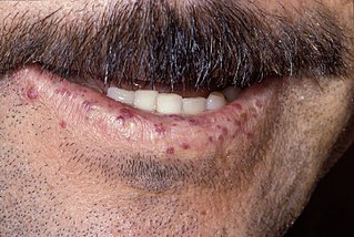
Yaws is a tropical infection of the skin, bones, and joints caused by the spirochete bacterium Treponema pallidum pertenue. The disease begins with a round, hard swelling of the skin, 2 to 5 cm in diameter. The center may break open and form an ulcer. This initial skin lesion typically heals after 3–6 months. After weeks to years, joints and bones may become painful, fatigue may develop, and new skin lesions may appear. The skin of the palms of the hands and the soles of the feet may become thick and break open. The bones may become misshapen. After 5 years or more, large areas of skin may die, leaving scars.

Chancroid is a bacterial sexually transmitted infection characterized by painful sores on the genitalia. Chancroid is known to spread from one individual to another solely through sexual contact. However, there have been reports of accidental infection through the hand.

Granuloma inguinale is a bacterial disease caused by Klebsiella granulomatis characterized by genital ulcers. It is endemic in many less-developed regions. It is also known as donovanosis, granuloma genitoinguinale, granuloma inguinale tropicum, granuloma venereum, granuloma venereum genitoinguinale, lupoid form of groin ulceration, serpiginous ulceration of the groin, ulcerating granuloma of the pudendum, and ulcerating sclerosing granuloma. Oral manifestations are also notably seen. The lesions of oral cavity are usually secondary to active genital lesions.

A granuloma is an aggregation of macrophages that forms in response to chronic inflammation. This occurs when the immune system attempts to isolate foreign substances that it is otherwise unable to eliminate. Such substances include infectious organisms including bacteria and fungi, as well as other materials such as foreign objects, keratin, and suture fragments.
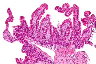
Whipple's disease is a rare systemic infectious disease caused by the bacterium Tropheryma whipplei. First described by George Hoyt Whipple in 1907 and commonly considered as a gastrointestinal disorder, Whipple's disease primarily causes malabsorption, but may affect any part of the human body, including the heart, brain, joints, skin, lungs and the eyes. Weight loss, diarrhea, joint pain, and arthritis are common presenting symptoms, but the presentation can be highly variable in certain individuals, and about 15% of patients do not have the standard signs and symptoms.

Infective endocarditis is an infection of the inner surface of the heart (endocardium), usually the valves. Signs and symptoms may include fever, small areas of bleeding into the skin, heart murmur, feeling tired, and low red blood cell count. Complications may include backward blood flow in the heart, heart failure – the heart struggling to pump a sufficient amount of blood to meet the body's needs, abnormal electrical conduction in the heart, stroke, and kidney failure.
Hereditary hemorrhagic telangiectasia (HHT), also known as Osler–Weber–Rendu disease and Osler–Weber–Rendu syndrome, is a rare autosomal dominant genetic disorder that leads to abnormal blood vessel formation in the skin, mucous membranes, and often in organs such as the lungs, liver, and brain.
Roth's spots, also known as Litten spots or the Litten sign, are non-specific red lesions with white or pale centres, seen on the retina of the eye and although traditionally associated with infective endocarditis, can occur in a number of other conditions including hypertension, diabetes, collagen vascular disease, extreme hypoxia, leukemia and HIV.

Osler's nodes are painful, red, raised lesions found on the hands and feet. They are associated with a number of conditions, including infective endocarditis, and are caused by immune complex deposition. Their presence is one definition of Osler's sign.
Reactive arthritis, previously known as Reiter's syndrome, is a form of inflammatory arthritis that develops in response to an infection in another part of the body (cross-reactivity). Coming into contact with bacteria and developing an infection can trigger the disease. By the time a person presents with symptoms, the "trigger" infection has often been cured or is in remission in chronic cases, thus making determination of the initial cause difficult.
Bartonellosis is an infectious disease caused by bacteria of the genus Bartonella. Bartonella species cause diseases such as Carrión's disease, trench fever, cat-scratch disease, bacillary angiomatosis, peliosis hepatis, chronic bacteremia, endocarditis, chronic lymphadenopathy, and neurological disorders.

Subacute bacterial endocarditis, abbreviated SBE, is a type of endocarditis. Subacute bacterial endocarditis can be considered a form of type III hypersensitivity.
Sporotrichosis, also known as rose handler's disease, is a fungal infection that may be localised to skin, lungs, bone and joint, or become systemic. It presents with firm painless nodules that later ulcerate. Following initial exposure to Sporothrix schenckii, the disease typically progresses over a period of a week to several months. Serious complications may develop in people who have a weakened immune system.
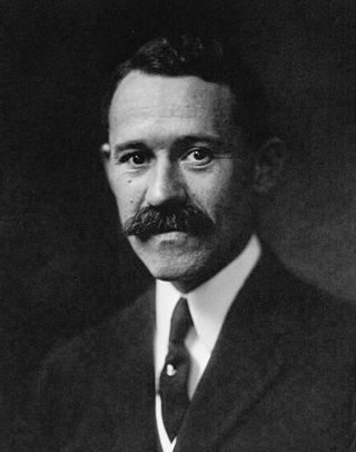
Theodore Caldwell Janeway was the first full-time professor of medicine at the Johns Hopkins University School of Medicine, recruited in 1914.
Bracht–Wachter bodies are a finding in infective endocarditis consisting of yellow-white miliary spots in the myocardium.

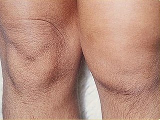
Lichen nitidus is a chronic inflammatory disease of unknown cause characterized by 1–2 mm, discrete and uniform, shiny, flat-topped, pale flesh-colored or reddish-brown papules that may appear as hypopigmented against dark skin. Occasionally, minimal scaling is present or can be induced by rubbing the surface of the papules. The disease usually affects children and young adults and is painless and usually nonpruritic, although protracted itching may occur in some cases. It is sometimes referred to by dermatologists as "mini lichen planus".

Austrian syndrome, also known as Osler's triad, is a medical condition that was named after Robert Austrian in 1957. The presentation of the condition consists of pneumonia, endocarditis, and meningitis, all caused by Streptococcus pneumoniae. It is associated with alcoholism due to hyposplenism and can be seen in males between the ages of 40 and 60 years old. Robert Austrian was not the first one to describe the condition, but Richard Heschl or William Osler were not able to link the signs to the bacteria because microbiology was not yet developed.

Progressive vaccinia is a rare cutaneous condition caused by the vaccinia virus, characterized by painless but progressive necrosis and ulceration.

Edward Gamaliel Janeway was an American physician who served as Health Commissioner of New York, and as president of the New York Medical Journal Association in the late nineteenth century. He was considered "one of America's premier internists in the late nineteenth and early twentieth centuries".
