Small conditional RNA

Last updated

A small conditional RNA (scRNA) is a small RNA molecule or complex (typically less than approximately 100 nt) engineered to interact and change conformation conditionally in response to cognate molecular inputs so as to perform signal transduction in vitro, in situ, or in vivo. [1]

Contents

In the absence of cognate input molecules, scRNAs are engineered to coexist metastably or stably without interacting. Detection of the cognate inputs initiates downstream conformational changes of one or more scRNAs leading to generation of the desired output signal. The output signal may be intended to read out the state of endogenous biological circuitry (e.g., mapping gene expression for biological research or medical diagnosis), [2] or to regulate the state of endogenous biological circuitry (e.g., perturbing gene expression for biological research or medical treatment). [1] scRNA sequences can be programmed to recognize different inputs or to activate different outputs, [1] [2] [3] achieving even single-nucleotide sequence selectivity. [3] scRNA signal transduction exploits principles from the emerging disciplines of dynamic RNA nanotechnology, molecular programming, and synthetic biology.

Figure 1. Fluorescent signal amplification using small conditional RNAs. Metastable fluorescent scRNAs self-assemble into fluorescent amplification polymers upon detection of a cognate RNA initiator. Initiator I nucleates with hairpin H1 via base pairing to single-stranded toehold '1', mediating a 3-way branch migration that opens the hairpin to form complex I*H1 containing single-stranded segment '3*-2*'. This complex nucleates with hairpin H2 by means of base pairing to toehold '3', mediating a branch migration that opens the hairpin to form complex I*H1*H2 containing single-stranded segment '2*-1*'. Thus, the initiator sequence is regenerated, providing the basis for a chain reaction of alternating H1 and H2 polymerization steps. Red stars denote fluorophores. Image from Choi et al. 2010; used with permission of the Nature Publishing Group. Hybridization Chain Reaction (HCR) mechanism.png
Figure 1. Fluorescent signal amplification using small conditional RNAs. Metastable fluorescent scRNAs self-assemble into fluorescent amplification polymers upon detection of a cognate RNA initiator. Initiator I nucleates with hairpin H1 via base pairing to single-stranded toehold '1', mediating a 3-way branch migration that opens the hairpin to form complex I·H1 containing single-stranded segment '3*-2*'. This complex nucleates with hairpin H2 by means of base pairing to toehold '3', mediating a branch migration that opens the hairpin to form complex I·H1·H2 containing single-stranded segment '2*-1*'. Thus, the initiator sequence is regenerated, providing the basis for a chain reaction of alternating H1 and H2 polymerization steps. Red stars denote fluorophores. Image from Choi et al. 2010; used with permission of the Nature Publishing Group.

Examples of scRNA signal transduction

Fluorophore-labeled scRNAs have been engineered to transduce between detection of mRNA targets and generation of bright fluorescent amplification polymers in situ (Figure 1). [2] In this context, scRNA signal transduction enables multiplexed mapping of mRNA expression within intact vertebrate embryos (Figure 2). [2] scRNAs have been engineered to perform shape and sequence transduction to conditionally produce a Dicer substrate targeting 'silencing target' mRNA Y upon detection of an independent 'detection target' mRNA X, with subsequent Dicer processing yielding a small interfering RNA (siRNA) targeting mRNA Y for destruction (Figure 3). [1] In this context, scRNA signal transduction provides a step towards implementing conditional RNA interference (Figure 4).

Figure 2. Simultaneous mapping of five different target mRNAs within an intact zebrafish embryo using scRNA signal amplification and confocal microscopy. RNA probes complementary to mRNA targets trigger chain reactions in which fluorophore- labeled scRNAs self-assemble into tethered fluorescent amplification polymers. The programmability and sequence selectivity of these amplification cascades enable five scRNA amplifiers to operate independently at the same time in the same sample, each staining for expression of one of the five target mRNAs. Scale bar: 50 mm. Image from Choi et al. 2010; used with permission of the Nature Publishing Group. Multiplexed mapping of mRNA expression within an intact zebrafish embryo.png
Figure 2. Simultaneous mapping of five different target mRNAs within an intact zebrafish embryo using scRNA signal amplification and confocal microscopy. RNA probes complementary to mRNA targets trigger chain reactions in which fluorophore- labeled scRNAs self-assemble into tethered fluorescent amplification polymers. The programmability and sequence selectivity of these amplification cascades enable five scRNA amplifiers to operate independently at the same time in the same sample, each staining for expression of one of the five target mRNAs. Scale bar: 50 μm. Image from Choi et al. 2010; used with permission of the Nature Publishing Group.
Figure 3. Conditional Dicer substrate formation via shape and sequence transduction with small conditional RNAs. scRNA A*B detects mRNA detection target X (containing subsequence 'a-b-c'), leading to production of Dicer substrate B*C targeting mRNA silencing target Y (containing independent subsequence 'w-x-y-z'). scRNAs A*B and C are stable in the absence of X. A swaps B for X (step 1) via toehold-mediated 3-way branch migration and spontaneous dissociation. B assembles with C (step 2) via loop/toehold nucleation and 3-way branch migration to form Dicer substrate B*C. Chemical modifications (2'OMe-RNA) prevent degradation: A and part of C (dashed backbone). Image from Hochrein et al. 2013; used with permission of the American Chemical Society. Conditional Dicer substrate formation via shape and sequence transduction with scRNAs.png
Figure 3. Conditional Dicer substrate formation via shape and sequence transduction with small conditional RNAs. scRNA A·B detects mRNA detection target X (containing subsequence 'a-b-c'), leading to production of Dicer substrate B·C targeting mRNA silencing target Y (containing independent subsequence 'w-x-y-z'). scRNAs A·B and C are stable in the absence of X. A swaps B for X (step 1) via toehold-mediated 3-way branch migration and spontaneous dissociation. B assembles with C (step 2) via loop/toehold nucleation and 3-way branch migration to form Dicer substrate B·C. Chemical modifications (2′OMe-RNA) prevent degradation: A and part of C (dashed backbone). Image from Hochrein et al. 2013; used with permission of the American Chemical Society.
Figure 4. Conditional RNAi (if gene X is transcribed, silence independent gene Y) provides a conceptual framework for exerting spatiotemporal control over gene knockdown. Toward this end, small conditional RNAs (scRNAs) interact and change conformation to transduce between binding of mRNA 'detection target' X and production of a Dicer substrate targeting independent mRNA 'silencing target' Y. Image from Hochrein et al. 2013; used with permission of the American Chemical Society. Molecular logic of conditional RNAi.png
Figure 4. Conditional RNAi (if gene X is transcribed, silence independent gene Y) provides a conceptual framework for exerting spatiotemporal control over gene knockdown. Toward this end, small conditional RNAs (scRNAs) interact and change conformation to transduce between binding of mRNA 'detection target' X and production of a Dicer substrate targeting independent mRNA 'silencing target' Y. Image from Hochrein et al. 2013; used with permission of the American Chemical Society.

Design elements

scRNAs can be engineered to exploit diverse design elements: [1]

Related Research Articles

Molecular biology is the branch of biology that seeks to understand the molecular basis of biological activity in and between cells, including biomolecular synthesis, modification, mechanisms, and interactions. The study of chemical and physical structure of biological macromolecules is known as molecular biology.

<span class="mw-page-title-main">Signal transduction</span> Cascade of intracellular and molecular events for transmission/amplification of signals

Signal transduction is the process by which a chemical or physical signal is transmitted through a cell as a series of molecular events, most commonly protein phosphorylation catalyzed by protein kinases, which ultimately results in a cellular response. Proteins responsible for detecting stimuli are generally termed receptors, although in some cases the term sensor is used. The changes elicited by ligand binding in a receptor give rise to a biochemical cascade, which is a chain of biochemical events known as a signaling pathway.

<span class="mw-page-title-main">Gene expression</span> Conversion of a genes sequence into a mature gene product or products

Gene expression is the process by which information from a gene is used in the synthesis of a functional gene product that enables it to produce end products, protein or non-coding RNA, and ultimately affect a phenotype, as the final effect. These products are often proteins, but in non-protein-coding genes such as transfer RNA (tRNA) and small nuclear RNA (snRNA), the product is a functional non-coding RNA. Gene expression is summarized in the central dogma of molecular biology first formulated by Francis Crick in 1958, further developed in his 1970 article, and expanded by the subsequent discoveries of reverse transcription and RNA replication.

<span class="mw-page-title-main">DNA microarray</span> Collection of microscopic DNA spots attached to a solid surface

A DNA microarray is a collection of microscopic DNA spots attached to a solid surface. Scientists use DNA microarrays to measure the expression levels of large numbers of genes simultaneously or to genotype multiple regions of a genome. Each DNA spot contains picomoles of a specific DNA sequence, known as probes. These can be a short section of a gene or other DNA element that are used to hybridize a cDNA or cRNA sample under high-stringency conditions. Probe-target hybridization is usually detected and quantified by detection of fluorophore-, silver-, or chemiluminescence-labeled targets to determine relative abundance of nucleic acid sequences in the target. The original nucleic acid arrays were macro arrays approximately 9 cm × 12 cm and the first computerized image based analysis was published in 1981. It was invented by Patrick O. Brown. An example of its application is in SNPs arrays for polymorphisms in cardiovascular diseases, cancer, pathogens and GWAS analysis. It is also used for the identification of structural variations and the measurement of gene expression.

<span class="mw-page-title-main">Fluorescent tag</span>

In molecular biology and biotechnology, a fluorescent tag, also known as a fluorescent label or fluorescent probe, is a molecule that is attached chemically to aid in the detection of a biomolecule such as a protein, antibody, or amino acid. Generally, fluorescent tagging, or labeling, uses a reactive derivative of a fluorescent molecule known as a fluorophore. The fluorophore selectively binds to a specific region or functional group on the target molecule and can be attached chemically or biologically. Various labeling techniques such as enzymatic labeling, protein labeling, and genetic labeling are widely utilized. Ethidium bromide, fluorescein and green fluorescent protein are common tags. The most commonly labelled molecules are antibodies, proteins, amino acids and peptides which are then used as specific probes for detection of a particular target.

<span class="mw-page-title-main">Biochip</span> Substrates performing biochemical reactions

In molecular biology, biochips are engineered substrates that can host large numbers of simultaneous biochemical reactions. One of the goals of biochip technology is to efficiently screen large numbers of biological analytes, with potential applications ranging from disease diagnosis to detection of bioterrorism agents. For example, digital microfluidic biochips are under investigation for applications in biomedical fields. In a digital microfluidic biochip, a group of (adjacent) cells in the microfluidic array can be configured to work as storage, functional operations, as well as for transporting fluid droplets dynamically.

<span class="mw-page-title-main">Morpholino</span> Chemical compound

A Morpholino, also known as a Morpholino oligomer and as a phosphorodiamidate Morpholino oligomer (PMO), is a type of oligomer molecule used in molecular biology to modify gene expression. Its molecular structure contains DNA bases attached to a backbone of methylenemorpholine rings linked through phosphorodiamidate groups. Morpholinos block access of other molecules to small specific sequences of the base-pairing surfaces of ribonucleic acid (RNA). Morpholinos are used as research tools for reverse genetics by knocking down gene function.

Fluorescence <i>in situ</i> hybridization Genetic testing technique

Fluorescence in situ hybridization (FISH) is a molecular cytogenetic technique that uses fluorescent probes that bind to only particular parts of a nucleic acid sequence with a high degree of sequence complementarity. It was developed by biomedical researchers in the early 1980s to detect and localize the presence or absence of specific DNA sequences on chromosomes. Fluorescence microscopy can be used to find out where the fluorescent probe is bound to the chromosomes. FISH is often used for finding specific features in DNA for use in genetic counseling, medicine, and species identification. FISH can also be used to detect and localize specific RNA targets in cells, circulating tumor cells, and tissue samples. In this context, it can help define the spatial-temporal patterns of gene expression within cells and tissues.

<i>In situ</i> hybridization

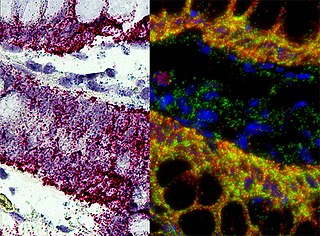
In situ hybridization (ISH) is a type of hybridization that uses a labeled complementary DNA, RNA or modified nucleic acids strand to localize a specific DNA or RNA sequence in a portion or section of tissue or if the tissue is small enough, in the entire tissue, in cells, and in circulating tumor cells (CTCs). This is distinct from immunohistochemistry, which usually localizes proteins in tissue sections.

Cis-regulatory elements (CREs) or Cis-regulatory modules (CRMs) are regions of non-coding DNA which regulate the transcription of neighboring genes. CREs are vital components of genetic regulatory networks, which in turn control morphogenesis, the development of anatomy, and other aspects of embryonic development, studied in evolutionary developmental biology.

A molecular logic gate is a molecule that performs a logical operation based on one or more physical or chemical inputs and a single output. The field has advanced from simple logic systems based on a single chemical or physical input to molecules capable of combinatorial and sequential operations such as arithmetic operations. Molecular logic gates work with input signals based on chemical processes and with output signals based on spectroscopic phenomena.

Biological computers use biologically derived molecules — such as DNA and/or proteins — to perform digital or real computations.

<span class="mw-page-title-main">Nucleic acid test</span> Group of techniques to detect a particular nucleic acid sequence

A nucleic acid test (NAT) is a technique used to detect a particular nucleic acid sequence and thus usually to detect and identify a particular species or subspecies of organism, often a virus or bacterium that acts as a pathogen in blood, tissue, urine, etc. NATs differ from other tests in that they detect genetic materials rather than antigens or antibodies. Detection of genetic materials allows an early diagnosis of a disease because the detection of antigens and/or antibodies requires time for them to start appearing in the bloodstream. Since the amount of a certain genetic material is usually very small, many NATs include a step that amplifies the genetic material—that is, makes many copies of it. Such NATs are called nucleic acid amplification tests (NAATs). There are several ways of amplification, including polymerase chain reaction (PCR), strand displacement assay (SDA), or transcription mediated assay (TMA).

A Riboprobe, abbreviation of RNA probe, is a segment of labelled RNA that can be used to detect a target mRNA or DNA during in situ hybridization. RNA probes can be produced by in vitro transcription of cloned DNA inserted in a suitable plasmid downstream of a viral promoter. Some bacterial viruses code for their own RNA polymerases, which are highly specific for the viral promoters. Using these enzymes, labeled NTPs, and inserts inserted in both forward and reverse orientations, both sense and antisense riboprobes can be generated from a cloned gene.

<span class="mw-page-title-main">Riboregulator</span>

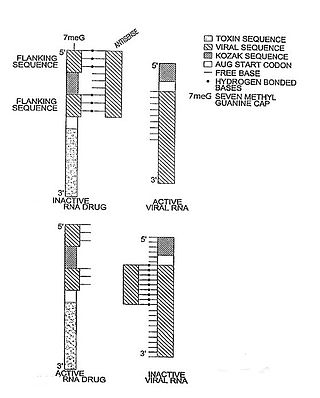
In molecular biology, a riboregulator is a ribonucleic acid (RNA) that responds to a signal nucleic acid molecule by Watson-Crick base pairing. A riboregulator may respond to a signal molecule in any number of manners including, translation of the RNA into a protein, activation of a ribozyme, release of silencing RNA (siRNA), conformational change, and/or binding other nucleic acids. Riboregulators contain two canonical domains, a sensor domain and an effector domain. These domains are also found on riboswitches, but unlike riboswitches, the sensor domain only binds complementary RNA or DNA strands as opposed to small molecules. Because binding is based on base-pairing, a riboregulator can be tailored to differentiate and respond to individual genetic sequences and combinations thereof.

The NAS Award in Molecular Biology is awarded by the U.S. National Academy of Sciences "for recent notable discovery in molecular biology by a young scientist who is a citizen of the United States." It has been awarded annually since its inception in 1962.

<span class="mw-page-title-main">RNA interference</span> Biological process of gene regulation

RNA interference (RNAi) is a biological process in which RNA molecules are involved in sequence-specific suppression of gene expression by double-stranded RNA, through translational or transcriptional repression. Historically, RNAi was known by other names, including co-suppression, post-transcriptional gene silencing (PTGS), and quelling. The detailed study of each of these seemingly different processes elucidated that the identity of these phenomena were all actually RNAi. Andrew Fire and Craig C. Mello shared the 2006 Nobel Prize in Physiology or Medicine for their work on RNAi in the nematode worm Caenorhabditis elegans, which they published in 1998. Since the discovery of RNAi and its regulatory potentials, it has become evident that RNAi has immense potential in suppression of desired genes. RNAi is now known as precise, efficient, stable and better than antisense therapy for gene suppression. Antisense RNA produced intracellularly by an expression vector may be developed and find utility as novel therapeutic agents.

Niles A. Pierce is an American mathematician, bioengineer, and professor at the California Institute of Technology. He is a leading researcher in the fields of molecular programming and dynamic nucleic acid nanotechnology. His research is focused on kinetically controlled DNA and RNA self-assembly. Pierce is working on applications in bioimaging.

<span class="mw-page-title-main">Spatial transcriptomics</span> Range of methods designed for assigning cell types

Spatial transcriptomics is a method for assigning cell types to their locations in the histological sections. This method can also be used to determine subcellular localization of mRNA molecules. The term is a variation of Spatial Genomics, first described by Doyle, et al., in 2000 and then expanded upon by Ståhl et al. in a technique developed in 2016, which has since undergone a variety of improvements and modifications.

This glossary of genetics is a list of definitions of terms and concepts commonly used in the study of genetics and related disciplines in biology, including molecular biology, cell biology, and evolutionary biology. It is intended as introductory material for novices; for more specific and technical detail, see the article corresponding to each term. For related terms, see Glossary of evolutionary biology.

References

  1. 1 2 3 4 5 6 7 Hochrein LM, Schwarzkopf M, Shahgholi M, Yin P, Pierce NA (2013). "Conditional Dicer substrate formation via shape and sequence transduction with small conditional RNAs". Journal of the American Chemical Society. 135 (46): 17322–17330. doi:10.1021/ja404676x. PMC   3842090 . PMID   24219616.
  2. 1 2 3 4 5 6 Choi HM, Chang JY, Trinh LA, Padilla JE, Fraser SE, Pierce NA (2010). "Programmable in situ amplification for multiplexed imaging of mRNA expression". Nature Biotechnology. 28 (11): 1208–1212. doi:10.1038/nbt.1692. PMC   3058322 . PMID   21037591.
  3. 1 2 Sternberg JB, Pierce NA (2014). "Exquisite sequence selectivity with small conditional RNAs". Nano Letters. 14 (8): 4568–4572. Bibcode:2014NanoL..14.4568S. doi:10.1021/nl501593r. PMC   4134187 . PMID   24979041.