Smear layer

Last updated

In dentistry, the smear layer is a layer found on root canal walls after root canal instrumentation. It consists of microcrystalline and organic particle debris. It was first described in 1975 and research has been performed since then to evaluate its importance in bacteria penetration into the dentinal tubules and its effects on endodontic treatment. More broadly, it is the organic layer found over all hard tooth surfaces.

Contents

Description

Early studies of dentinal walls after cavity preparation performed by Brännström and Johnson (1974) showed the presence of a thin layer of debris that was 2 to 5 micrometres thick. [1]

In 1975 McComb and Smith first described the smear layer. They observed an amorphous layer of debris, with an irregular and granular surface, on instrumented dentinal walls using a scanning electron microscope (SEM). The thin, granular microcrystalline layer of debris was 2-5 micrometres thick and was found packed onto the canal wall. [1] [2] The authors stated that “most standard instrumentation techniques produced a canal wall that was smeared and packed with debris.” [3]

In the same year Mader et al. studied the morphological characteristics of the smear layer in teeth that were endodontically instrumented with k type files and irrigated with 5.25% NaOCl. [4] The smear layer was examined from two aspects; the first aspect looked “down onto” the smear layer and the second from the side. Photomicrographs obtained by SEM showed that the smear layer consists of two confluent components. These were described as a thin superficial layer 1-2 micrometres thick overlying a densely packed layer and a second that penetrated into the dentinal tubules for distances of up to 40 micrometres. The packed material showed finger like structures projecting into the tubules from the canal wall. [4]

Contents

Composition

In 1984 Pashely described the smear layer as being composed of two phases; an organic phase, composed of collagen residues and glycosaminoglycans from extracellular matrix of pulp cells, which acts as a matrix for an inorganic phase. This organo-mineral content is composed of two distinct superimposed layers. The first layer covers the canal wall and is loosely adherent and easy to remove. The second layer however occludes the dentinal tubules and strongly adheres to the canal walls. [4]

Contents of the smear layer

  • Dentine particles
  • Residual vital pulp tissue
  • Residual necrotic pulp tissue
  • Erythrocytes
  • Remnant of odontoblast process
  • Saliva
  • Bacterial components [5]

Thickness of the smear layer

The smear layer is a physical barrier that decreases the penetration of disinfecting agents into dentinal tubules, and consequently, their efficacy. [6] The most important cause of endodontic failure is the residual microorganisms that are harboured within the root canal system and hard-to-reach areas. Studies were conducted into the thickness of smear layer created by different instruments, to enhance the understanding and aid the removal of the smear layer, and therefore aid the removal of any bacteria that may otherwise have been entombed by the smear layer. [6] [7] Results of the study showed that the Protaper series of rotary instruments caused the maximum amount of smear layer, followed by the Profile series of rotary instruments. [7] The hand instruments caused the least amount of smear layer. [7] Increasing the roughness of instruments has been found to increase the thickness of the smear layer as well. [7]

Bacterial Penetration

Olgart et al. (1974) examined the penetration of bacteria into dentinal tubules of ground, fractured and acid treated dentin surfaces. In vitro the penetration of bacteria into tubules of intact dentin exposed by fracture was compared in pairs of teeth, one of which in each pair was mounted with intrapulpal hydrostatic pressure (30mmHg). In vivo, intra pair comparisons of bacterial invasion into dentinal tubules beneath ground, fractured and acid treated surfaces were made. They observed that an outward flow of fluids into the tubules due to intrapulpal pressure mechanically hindered bacterial growth and that the debris and smear layer produced from grinding obstructed the bacterial invasion into tubules. However this barrier seemed to be removed after a few days which allowed bacterial growth into intact dentin. Olgart came to a conclusion that acid produced by microorganisms may dissolve the smear layer allowing bacteria to pass into dentinal tubules. [8]

However, when Pashley et al. (1981) studied the scanning electron microscope (SEM) appearance of dentin before and after removing successive layers of the smear layer they came to a different conclusion. Twenty dentin disks were cut from human extracted third molars. The dentin surface of the disks was etched with 6% citric acid for 5, 15, 30, 45 and 60 seconds. SEM examination showed that citric acid was able to remove smear layer in successive layers according to etching time finally exposing the dentinal tubules. Pashley concluded that the maintenance of the smear layer established a protective diffusion barrier. [9]

Gettleman et al. (1991) assessed the influence of a smear layer on the adhesion of sealer cements to dentin. A total of 120 teeth was tested, 40 per sealer namely AH26, Sultan, and Sealapex; 20 each with and without the smear layer. The teeth were split longitudinally, and the internal surfaces were ground flat. In the smear layer-free specimens the smear layer was removed by washing for 3 minutes with 17% EDTA followed by 5.25% NaOCl. Using a specially designed jig, the sealer was placed into a 4-mm wide × 4-mm deep well which was then set onto the tooth at a 90-degree angle and allowed to set for 7 days. This set-up was then placed into a mounting jig which was designed for the Instron Universal Testing Machine so that only a tensile load was applied without shearing. The set-up was subjected to a tensile load at a crosshead speed of 1 mm per min. The only significant difference with regard to the presence or absence of the smear layer was found with AH26, which had a stronger bond when the smear layer was removed. [10]

Removal of the smear layer

Why is the smear layer removed?

The smear layer can affect bonding, disinfection as well as obturation hence why it is considered important to remove. As discussed earlier this is a result of the fact that bacteria can be left entombed within the smear layer, if not removed.

How is the smear layer removed?

Endodontic Irrigation

Because the smear layer produced during endodontic instrumentation contains both inorganic and organic material, it cannot be removed by any of the presently available root canal irrigants alone. Therefore, the recommended protocol for smear layer removal is NaOCl followed by EDTA (ethylenediaminetetraacetic acid) or citric acid. Water, saline, chlorhexidine (CHX), or iodine compounds have no dissolving effect on the smear layer. [16]

Paste Lubricants

Gel based lubricants can be placed on the instrument before insertion into the root canal to reduce friction. Examples include “Glyde” and “Fileze” which both contain the chelating agent EDTA which can help enlarge narrow root canals by softening the canal walls. [19]

Dentine conditioners

These are generally acidic solutions which dissolve or at least solubilize the smear layer in attempt to expose the underlying dentine to the bonding agent. Examples include: phosphoric acid, nitric acid, maleic acid, citric acid, EDTA. Most manufacturers now supply a single agent to simultaneously etch enamel and condition the dentine. [21]

Further research

Clark-Holke et al. (2003) focused on determining the effect of the smear layer on the magnitude of bacterial penetration through the apical foramen around obturating materials. Thirty extracted teeth were classified into two test groups; the first group had the smear layer removed by rinsing with 17% EDTA while in the second group the smear layer was left intact. Canal preparation and obturation using lateral condensation, gutta-percha, and AH 26 sealer was performed on all of the teeth. The model systems consisted of an upper chamber attached to the cemento-enamel junction and a lower chamber at the apices of the teeth. Standardized bacterial suspensions containing Fusobacterium nucleatum , Campylobacter rectus and Peptostreptococcus micros were inoculated into the upper chambers. Models were incubated anaerobically at 37 °C. Leakage results were as follows: In the first group 6 teeth showed bacterial leakage, the second group and third groups showed no bacterial leakage. This study indicated that removal of the smear layer reduced the leakage of bacteria through the root canal system. [22]

Kokkas et al. (2004) examined the effect of the smear layer on the penetration depth of three different sealers (AH Plus, Apexit, and a Grossman type-Roth 811) into the dentinal tubules. Sixty four extracted human single-rooted teeth were used and divided into two groups. The smear layer remained intact in all the roots of group A. Complete removal of the smear layer in group B was achieved after irrigation with 3 ml of 17% EDTA for 3 min, followed by 3 ml of 1% NaOCl solution. Ten roots from each group were obturated with AH Plus and laterally condensed gutta-percha points. The same process was repeated for the remaining roots by using sealers Apexit and Roth 811 correspondingly. After complete setting, the maximum penetration depth of the sealers into the dentinal tubules was examined in upper, middle, and lower levels. The smear layer prevented all the sealers from penetrating dentinal tubules. In contrast, in smear layer–free root canals, all the sealers penetrated dentinal tubules, although the depth of penetration varied between the sealers. [23] Furthermore, smear layer adversely affected the coronal and apical sealing ability of sealers.

Çobankara et al. (2004) determined the effect of the smear layer on apical and coronal leakage in root canals obturated with AH26 or RoekoSeal sealers. A total of 160 maxillary anterior teeth were used. Eight groups were created by all possible combinations of three factors: smear layer (present/absent), leakage assessment (apical/coronal), and sealer used (AH26/Roeko-Seal). All teeth were obturated using lateral condensation technique of gutta-percha. A fluid filtration method was used to test apical or coronal leakage. According to the results of this study, the smear (+) groups displayed higher apical and coronal leakage than those smear (-) groups for both root canal sealers. Apical leakage was significantly higher than coronal leakage for both root canal sealers used in this study. It was determined that that removal of the smear layer has a positive effect in reducing apical and coronal leakage for both AH26 and RoekoSeal root canal sealers. [24]

However Bertacci et al. (2007) evaluated the ability of a warm gutta-percha obturation system Thermafil to fill lateral channels in the presence or absence of the smear layer. Forty single-rooted extracted human teeth were randomly divided into two groups one of which had the smear layer removed by 5 ml of 5% NaOCl followed by 2.5 ml of 17% EDTA. Obturation was performed using AH Plus sealer and Thermafil. Specimens were cleared in methyl salicylate and analyzed under a stereomicroscope to evaluate the number, length, and diameter of lateral channels. All lateral channels were found to be filled in both groups. No statistically significant differences regarding number, length, and diameter were observed between the two groups. It was concluded that the smear layer did not prevent the sealing of lateral channels. [25]

Yildirim et al. (2008) investigated the effect of the smear layer on apical microleakage in teeth obturated with MTA. Fifty single-rooted central maxillary teeth were used in this study. The selected teeth were instrumented and randomly divided into 2 groups. In the first group (smear [+]), the teeth were irrigated with only 5.25% NaOCl. In the second group (smear [-]), the teeth were irrigated with EDTA (17%) and NaOCl (5.25%) to remove the smear layer. The teeth were then filled with MTA. The computerized fluid filtration method was used for evaluation of apical microleakage. The quantitative apical leakage of each tooth was measured after 2, 30, and 180 days. It was found that there was no difference between the groups after 2 days but removal of the smear layer caused significantly more apical microleakage than when the smear layer was left intact after 30 and 180 days. It was concluded that the apical microleakage of MTA is less when the smear layer is present than when it is absent. [26]

Saleh et al. (2008) studied the effect of the smear layer on the penetration of bacteria along different root canal filling materials. A total of 110 human root segments were instrumented to size 80 under irrigation with 1% sodium hypochlorite. Half of the roots were irrigated with a 5-mL rinse of 17% EDTA to remove the smear layer. Roots were filled with gutta-percha (GP) and AH Plus sealer (AH), GP and Apexit sealer (AP), or RealSeal cones and sealer (RS). Following storage in humid conditions at 37 °C for 7 days, the specimens were mounted into a bacterial leakage test model for 135 days. Survival analyses were performed to calculate the median time of leakage and log-rank test was used for pairwise comparisons of groups. Selected specimens were longitudinally sectioned and inspected by scanning electron microscopy for the presence of bacteria at the interfaces. In the presence of the smear layer, RS and AP leaked significantly more slowly than in its absence. In the absence of the smear layer, AH leaked significantly more slowly than RS. It was concluded that removal of the smear layer did not impair bacterial penetration along root canal fillings. A comparison of the sealers revealed no difference except that AH performed better than RS in the absence of the smear layer. [27]

Fachin et al.(2009) evaluated whether smear layer removal has any influence on the filling of the root canal system, by examining the obturation of lateral canals, secondary canals and apical deltas. Eighty canines were randomly divided into two groups, according to their irrigation regimen. Both groups were irrigated with 1% NaOCl during canal shaping, but only the teeth in Group II received a final irrigation with 17% EDTA for smear layer removal. The root canals were obturated with lateral condensation of gutta-percha and the specimens were cleared, allowing for observation under the microscope. The results showed that In Groups I and II, 42.5% and 37.5% of the teeth, respectively, presented at least one filled canal ramification. In conclusion, smear layer removal under the conditions tested in this study did not affect the obturation of root canal ramifications when lateral condensation of gutta-percha was the technique used for root canal filling. [28]

Related Research Articles

<span class="mw-page-title-main">Dentin</span> Calcified tissue of the body; one of the four major components of teeth

Dentin or dentine is a calcified tissue of the body and, along with enamel, cementum, and pulp, is one of the four major components of teeth. It is usually covered by enamel on the crown and cementum on the root and surrounds the entire pulp. By volume, 45% of dentin consists of the mineral hydroxyapatite, 33% is organic material, and 22% is water. Yellow in appearance, it greatly affects the color of a tooth due to the translucency of enamel. Dentin, which is less mineralized and less brittle than enamel, is necessary for the support of enamel. Dentin rates approximately 3 on the Mohs scale of mineral hardness. There are two main characteristics which distinguish dentin from enamel: firstly, dentin forms throughout life; secondly, dentin is sensitive and can become hypersensitive to changes in temperature due to the sensory function of odontoblasts, especially when enamel recedes and dentin channels become exposed.

<span class="mw-page-title-main">Endodontics</span> Field of dentistry

Endodontics is the dental specialty concerned with the study and treatment of the dental pulp.

<span class="mw-page-title-main">Pulp (tooth)</span> Part in the center of a tooth made up of living connective tissue and cells called odontoblasts

The pulp is the connective tissue, nerves, blood vessels, and odontoblasts that comprise the innermost layer of a tooth. The pulp's activity and signalling processes regulate its behaviour.

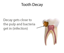
Pulpitis is inflammation of dental pulp tissue. The pulp contains the blood vessels, the nerves, and connective tissue inside a tooth and provides the tooth's blood and nutrients. Pulpitis is mainly caused by bacterial infection which itself is a secondary development of caries. It manifests itself in the form of a toothache.

<span class="mw-page-title-main">Dentine bonding agents</span>

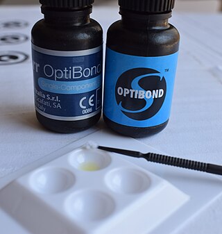
Also known as a "bonderizer" bonding agents are resin materials used to make a dental composite filling material adhere to both dentin and enamel.

<span class="mw-page-title-main">Temporary crown</span>

A temporary crown is a temporary (short-term) crown used in dentistry. Like other interim restorations, it serves until a final (definitive) restoration can be inserted. Usually the temporary crown is constructed from acrylic resins (monomethacrylate-based/polymethacrylate-based) or, chemical-cure/light cure composite (dimethacrylate-based), although alternative systems using aluminium crown forms are occasionally used. Temporary crowns function to protect the tooth, prevent teeth shifting, provide cosmetics, shape the gum tissue properly, and prevent sensitivity.

<span class="mw-page-title-main">Dentinogenesis imperfecta</span> Medical condition

Dentinogenesis imperfecta (DI) is a genetic disorder of tooth development. It is inherited in an autosomal dominant pattern, as a result of mutations on chromosome 4q21, in the dentine sialophosphoprotein gene (DSPP). It is one of the most frequently occurring autosomal dominant features in humans. Dentinogenesis imperfecta affects an estimated 1 in 6,000-8,000 people.

Dentin hypersensitivity is dental pain which is sharp in character and of short duration, arising from exposed dentin surfaces in response to stimuli, typically thermal, evaporative, tactile, osmotic, chemical or electrical; and which cannot be ascribed to any other dental disease.

<span class="mw-page-title-main">Dentin dysplasia</span> Medical condition

Dentin dysplasia (DD) is a rare genetic developmental disorder affecting dentine production of the teeth, commonly exhibiting an autosomal dominant inheritance that causes malformation of the root. It affects both primary and permanent dentitions in approximately 1 in every 100,000 patients. It is characterized by presence of normal enamel but atypical dentin with abnormal pulpal morphology. Witkop in 1972 classified DD into two types which are Type I (DD-1) is the radicular type, and type II (DD-2) is the coronal type. DD-1 has been further divided into 4 different subtypes (DD-1a,1b,1c,1d) based on the radiographic features.

<span class="mw-page-title-main">Root canal treatment</span> Dental treatment

Root canal treatment is a treatment sequence for the infected pulp of a tooth which is intended to result in the elimination of infection and the protection of the decontaminated tooth from future microbial invasion. Root canals, and their associated pulp chamber, are the physical hollows within a tooth that are naturally inhabited by nerve tissue, blood vessels and other cellular entities. Together, these items constitute the dental pulp.

In dentistry, the hydrodynamic or fluid movement theory is one of three main theories developed to explain dentine hypersensitivity, which is a sharp, transient pain arising from stimuli exposure. It states that different types of stimuli act on exposed dentine, causing increased fluid flow through the dentinal tubules. In response to this movement, mechanoreceptors on the pulp nerves trigger the acute, temporary pain of dentine hypersensitivity.

Mineral trioxide aggregate (MTA) was developed for use as a dental root repair material by Mahmoud Torabinejad. It is formulated from commercial Portland cement, combined with bismuth oxide powder for radio-opacity. MTA is used for creating apical plugs during apexification, repairing root perforations during root canal therapy, and treating internal root resorption. This can be used for root-end filling material and as pulp capping material. Originally, MTA was dark gray in color, but white versions have been on the market since 2002.

<span class="mw-page-title-main">Pulpotomy</span>

Pulpotomy is a minimally invasive procedure performed in children on a primary tooth with extensive caries but without evidence of root pathology. The minimally invasive endodontic techniques of vital pulp therapy (VPT) are based on improved understanding of the capacity of pulp (nerve) tissues to heal and regenerate plus the availability of advanced endodontic materials. During the caries removal, this results in a carious or mechanical pulp exposure (bleeding) from the cavity. During pulpotomy, the inflamed/diseased pulp tissue is removed from the coronal pulp chamber of the tooth leaving healthy pulp tissue which is dressed with a long-term clinically-successful medicament that maintains the survival of the pulp and promotes repair. There are various types of medicament placed above the vital pulp such as Buckley's Solution of formocresol, ferric sulfate, calcium hydroxide or mineral trioxide aggregate (MTA). MTA is a more recent material used for pulpotomies with a high rate of success, better than formocresol or ferric sulfate. It is also recommended to be the preferred pulpotomy agent in the future. After the coronal pulp chamber is filled, the tooth is restored with a filling material that seals the tooth from microleakage, such as a stainless steel crown which is the most effective long-term restoration. However, if there is sufficient remaining supporting tooth structure, other filling materials such as amalgam or composite resin can provide a functional alternative when the primary tooth has a life span of two years or less. The medium- to long-term treatment outcomes of pulpotomy in symptomatic permanent teeth with caries, especially in young people, indicate that pulpotomy can be a potential alternative to root canal therapy (RCT).

<span class="mw-page-title-main">Tooth resorption</span> Medical condition

Resorption of the root of the tooth, or root resorption, is the progressive loss of dentin and cementum by the action of odontoclasts. Root resorption is a normal physiological process that occurs in the exfoliation of the primary dentition. However, pathological root resorption occurs in the permanent or secondary dentition and sometimes in the primary dentition.

Endodontic files and reamers are surgical instruments used by dentists when performing root canal treatment. These tools are used to clean and shape the root canal, with the concept being to perform complete chemomechanical debridement of the root canal to the length of the apical foramen. Preparing the canal in this way facilitates the chemical disinfection to a satisfactory length but also provides a shape conducive to obturation.

<span class="mw-page-title-main">Pulp stone</span>

Pulp stones are nodular, calcified masses appearing in either or both the coronal and root portion of the pulp organ in teeth. Pulp stones are not painful unless they impinge on nerves.

<span class="mw-page-title-main">Regenerative endodontics</span> Dental specialty

Regenerative endodontic procedures is defined as biologically based procedures designed to replace damaged structures such as dentin, root structures, and cells of the pulp-dentin complex. This new treatment modality aims to promote normal function of the pulp. It has become an alternative to heal apical periodontitis. Regenerative endodontics is the extension of root canal therapy. Conventional root canal therapy cleans and fills the pulp chamber with biologically inert material after destruction of the pulp due to dental caries, congenital deformity or trauma. Regenerative endodontics instead seeks to replace live tissue in the pulp chamber. The ultimate goal of regenerative endodontic procedures is to regenerate the tissues and the normal function of the dentin-pulp complex.

<span class="mw-page-title-main">Pulp capping</span>

Pulp capping is a technique used in dental restorations to prevent the dental pulp from necrosis, after being exposed, or nearly exposed during a cavity preparation, from a traumatic injury, or by a deep cavity that reaches the center of the tooth causing the pulp to die. When dental caries is removed from a tooth, all or most of the infected and softened enamel and dentin are removed. This can lead to the pulp of the tooth either being exposed or nearly exposed which causes pulpitis (inflammation). Pulpitis, in turn, can become irreversible, leading to pain and pulp necrosis, and necessitating either root canal treatment or extraction. The ultimate goal of pulp capping or stepwise caries removal is to protect a healthy dental pulp and avoid the need for root canal therapy.

Biofilling, also known as orthograde canal grafting technique or 4D sealing, is an endodontic root canal obturation technique with a Bioceramic material after root canal preparation and enlargement procedure.

Apexification is a method of dental treatment to induce a calcific barrier in a root with incomplete formation or open apex of a tooth with necrotic pulp. Pulpal involvement usually occurs as a consequence of trauma or caries involvement of young or immature permanent teeth. As a sequelae of untreated pulp involvement, loss of pulp vitality or necrotic pulp took place for the involved teeth.

References

  1. 1 2 Brännström, Martin; Johnson, Gunilla (April 1974). "Effects of various conditioners and cleaning agents on prepared dentin surfaces: A scanning electron microscopic investigation". The Journal of Prosthetic Dentistry. 31 (4): 422–430. doi:10.1016/0022-3913(74)90152-8. ISSN   0022-3913. PMID   4592745.
  2. Watts, D.; Silikas, N. (2005), "In Situ Photo-Polymerisation and Polymerisation-Shrinkage Phenomena", Dental Hard Tissues and Bonding, Springer Berlin Heidelberg, pp. 123–154, doi:10.1007/3-540-28559-8_6, ISBN   978-3-540-23408-1
  3. McComb, Dorothy; Smith, Dennis C. (July 1975). "A preliminary scanning electron microscopic study of root canals after endodontic procedures". Journal of Endodontics. 1 (7): 238–242. doi:10.1016/s0099-2399(75)80226-3. ISSN   0099-2399. PMID   1061799.
  4. 1 2 3 Mader, Carson L.; Baumgartner, J. Craig; Peters, Donald D. (October 1984). "Scanning electron microscopic investigation of the smeared layer on root canal walls". Journal of Endodontics. 10 (10): 477–483. doi:10.1016/s0099-2399(84)80204-6. ISSN   0099-2399. PMID   6593410.
  5. Şen, B. H.; Wesselink, P. R.; Türkün, M. (1995). "The smear layer: a phenomenon in root canal therapy". International Endodontic Journal. 28 (3): 141–148. doi:10.1111/j.1365-2591.1995.tb00289.x. ISSN   1365-2591. PMID   8626198.
  6. 1 2 Zargar, Nazanin; Dianat, Omid; Asnaashari, Mohammad; Ganjali, Mojtaba; Zadsirjan, Saeede (2015). "The Effect of Smear Layer on Antimicrobial Efficacy of Three Root Canal Irrigants". Iranian Endodontic Journal. 10 (3): 179–183. doi:10.7508/iej.2015.03.007. ISSN   1735-7497. PMC   4509126 . PMID   26213540.
  7. 1 2 3 4 Bindra. "A scanning electron microscope evaluation of smear layer removal from root canals prepared by manual or rotary instrumentation using three different irrigating systems: An in vitro study". www.endodontologyonweb.org. Retrieved 2020-03-03.
  8. Olgart L, Brännström M, Johnson G. Invasion of bacteria into dentinal tubules: Experiments in vivo and Invitro. Acta Odontologica Scandinavica 1974; 32:61-70.
  9. Pashley DH, Michelich V, Kehl T. Dentin permeability: effects of smear layer removal. J Prosthet Dent 1981; 46:531-7.
  10. Gettleman BH, Messer HH, ElDeeb ME. Adhesion of sealer cements to dentin with and without the smear layer. J Endod 1991; 17:15-20.
  11. 1 2 3 McCabe, J (2008). Applied Dental Materials 9th Edition. pp. Chapter 30.
  12. Goldberg, Fernando; Abramovich, Abraham (1977-03-01). "Analysis of the effect of EDTAC on the dentinal walls of the root canal". Journal of Endodontics. 3 (3): 101–105. doi:10.1016/S0099-2399(77)80203-3. ISSN   0099-2399. PMID   404380.
  13. Pashley, D. H. (1996). "Dynamics of the pulpo-dentin complex". Critical Reviews in Oral Biology and Medicine. 7 (2): 104–133. doi:10.1177/10454411960070020101. ISSN   1045-4411. PMID   8875027.
  14. Love, R. M.; Jenkinson, H. F. (2002). "Invasion of dentinal tubules by oral bacteria". Critical Reviews in Oral Biology and Medicine. 13 (2): 171–183. doi: 10.1177/154411130201300207 . ISSN   1045-4411. PMID   12097359.
  15. McComb, D.; Smith, D. C. (July 1975). "A preliminary scanning electron microscopic study of root canals after endodontic procedures". Journal of Endodontics. 1 (7): 238–242. doi:10.1016/S0099-2399(75)80226-3. ISSN   0099-2399. PMID   1061799.
  16. Saunders, W. P.; Saunders, E. M. (September 1992). "The effect of smear layer upon the coronal leakage of gutta-percha fillings and a glass ionomer sealer". International Endodontic Journal. 25 (5): 245–249. doi:10.1111/j.1365-2591.1992.tb01157.x. ISSN   0143-2885. PMID   1291521.
  17. Saunders, Saunders, WP, WE (1992). A Concise Guide to Endodontic Procedures. pp. Chapter 7.
  18. Haapasalo, M.; Shen, Y.; Wang, Z.; Gao, Y. (March 2014). "Irrigation in endodontics". British Dental Journal. 216 (6): 299–303. doi:10.1038/sj.bdj.2014.204. ISSN   1476-5373. PMID   24651335. S2CID   82082.
  19. 1 2 3 Chandler, Nicholas; Chellappa, Deepak (2019). "Lubrication during root canal treatment". Australian Endodontic Journal. 45 (1): 106–110. doi: 10.1111/aej.12282 . ISSN   1747-4477. PMID   30105836.
  20. Haapasalo, Markus; Qian, Wei; Shen, Ya (2012). "Irrigation: beyond the smear layer". Endodontic Topics. 27 (1): 35–53. doi:10.1111/etp.12030. ISSN   1601-1546.
  21. 1 2 3 4 Gokce, Kagan; Aykor, Arzu; Ersoy, Mustafa; Ozel, Emre; Soyman, Mubin (October 2008). "Effect of phosphoric acid etching and self-etching primer application methods on dentinal shear bond strength". The Journal of Adhesive Dentistry. 10 (5): 345–349. ISSN   1461-5185. PMID   19058679.
  22. Clark-Holke D, Drake D, Walton R, Rivera E, Guthmiller JM. Bacterial penetration through canals of endodontically treated teeth in the presence or absence of the smear layer. J Dent 2003; 31:275-281.
  23. Kokkas AB, Boutsioukis ACh, Vassiliadis LP, Stavrianos CK. The influence of the smear layer on dentinal tubule penetration depth by three different root canal sealers. J Endod 2004; 30:100-102.
  24. Çobankara FK, Adanir N, Sema Belli. Evaluation of the influence of smear layer on the apical and coronal sealing ability of two sealers. J Endod 2004; 30:406-409.
  25. Bertacci A, Baroni C, Breschi L, Venturi M, Prati C. The influence of smear layer in lateral channels filling. Clin Oral Investig 2007; 11:353-359.
  26. Yildirim T, Oruçoğlu H, Cobankara FK. Long-term evaluation of the influence of smear layer on the apical sealing ability of MTA. J Endod 2008; 34:1537-1540.
  27. Saleh IM, Ruyter IE, Haapasalo M, Ørstavik D. Bacterial penetration along different root canal filling materials in the presence or absence of smear layer. Int Endod J. 2008; 41:32-40.
  28. Fachin EV, Scarparo RK, Massoni LI. Influence of smear layer removal on the obturation of root canal ramifications. J Appl Oral Sci 2009; 17:240-243.