Spinal cord injury research

Last updated

Spinal cord injury research seeks new ways to cure or treat spinal cord injury in order to lessen the debilitating effects of the injury in the short or long term. There is no cure for SCI, and current treatments are mostly focused on spinal cord injury rehabilitation and management of the secondary effects of the condition. [1] Two major areas of research include neuroprotection, ways to prevent damage to cells caused by biological processes that take place in the body after the injury, and neuroregeneration, regrowing or replacing damaged neural circuits.

Contents

Pathophysiology

Secondary injury takes place minutes to weeks after the initial insult and includes a number of cascading processes that further harm tissues already damaged by the primary injury. [2] It results in formation of a glial scar, which impedes axonal growth. [2] Secondary injuries can occur from different forms of stress added to the spinal cord in forms such as additional contusions, compressions, kinking, or stretching of the spinal cord.

[3]

Complications from a secondary SCI are a result of a homeostatic imbalance potentially leading to metabolic and hemostatic changes from an inflammatory response. Potential immediate effects of secondary SCI include neuronal injury, neuroinflammation, breakdown of blood-spinal cord barrier (BSCB), ischemic dysfunction, oxidative stress, and daily-life function complications.

Animal models

Animals used as SCI model organisms in research include mice, rats, cats, dogs, pigs, and non-human primates; the latter are close to humans but raise ethical concerns about primate experimentation. [1] Special devices exist to deliver blows of specific, monitored force to the spinal cord of an experimental animal. [1] There are various mechanical impact classifications of these injuries that can be replicated in an animal model. This includes contusion, compression, collagenase and ischemia reperfusion, distraction, dislocation, and transection.

Limitations of these model experiments are common. For instance, ischemia-reperfusion SCI involves the interruption of blood flow to the spinal cord. Complications have been observed to arise in animal models from the need to cross clamp the aorta.

Epidural cooling saddles, surgically placed over acutely traumatized spinal cord tissue, have been used to evaluate potentially beneficial effects of localized hypothermia, with and without concomitant glucocorticoids. [4] [5]

Surgery

Surgery is currently used to provide stability to the injured spinal column or to relieve pressure from the spinal cord. [1] [6] How soon after injury to perform decompressive surgery is a controversial topic, and it has been difficult to prove that earlier surgery provides better outcomes in human trials. [1] Some argue that early surgery might further deprive an already injured spinal cord of oxygen, but most studies show no difference in outcomes between early (within three days) and late surgery (after five days), and some show a benefit to earlier surgery. [7]

In 2014 Darek Fidyka underwent pioneering spinal surgery that used nerve grafts, from his ankle, to 'bridge the gap' in his severed spinal cord and olfactory ensheathing cells (OECs) to stimulate the spinal cord cells. The surgery was performed in Poland in collaboration with Prof. Geoff Raisman, chair of neural regeneration at University College London's Institute of Neurology, and his research team. The OECs were taken from the patient's olfactory bulbs in his brain and then grown in the lab, these cells were then injected above and below the impaired spinal tissue. [8]

Neuroprotection

Neuroprotection aims to prevent the harm that occurs from secondary injury. [2] One example is to target the protein calpain which appears to be involved in apoptosis; inhibiting the protein has produced improved outcomes in animal trials. [2] Iron from blood damages the spinal cord through oxidative stress, so one option is to use a chelation agent to bind the iron; animals treated this way have shown improved outcomes. [2] Free radical damage by reactive oxygen species (ROS) is another therapeutic target that has shown improvement when targeted in animals. [2] One antibiotic, minocycline, is under investigation in human trials for its ability to reduce free radical damage, excitotoxicity, disruption of mitochondrial function, and apoptosis. [2] Riluzole, an anticonvulsant, is also being investigated in clinical trials for its ability to block sodium channels in neurons, which could prevent damage by excitotoxicity. [2] Other potentially neuroprotective agents under investigation in clinical trials include cethrin, erythropoietin, and dalfampridine. [2]

Hypothermia

One experimental treatment, therapeutic hypothermia, is used in treatment but there is no evidence that it improves outcomes. [9] [10] Some experimental treatments, including systemic hypothermia, have been performed in isolated cases in order to draw attention to the need for further preclinical and clinical studies to help clarify the role of hypothermia in acute spinal cord injury. [11] Despite limited funding, a number of experimental treatments such as local spine cooling and oscillating field stimulation have reached controlled human trials. [12] [13]

Methylprednisolone

Inflammation and glial scar are considered important inhibitory factors to neuroregeneration after SCI. However, aside from methylprednisolone, none of these developments have reached even limited use in the clinical care of human spinal cord injury in the US. [14] Methylprednisolone can be given shortly after the injury but evidence for harmful side effects outweighs that for a benefit. [6] Research is being done into more efficient delivery mechanisms for methylprednisolone that would reduce its harmful effects. [1]

Neuroregeneration

Neuroregeneration aims to reconnect the broken circuits in the spinal cord to allow function to return. [2] One way is to regrow axons, which occurs spontaneously in the peripheral nervous system. However, myelin in the central nervous system contains molecules that impede axonal growth; thus, these factors are a target for therapies to create an environment conducive to growth. [2] One such molecule is Nogo-A, a protein associated with myelin. When this protein is targeted with inhibitory antibodies in animal models, axons grow better and functional recovery is improved. [2]

Stem cells

Stem cells are cells that can differentiate to become different types of cells. [15] The hope is that stem cells transplanted into an injured area of the spinal cord will allow neuroregeneration. [6] Types of cells being researched for use in SCI include embryonic stem cells, neural stem cells, mesenchymal stem cells, olfactory ensheathing cells, Schwann cells, activated macrophages, and induced pluripotent stem cells. [1] When stem cells are injected in the area of damage in the spinal cord, they secrete neurotrophic factors, and these factors help neurons and blood vessels to grow, thus helping repair the damage. [16] [17] [18] It is also necessary to recreate an environment in which stem cells will grow. [19]

An ongoing Phase 2 trial in 2016 presented data [20] showing that after 90 days of treatment with oligodendrocyte progenitor cells derived from embryonic stem cells, 4 out of 4 subjects with complete cervical injuries had improved motor levels, with 2 of 4 improving two motor levels (on at least one side, with one patient improving two motor levels on both sides). The trial's original endpoint had been 2/5 patients improving two levels on one side within 6–12 months. All 8 cervical subjects in this Phase 1–2 trial had exhibited improved upper extremity motor scores (UEMS) relative to baseline with no serious adverse side effects, and a 2010 Phase 1 trial in 5 thoracic patients has found no safety issues after 5–6 years of follow-up.

Six-month efficacy data is expected in January 2017; meanwhile, a higher dose is being investigated and the study is now also recruiting patients with incomplete injuries. [21]

In 2022, a team reported the first [22] engineered functional human (motor-)neuronal networks derived from induced pluripotent stem cells (iPSCs) from the patient for implantation to regenerate injured spinal cord that shows success in tests with mice. [23] [24]

Embryonic stem cells

Human embryonic stem cells in cell culture Humanstemcell.JPG
Human embryonic stem cells in cell culture

Embryonic stem cells (ESCs) are pluripotent; they can develop into every type of cell in an organism, such as oligodendrocytes. . [6] Oligodendrocytes and motor neurons have been predicted to be a favorable target for ESCs regarding treating neurological disorders and traumas. [25] After a SCI takes place there is evidence of oligodendrocytes degradation, eventually leading to cell death. This results in a lack of myelination, which enhances signals sent amongst neurons, causing disfunction in signaling. A potential solution could be hESC-derived oligodendrocyte transplantation; however, the success of this process depends off the cell’s ability to differentiate towards neural cell types in vitro. This is where more testing and research is being conducted using animal models.   

Neural stem cells

Neural stem cells (NSCs) are multipotent; they can differentiate into different kinds of neural cells, either neurons or glia, namely oligodendrocytes and astrocytes. [15] The hope is that these cells when injected into an injured spinal cord will replace killed neurons and oligodendrocytes and secrete factors that support growth. [1] However they may fail to differentiate into neurons when transplanted, either remaining undifferentiated or becoming glia. [15] A phase I/II clinical trials implanting NSCs into humans with SCI began in 2011 [1] and ended in June 2015. [26]

Mesenchymal stem cells

Mesenchymal stem cells do not need to come from fetuses, so avoid difficulties around ethics; they come from tissues including bone marrow, adipose tissue, the umbilical cord. [1] Unlike other types of stem cells, mesenchymal cells do not present the threat of tumor formation or triggering an immune system response. [1] Animal studies with injection of bone marrow stem cells have shown improvement in motor function; however not so in a human trial a year post-injury. [1] More trials are underway. [1] Adipose and umbilical tissue stem cells need further study before human trials can be performed, but two Korean studies were begun to investigate adipose cells in SCI patients. [1]

Olfactory ensheathing cells

Transplantation of tissues such as olfactory ensheathing cells from the olfactory bulbs has been shown to produce beneficial effects in spinal cord injured rats. [27] Trials have also begun to show success when olfactory ensheathing cells are transplanted into humans with severed spinal cords. [28] People have recovered sensation, use of formerly paralysed muscles, and bladder and bowel function after the surgeries, [29] eg Darek Fidyka.

Induced pluripotent stem cells

Japanese researchers in 2006 discovered that adding certain transcription factors to cells caused them to become pluripotent and able to differentiate into multiple cell types. [6] This way a patient's own tissues could be used, theoretically because of a reduced chance of transplant rejection. [6]

Engineering approaches

Recent approaches have used various engineering techniques to improve spinal cord injury repair. Use of biomaterials is an engineering approach to SCI treatment that can be combined with stem cell transplantation. [6] They can help deliver cells to the injured area and create an environment that fosters their growth. [6] The general hypothesis behind engineered biomaterials is that bridging the lesion site using a growth permissive scaffold may help axons grow and thereby improve function. The biomaterials used must be strong enough to provide adequate support but soft enough not to compress the spinal cord. [2] They must degrade over time to make way for the body to regrow tissue. [2] Engineered treatments do not induce an immune response as biological treatments may, and they are easily tunable and reproducible. In-vivo administration of hydrogels or self-assembling nanofibers has been shown to promote axonal sprouting and partial functional recovery. [30] [31] In addition, administration of carbon nanotubes has shown to increase motor axon extension and decrease the lesion volume, without inducing neuropathic pain. [32] In addition, administration of poly-lactic acid microfibers has shown that topographical guidance cues alone can promote axonal regeneration into the injury site. [33] However, all of these approaches induced modest behavioral or functional recovery suggesting that further investigation is necessary.

Hydrogels

Hydrogels are structures made of polymers that are designed to be similar to the natural extracellular matrix around cells. [2] They can be used to help deliver drugs more efficiently to the spinal cord and to support cells, and they can be injected into an injured area to fill a lesion. [2] They can be implanted into a lesion site with drugs or growth factors in them to give the chemicals the best access to the damaged area and to allow sustained release. [2]

Bionanoengineered scaffolds

In November 2021, a novel therapy for spinal cord injury was reported – an injectable gel of nanofibers that mimic the matrix around cells and contain molecules that were engineered to wiggle. These moving molecules connect with receptors of cells, causing repair signals inside – in particular, leading to relatively higher vascular growth, axonal regeneration, myelination, survival of motor neurons, reduced gliosis, and functional recovery – enabling paralyzed mice to walk again. [34] [35] [36]

Exoskeletons

The technology for creating powered exoskeletons, wearable machinery to assist with walking movements, is currently making significant advances. There are products available, such as the Ekso, which allows individuals with up to a C7 complete (or any level of incomplete) spinal injury to stand upright and make technologically assisted steps. [37] The initial purpose for this technology is for functional based rehabilitation, but as the technology develops, so will its uses. [37]

Functional electrical stimulation (FES) uses coordinated electric shocks to muscles to cause them to contract in a walking pattern. [38] While it can strengthen muscles, a significant downside for the users of FES is that their muscles tire after a short time and distance. [38] One research direction combines FES with exoskeletons to minimize the downsides of both technologies, supporting the person's joints and using the muscles to reduce the power needed from the machine, and thus its weight. [38] A research team at the McKelvey School of Engineering at Washington University in St. Louis, led by assistant professor of biomedical engineering Ismael Seáñez, is launching a clinical trial of electrical spinal cord stimulation for helping restore movement in movement-impaired or paralyzed patients. [39]

Brain–computer interface

Recent research shows that combining brain–computer interface and functional electrical stimulation can restore voluntary control of paralyzed muscles. A study with monkeys showed that it is possible to directly use commands from the brain, bypassing the spinal cord and enable limited hand control and function. [40]

Brain-machine interface

A 2016 study developed by the Walk Again Project with eight paraplegics demonstrated neurological recovernment with the use of therapies based upon BMI, virtual reality an the use of robots. One patient became able to walk with supports after a decade paralised and another became able to carry out a pregnancy. [41] [42] [43] [44] [45]

Spinal cord implants

Spinal cord implants, such as e-dura implants, designed for implantation on the surface of the spinal cord, are being studied for paralysis following a spinal cord injury. [46]

E-dura implants are designed using methods of soft neurotechnology, in which electrodes and a microfluidic delivery system are distributed along the spinal implant. [47] Chemical stimulation of the spinal cord is administered through the microfluidic channel of the e-dura. The e-dura implants, unlike previous surface implants, closely mimic the physical properties of living tissue and can deliver electric impulses and pharmacological substances simultaneously. Artificial dura mater was constructed through the utilization of PDMS and gelatin hydrogel. [47] The hydrogel simulates spinal tissue and a silicone membrane simulates the dura mater. These properties allow the e-dura implants to sustain long-term application to the spinal cord and brain without leading to inflammation, scar tissue buildup, and rejection normally caused by surface implants rubbing against nerve tissue.

In 2018 two distinct research teams from Minnesota's Mayo Clinic and Kentucky's University of Louisville managed to restore some mobility to patients suffering from paraplegia with an electronic spinal cord stimulator. The theory behind the new spinal cord stimulator is that in certain cases of spinal cord injury the spinal nerves between the brain and the legs are still alive, but just dormant. [48] On 1 November 2018 a third distinct research team from the University of Lausanne published similar results with a similar stimulation technique in the journal Nature . [49] [50] In 2022, researchers demonstrated a spinal cord stimulator that enabled patients with spinal cord injury to walk again via epidural electrical stimulation (EES) with substantial neurorehabilitation-progress during the first day. [51] [52] In a study published in May 2023 in the journal Nature , researchers in Switzerland described implants which allowed a 40-year old man, paralyzed from the hips down for 12 years, to stand, walk and ascend a steep ramp with only the assistance of a walker. More than a year after the implant was inserted, he has retained these abilities and was walking with crutches even when the implant was switched off. [53]

Related Research Articles

<span class="mw-page-title-main">Central nervous system</span> Brain and spinal cord

The central nervous system (CNS) is the part of the nervous system consisting of the brain and spinal cord, the retina and optic nerve, and the olfactory nerve and epithelia. The CNS is so named because the brain integrates the received information and coordinates and influences the activity of all parts of the bodies of bilaterally symmetric and triploblastic animals—that is, all multicellular animals except sponges and diploblasts. It is a structure composed of nervous tissue positioned along the rostral to caudal axis of the body and may have an enlarged section at the rostral end which is a brain. Only arthropods, cephalopods and vertebrates have a true brain, though precursor structures exist in onychophorans, gastropods and lancelets.

<span class="mw-page-title-main">Schwann cell</span> Glial cell type

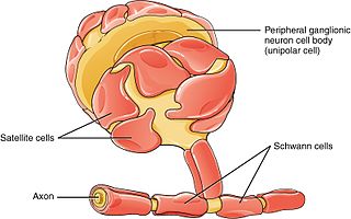
Schwann cells or neurolemmocytes are the principal glia of the peripheral nervous system (PNS). Glial cells function to support neurons and in the PNS, also include satellite cells, olfactory ensheathing cells, enteric glia and glia that reside at sensory nerve endings, such as the Pacinian corpuscle. The two types of Schwann cells are myelinating and nonmyelinating. Myelinating Schwann cells wrap around axons of motor and sensory neurons to form the myelin sheath. The Schwann cell promoter is present in the downstream region of the human dystrophin gene that gives shortened transcript that are again synthesized in a tissue-specific manner.

Myelitis is inflammation of the spinal cord which can disrupt the normal responses from the brain to the rest of the body, and from the rest of the body to the brain. Inflammation in the spinal cord can cause the myelin and axon to be damaged resulting in symptoms such as paralysis and sensory loss. Myelitis is classified to several categories depending on the area or the cause of the lesion; however, any inflammatory attack on the spinal cord is often referred to as transverse myelitis.

<span class="mw-page-title-main">Paraplegia</span> Impairment of motor and sensory functions in the lower limbs

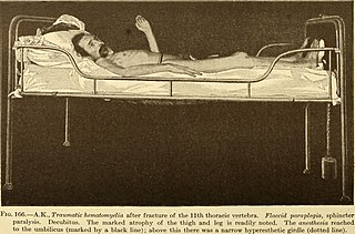
Paraplegia, or paraparesis, is an impairment in motor or sensory function of the lower extremities. The word comes from Ionic Greek (παραπληγίη) "half-stricken". It is usually caused by spinal cord injury or a congenital condition that affects the neural (brain) elements of the spinal canal. The area of the spinal canal that is affected in paraplegia is either the thoracic, lumbar, or sacral regions. If four limbs are affected by paralysis, tetraplegia or quadriplegia is the correct term. If only one limb is affected, the correct term is monoplegia. Spastic paraplegia is a form of paraplegia defined by spasticity of the affected muscles, rather than flaccid paralysis.

<span class="mw-page-title-main">Functional electrical stimulation</span> Technique that uses low-energy electrical pulses

Functional electrical stimulation (FES) is a technique that uses low-energy electrical pulses to artificially generate body movements in individuals who have been paralyzed due to injury to the central nervous system. More specifically, FES can be used to generate muscle contraction in otherwise paralyzed limbs to produce functions such as grasping, walking, bladder voiding and standing. This technology was originally used to develop neuroprostheses that were implemented to permanently substitute impaired functions in individuals with spinal cord injury (SCI), head injury, stroke and other neurological disorders. In other words, a person would use the device each time he or she wanted to generate a desired function. FES is sometimes also referred to as neuromuscular electrical stimulation (NMES).

<span class="mw-page-title-main">Embryonic stem cell</span> Type of pluripotent blastocystic stem cell

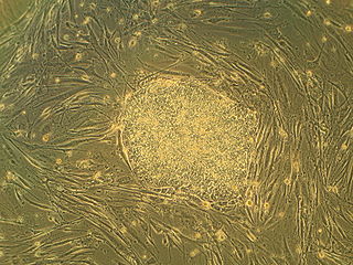
Embryonic stem cells (ESCs) are pluripotent stem cells derived from the inner cell mass of a blastocyst, an early-stage pre-implantation embryo. Human embryos reach the blastocyst stage 4–5 days post fertilization, at which time they consist of 50–150 cells. Isolating the inner cell mass (embryoblast) using immunosurgery results in destruction of the blastocyst, a process which raises ethical issues, including whether or not embryos at the pre-implantation stage have the same moral considerations as embryos in the post-implantation stage of development.

<span class="mw-page-title-main">Spinal cord injury</span> Injury to the main nerve bundle in the back of humans

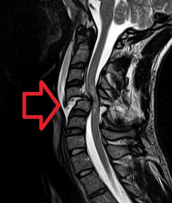
A spinal cord injury (SCI) is damage to the spinal cord that causes temporary or permanent changes in its function. It is a destructive neurological and pathological state that causes major motor, sensory and autonomic dysfunctions.

Neural engineering is a discipline within biomedical engineering that uses engineering techniques to understand, repair, replace, or enhance neural systems. Neural engineers are uniquely qualified to solve design problems at the interface of living neural tissue and non-living constructs.

<span class="mw-page-title-main">Adult stem cell</span> Multipotent stem cell in the adult body

Adult stem cells are undifferentiated cells, found throughout the body after development, that multiply by cell division to replenish dying cells and regenerate damaged tissues. Also known as somatic stem cells, they can be found in juvenile, adult animals, and humans, unlike embryonic stem cells.

Stem-cell therapy uses stem cells to treat or prevent a disease or condition. As of 2016, the only established therapy using stem cells is hematopoietic stem cell transplantation. This usually takes the form of a bone marrow transplantation, but the cells can also be derived from umbilical cord blood. Research is underway to develop various sources for stem cells as well as to apply stem-cell treatments for neurodegenerative diseases and conditions such as diabetes and heart disease.

Neuroepithelial cells, or neuroectodermal cells, form the wall of the closed neural tube in early embryonic development. The neuroepithelial cells span the thickness of the tube's wall, connecting with the pial surface and with the ventricular or lumenal surface. They are joined at the lumen of the tube by junctional complexes, where they form a pseudostratified layer of epithelium called neuroepithelium.

Neural stem cells (NSCs) are self-renewing, multipotent cells that firstly generate the radial glial progenitor cells that generate the neurons and glia of the nervous system of all animals during embryonic development. Some neural progenitor stem cells persist in highly restricted regions in the adult vertebrate brain and continue to produce neurons throughout life. Differences in the size of the central nervous system are among the most important distinctions between the species and thus mutations in the genes that regulate the size of the neural stem cell compartment are among the most important drivers of vertebrate evolution.

<span class="mw-page-title-main">Floor plate</span> Embryonic structure

The floor plate is a structure integral to the developing nervous system of vertebrate organisms. Located on the ventral midline of the embryonic neural tube, the floor plate is a specialized glial structure that spans the anteroposterior axis from the midbrain to the tail regions. It has been shown that the floor plate is conserved among vertebrates, such as zebrafish and mice, with homologous structures in invertebrates such as the fruit fly Drosophila and the nematode C. elegans. Functionally, the structure serves as an organizer to ventralize tissues in the embryo as well as to guide neuronal positioning and differentiation along the dorsoventral axis of the neural tube.

Neuroregeneration involves the regrowth or repair of nervous tissues, cells or cell products. Neuroregenerative mechanisms may include generation of new neurons, glia, axons, myelin, or synapses. Neuroregeneration differs between the peripheral nervous system (PNS) and the central nervous system (CNS) by the functional mechanisms involved, especially in the extent and speed of repair. When an axon is damaged, the distal segment undergoes Wallerian degeneration, losing its myelin sheath. The proximal segment can either die by apoptosis or undergo the chromatolytic reaction, which is an attempt at repair. In the CNS, synaptic stripping occurs as glial foot processes invade the dead synapse.

Neural tissue engineering is a specific sub-field of tissue engineering. Neural tissue engineering is primarily a search for strategies to eliminate inflammation and fibrosis upon implantation of foreign substances. Often foreign substances in the form of grafts and scaffolds are implanted to promote nerve regeneration and to repair damage caused to nerves of both the central nervous system (CNS) and peripheral nervous system (PNS) by an injury.

<span class="mw-page-title-main">Olfactory ensheathing cell</span> Type of macroglia that ensheath unmyelinated olfactory neurons

Olfactory ensheathing cells (OECs), also known as olfactory ensheathing glia or olfactory ensheathing glial cells, are a type of macroglia found in the nervous system. They are also known as olfactory Schwann cells, because they ensheath the non-myelinated axons of olfactory neurons in a similar way to which Schwann cells ensheath non-myelinated peripheral neurons. They also share the property of assisting axonal regeneration.

Regeneration in humans is the regrowth of lost tissues or organs in response to injury. This is in contrast to wound healing, or partial regeneration, which involves closing up the injury site with some gradation of scar tissue. Some tissues such as skin, the vas deferens, and large organs including the liver can regrow quite readily, while others have been thought to have little or no capacity for regeneration following an injury.

Paweł Tabakow is a Polish neurosurgeon who is known for prepared and performing the operation that allowed Darek Fidyka to recover sensory and motor function after the complete severing of his spinal cord. Tabakow has claimed that an Indian ambassador and other people from round the world have contacted him about performing similar treatments.

Mary Elizabeth Bartlett Bunge was an American neuroscientist who researched a cure for paralysis at the University of Miami, where she was a professor of cell biology.

Nano neuro knitting is an emerging technology for repairing nervous system tissues via nano scaffolding techniques. Currently being explored in numerous research endeavors, nano neuro knitting has been shown to allow partial reinnervation in damaged areas of the nervous system through the interactions between potentially regenerative axons and peptide scaffolds. This interaction has been shown to lead to sufficient axon density renewal to the point that functionality is restored. While nano neuro knitting shows promise, the uncertainty of the effects in human subjects warrants further investigation before clinical trials initiate.

References

  1. 1 2 3 4 5 6 7 8 9 10 11 12 13 14 Silva, N.A.; Sousa, N.; Reis, R.L.; Salgado, A.J. (2014). "From basics to clinical: A comprehensive review on spinal cord injury". Progress in Neurobiology. 114: 25–57. doi:10.1016/j.pneurobio.2013.11.002. PMID   24269804. S2CID   23121381.
  2. 1 2 3 4 5 6 7 8 9 10 11 12 13 14 15 16 17 Kabu, S.; Gao, Y.; Kwon, B.K.; Labhasetwar, V. (2015). "Drug delivery, cell-based therapies, and tissue engineering approaches for spinal cord injury". Journal of Controlled Release. 219: 141–54. doi:10.1016/j.jconrel.2015.08.060. PMC   4656085 . PMID   26343846.
  3. Anwar, M. Akhtar; Al Shehabi, Tuqa S.; Eid, Ali H. (2016). "Inflammogenesis of Secondary Spinal Cord Injury". Frontiers in Cellular Neuroscience. 10: 98. doi: 10.3389/fncel.2016.00098 . ISSN   1662-5102. PMC   4829593 . PMID   27147970.
  4. Kuchner, E. F.; Hansebout, R. R.; Pappius, H. M. (1 October 2000). "Effects of dexamethasone and of local hypothermia on early and late tissue electrolyte changes in experimental spinal cord injury". Journal of Spinal Disorders. 13 (5): 391–398. doi:10.1097/00002517-200010000-00004. ISSN   0895-0385. PMID   11052347.
  5. Kuchner, E. F.; Hansebout, R. R. (1 December 1976). "Combined steroid and hypothermia treatment of experimental spinal cord injury". Surgical Neurology. 6 (6): 371–376. ISSN   0090-3019. PMID   1006512.
  6. 1 2 3 4 5 6 7 8 Assunção-Silva, R.C.; Gomes, E.D.; Sousa, N.; Silva, N.A.; Salgado, A.J. (2015). "Hydrogels and Cell Based Therapies in Spinal Cord Injury Regeneration". Stem Cells International. 2015: 1–24. doi: 10.1155/2015/948040 . PMC   4466497 . PMID   26124844.
  7. Bigelow & Medzon 2011, pp. 176–77.
  8. "Paralysed man Darek Fidyka walks again after pioneering surgery". TheGuardian.com . 20 October 2014.
  9. "Therapeutic Hypothermia: eMedicine Clinical Procedures" . Retrieved 21 February 2011.
  10. "Hypothermia" . Retrieved 21 February 2011.
  11. Cappuccino, Andrew; Bisson, Leslie J.; Carpenter, Bud; Marzo, John; Dietrich Wd, W Dalton; Cappuccino, Helen (2010). "The Use of Systemic Hypothermia for the Treatment of an Acute Cervical Spinal Cord Injury in a Professional Football Player". Spine. 35 (2): E57–62. doi:10.1097/BRS.0b013e3181b9dc28. PMID   20081503. S2CID   12799582.
  12. Hansebout, RR; Tanner, JA; Romero-Sierra, C (1984). "Current status of spinal cord cooling in the treatment of acute spinal cord injury". Spine. 9 (5): 508–11. doi:10.1097/00007632-198407000-00020. PMID   6495017. S2CID   39978864.
  13. Shapiro, Scott; Borgens, Richard; Pascuzzi, Robert; Roos, Karen; Groff, Michael; Purvines, Scott; Rodgers, Richard Ben; Hagy, Shannon; Nelson, Paul (2005). "Oscillating field stimulation for complete spinal cord injury in humans: A Phase 1 trial". Journal of Neurosurgery: Spine. 2 (1): 3–10. doi: 10.3171/spi.2005.2.1.0003 . PMID   15658119.
  14. Cadotte, DW; Fehlings, MG (2011). "Spinal cord injury: A systematic review of current treatment options". Clinical Orthopaedics and Related Research. 469 (3): 732–41. doi:10.1007/s11999-010-1674-0. PMC   3032846 . PMID   21080129.
  15. 1 2 3 Yu, W.Y.; He, D.W. (2015). "Current trends in spinal cord injury repair" (PDF). European Review for Medical and Pharmacological Sciences. 19 (18): 3340–44. PMID   26439026.
  16. Abraham S (March 2008). "Autologous Stem Cell Injections for Spinal Cord Injury – A multicentric Study with 6 month follow up of 108 patients". 7th Annual Meeting of Japanese Society of Regenerative Medicine, Nagoya, Japan.[ verification needed ]
  17. R Ravikumar, S Narayanan and S Abraham (November 2007). "Autologous stem cells for spinal cord injury". Regenerative Medicine. 2 (6): 53–61.[ verification needed ]
  18. Abraham S (June 2007). "Autologous Bone Marrow Mononuclear Cells for spinal cord injury: A case report". Cytotherapy. 9 (1).[ verification needed ]
  19. Office of Communications and Public Liaison, National Institute of Neurological Disorders and Stroke, ed. (2013). Spinal Cord Injury: Hope Through Research. Bethesda, MD: National Institutes of Health. Archived from the original on 19 November 2015.
  20. Wirth, Edward (14 September 2016). "Initial Clinical Trials of hESC-Derived Oligodendrocyte Progenitor Cells in Subacute Spinal Cord Injury" (PDF). ISCoS Meeting presentation. Asterias Biotherapeutics. Retrieved 14 September 2016.
  21. "Asterias Biotherapeutics Announces Positive Efficacy Data in Patients with Complete Cervical Spinal Cord Injuries Treated with AST-OPC1". asteriasbiotherapeutics.com. Retrieved 15 September 2016.
  22. "In world-first, researchers engineer human spinal cord implants for treating paralysis". Tel-Aviv University . Retrieved 10 March 2022.
  23. "Engineered spinal cord implants restore movement to paralysed mice". Physics World. 23 February 2022. Retrieved 10 March 2022.
  24. Wertheim, Lior; Edri, Reuven; Goldshmit, Yona; Kagan, Tomer; Noor, Nadav; Ruban, Angela; Shapira, Assaf; Gat-Viks, Irit; Assaf, Yaniv; Dvir, Tal (7 February 2022). "Regenerating the Injured Spinal Cord at the Chronic Phase by Engineered iPSCs-Derived 3D Neuronal Networks". Advanced Science. 9 (11): 2105694. doi:10.1002/advs.202105694. PMC   9008789 . PMID   35128819. S2CID   246633147.
  25. Ronaghi, Mohammad; Erceg, Slaven; Moreno-Manzano, Victoria; Stojkovic, Miodrag (1 January 2010). "Challenges of Stem Cell Therapy for Spinal Cord Injury: Human Embryonic Stem Cells, Endogenous Neural Stem Cells, or Induced Pluripotent Stem Cells?". Stem Cells . 28 (1). Oxford University Press: 93–99. doi: 10.1002/stem.253 . PMID   19904738. S2CID   206516299.
  26. "CTG Labs - NCBI". 16 June 2015.
  27. Iwatsuki, K.; Yoshimine, T.; Kishima, H.; Aoki, M.; Yoshimura, K.; Ishihara, M.; Ohnishi, Y.; Lima, C. (2008). "Transplantation of olfactory mucosa following spinal cord injury promotes recovery in rats". NeuroReport. 19 (13): 1249–52. doi:10.1097/WNR.0b013e328305b70b. PMID   18695502. S2CID   30934447.
  28. Tabakow, P; Jarmundowicz, W; Czapiga, B; Fortuna, W; Miedzybrodzki, R; Czyz, M; Huber, J; Szarek, D; Okurowski, S; Szewczyk, P; Gorski, A; Raisman, G (2013). "Transplantation of autologous olfactory ensheathing cells in complete human spinal cord injury". Cell Transplantation. 22 (9): 1591–612. doi:10.3727/096368912X663532. PMID   24007776. S2CID   35220044.
  29. Mariano, E.D.; Teixeira, M.J.; Marie, S.K.; Lepski, G. (2015). "Adult stem cells in neural repair: Current options, limitations and perspectives". World Journal of Stem Cells. 7 (2): 477–82. doi: 10.4252/wjsc.v7.i2.477 . PMC   4369503 . PMID   25815131.
  30. Piantino, J.; Burdick, J.; Goldberg, D.; Langer, R.; Benowitz, L. (2006). "An injectable, biodegradable hydrogel for trophic factor delivery enhances axonal rewiring and improves performance after spinal cord injury". Experimental Neurology. 201 (2): 359–67. doi:10.1016/j.expneurol.2006.04.020. PMID   16764857. S2CID   10199210.
  31. Tysseling-Mattiace, V. M.; Sahni, V.; Niece, K. L.; Birch, D.; Czeisler, C.; Fehlings, M. G.; Stupp, S. I.; Kessler, J. A. (2008). "Self-Assembling Nanofibers Inhibit Glial Scar Formation and Promote Axon Elongation after Spinal Cord Injury". Journal of Neuroscience. 28 (14): 3814–23. doi:10.1523/JNEUROSCI.0143-08.2008. PMC   2752951 . PMID   18385339.
  32. Roman, Jose A.; Niedzielko, Tracy L.; Haddon, Robert C.; Parpura, Vladimir; Floyd, Candace L. (2011). "Single-Walled Carbon Nanotubes Chemically Functionalized with Polyethylene Glycol Promote Tissue Repair in a Rat Model of Spinal Cord Injury". Journal of Neurotrauma. 28 (11): 2349–62. doi:10.1089/neu.2010.1409. PMC   3218389 . PMID   21303267.
  33. Hurtado, Andres; Cregg, Jared M.; Wang, Han B.; Wendell, Dane F.; Oudega, Martin; Gilbert, Ryan J.; McDonald, John W. (2011). "Robust CNS regeneration after complete spinal cord transection using aligned poly-l-lactic acid microfibers". Biomaterials. 32 (26): 6068–79. doi:10.1016/j.biomaterials.2011.05.006. PMC   4163047 . PMID   21636129.
  34. "Therapy used on mice may transform spinal injury treatments, say scientists". The Guardian. 11 November 2021. Retrieved 11 December 2021.
  35. University. "'Dancing molecules' successfully repair severe spinal cord injuries in mice". Northwestern University . Retrieved 11 December 2021.
  36. Álvarez, Z.; Kolberg-Edelbrock, A. N.; Sasselli, I. R.; Ortega, J. A.; Qiu, R.; Syrgiannis, Z.; Mirau, P. A.; Chen, F.; Chin, S. M.; Weigand, S.; Kiskinis, E.; Stupp, S. I. (12 November 2021). "Bioactive scaffolds with enhanced supramolecular motion promote recovery from spinal cord injury". Science. 374 (6569): 848–856. Bibcode:2021Sci...374..848A. doi:10.1126/science.abh3602. PMC   8723833 . PMID   34762454.
  37. 1 2 "Ekso Bionics - Pioneers in Wearable Bionic Exoskeleton Suits Since 2005". 26 July 2021.
  38. 1 2 3 del-Ama, A.J.; Koutsou, A.D.; Moreno J.C.; de-los-Reyes, A.; Gil-Agudo, A.; Pons, J.L. (2012). "Review of hybrid exoskeletons to restore gait following spinal cord injury". Journal of Rehabilitation Research and Development. 49 (4): 497–514. doi: 10.1682/JRRD.2011.03.0043 . PMID   22773254.
  39. "New research focuses on restoring movement after spinal cord injury". News-Medical.net. 6 September 2022. Retrieved 7 September 2022.
  40. Ethier, C.; Oby, E.R.; Bauman, M.J.; Miller, L.E. (2012). "Restoration of grasp following paralysis through brain-controlled stimulation of muscles". Nature. 485 (7398): 368–71. Bibcode:2012Natur.485..368E. doi:10.1038/nature10987. PMC   3358575 . PMID   22522928.
  41. Lenharo, Mariana (11 August 2016). "Treino com exoesqueleto levou a recuperação parcial de paraplégicos". Bem Estar (in Brazilian Portuguese). Retrieved 15 May 2024.
  42. Donati, Ana R. C.; Shokur, Solaiman; Morya, Edgard; Campos, Debora S. F.; Moioli, Renan C.; Gitti, Claudia M.; Augusto, Patricia B.; Tripodi, Sandra; Pires, Cristhiane G.; Pereira, Gislaine A.; Brasil, Fabricio L.; Gallo, Simone; Lin, Anthony A.; Takigami, Angelo K.; Aratanha, Maria A. (11 August 2016). "Long-Term Training with a Brain-Machine Interface-Based Gait Protocol Induces Partial Neurological Recovery in Paraplegic Patients". Scientific Reports. 6 (1): 30383. Bibcode:2016NatSR...630383D. doi:10.1038/srep30383. ISSN   2045-2322. PMC   4980986 . PMID   27513629.
  43. Chamary, J. V. (19 August 2016). "Technology Rewires The Brain To Overcome Complete Paralysis". Forbes. Retrieved 15 May 2024.
  44. "Paraplégicos recuperam mobilidade e vida sexual após tratamento com Nicolelis". www.uol.com.br (in Brazilian Portuguese). 11 August 2016. Retrieved 15 May 2024.
  45. Radford, Tim (11 August 2016). "'Brain training' technique restores feeling and movement to paraplegic patients". The Guardian. ISSN   0261-3077 . Retrieved 15 May 2024.
  46. Paddock, Catharine (2015). Soft spinal implants show promise as long-term solution paralysis. Medical News Today . Retrieved 03/09/2015.
  47. 1 2 Minev, I.; Musienko, P.; Hirsch, A.; Barraud, Q.; Wenger, N.; Moraud, E.; Gandar, J.; Capogrosso, M.; Milekovic, T.; Asboth, L.; Torres, R.; Vachicouras, N.; Liu, Q.; Pavlova, N.; Duis, S.; Larmagnac, A.; Voros, J.; Micera, S.; Suo, Z.; Courtine, G.; Lacour, S. (2015). "Electronic dura mater for long-term multimodal neural interfaces" (PDF). Science . 347 (6218): 159–63. Bibcode:2015Sci...347..159M. doi:10.1126/science.1260318. PMID   25574019. S2CID   1941485.
  48. "Spinal implant helps paralyzed patients walk". Deutsche Welle. 24 September 2018. Retrieved 4 October 2018. Spinal cord stimulators and intense physical therapy are helping paraplegic patients relearn how to walk. Spinal cord stimulators can potentially help "wake up" dormant nerves.
  49. Chen, Angus (31 October 2018). "Spinal Stimulator Implant Gives Paralytic Patients a Chance to Regain Movement". Scientific American . Springer Nature . Retrieved 1 November 2018. A new therapy that amplifies nerve impulses may also help the body heal
  50. Wagner, Fabien B. (1 November 2018). "Targeted neurotechnology restores walking in humans with spinal cord injury". Nature . 563 (7729). United Kingdom: Springer Nature: 65–71. Bibcode:2018Natur.563...65W. doi:10.1038/s41586-018-0649-2. PMID   30382197. S2CID   53148162.
  51. "Paralysed man with severed spine walks thanks to implant". BBC News. 7 February 2022. Retrieved 10 March 2022.
  52. Rowald, Andreas; Komi, Salif; Demesmaeker, Robin; Baaklini, Edeny; Hernandez-Charpak, Sergio Daniel; Paoles, Edoardo; Montanaro, Hazael; Cassara, Antonino; Becce, Fabio; Lloyd, Bryn; Newton, Taylor; Ravier, Jimmy; Kinany, Nawal; D’Ercole, Marina; Paley, Aurélie; Hankov, Nicolas; Varescon, Camille; McCracken, Laura; Vat, Molywan; Caban, Miroslav; Watrin, Anne; Jacquet, Charlotte; Bole-Feysot, Léa; Harte, Cathal; Lorach, Henri; Galvez, Andrea; Tschopp, Manon; Herrmann, Natacha; Wacker, Moïra; Geernaert, Lionel; Fodor, Isabelle; Radevich, Valentin; Van Den Keybus, Katrien; Eberle, Grégoire; Pralong, Etienne; Roulet, Maxime; Ledoux, Jean-Baptiste; Fornari, Eleonora; Mandija, Stefano; Mattera, Loan; Martuzzi, Roberto; Nazarian, Bruno; Benkler, Stefan; Callegari, Simone; Greiner, Nathan; Fuhrer, Benjamin; Froeling, Martijn; Buse, Nik; Denison, Tim; Buschman, Rik; Wende, Christian; Ganty, Damien; Bakker, Jurriaan; Delattre, Vincent; Lambert, Hendrik; Minassian, Karen; van den Berg, Cornelis A. T.; Kavounoudias, Anne; Micera, Silvestro; Van De Ville, Dimitri; Barraud, Quentin; Kurt, Erkan; Kuster, Niels; Neufeld, Esra; Capogrosso, Marco; Asboth, Leonie; Wagner, Fabien B.; Bloch, Jocelyne; Courtine, Grégoire (February 2022). "Activity-dependent spinal cord neuromodulation rapidly restores trunk and leg motor functions after complete paralysis" . Nature Medicine. 28 (2): 260–271. doi:10.1038/s41591-021-01663-5. ISSN   1546-170X. PMID   35132264. S2CID   246651655.
  53. Whang, Oliver (24 May 2023). "Brain Implants Allow Paralyzed Man to Walk Using His Thoughts". The New York Times. Archived from the original on 26 July 2023.

Bibliography