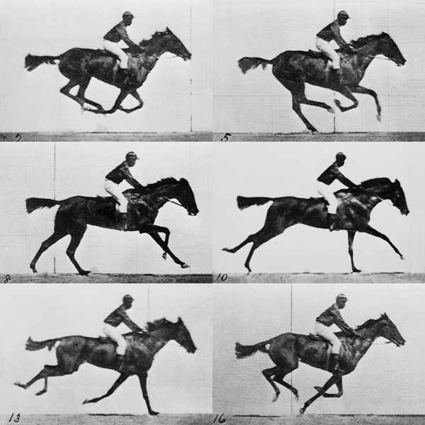
A streak camera is an instrument for measuring the variation in a pulse of light's intensity with time. They are used to measure the pulse duration of some ultrafast laser systems and for applications such as time-resolved spectroscopy and LIDAR.

A streak camera is an instrument for measuring the variation in a pulse of light's intensity with time. They are used to measure the pulse duration of some ultrafast laser systems and for applications such as time-resolved spectroscopy and LIDAR.
Mechanical streak cameras use a rotating mirror or moving slit system to deflect the light beam. They are limited in their maximum scan speed and thus temporal resolution. [1]
Optoelectronic streak cameras work by directing the light onto a photocathode, which when hit by photons produces electrons via the photoelectric effect. The electrons are accelerated in a cathode-ray tube and pass through an electric field produced by a pair of plates, which deflects the electrons sideways. By modulating the electric potential between the plates, the electric field is quickly changed to give a time-varying deflection of the electrons, sweeping the electrons across a phosphor screen at the end of the tube. [2] A linear detector, such as a charge-coupled device (CCD) array is used to measure the streak pattern on the screen, and thus the temporal profile of the light pulse. [3]
The time-resolution of the best optoelectronic streak cameras is around 180 femtoseconds. [4] Measurement of pulses shorter than this duration requires other techniques such as optical autocorrelation and frequency-resolved optical gating (FROG). [5]
In December 2011, a team at MIT released images combining the use of a streak camera with repeated laser pulses to simulate a movie with a frame rate of one trillion frames per second. [6] This was surpassed in 2020 by a team from Caltech that achieved frame rates of 70 trillion fps. [7]
Mode locking is a technique in optics by which a laser can be made to produce pulses of light of extremely short duration, on the order of picoseconds (10−12 s) or femtoseconds (10−15 s). A laser operated in this way is sometimes referred to as a femtosecond laser, for example, in modern refractive surgery. The basis of the technique is to induce a fixed phase relationship between the longitudinal modes of the laser's resonant cavity. Constructive interference between these modes can cause the laser light to be produced as a train of pulses. The laser is then said to be "phase-locked" or "mode-locked".
In physics and physical chemistry, time-resolved spectroscopy is the study of dynamic processes in materials or chemical compounds by means of spectroscopic techniques. Most often, processes are studied after the illumination of a material occurs, but in principle, the technique can be applied to any process that leads to a change in properties of a material. With the help of pulsed lasers, it is possible to study processes that occur on time scales as short as 10−16 seconds. All time-resolved spectra are suitable to be analyzed using the two-dimensional correlation method for a correlation map between the peaks.

Kerr-lens mode-locking (KLM) is a method of mode-locking lasers via the nonlinear optical Kerr effect. This method allows the generation of pulses of light with a duration as short as a few femtoseconds.
Photoemission electron microscopy is a type of electron microscopy that utilizes local variations in electron emission to generate image contrast. The excitation is usually produced by ultraviolet light, synchrotron radiation or X-ray sources. PEEM measures the coefficient indirectly by collecting the emitted secondary electrons generated in the electron cascade that follows the creation of the primary core hole in the absorption process. PEEM is a surface sensitive technique because the emitted electrons originate from a shallow layer. In physics, this technique is referred to as PEEM, which goes together naturally with low-energy electron diffraction (LEED), and low-energy electron microscopy (LEEM). In biology, it is called photoelectron microscopy (PEM), which fits with photoelectron spectroscopy (PES), transmission electron microscopy (TEM), and scanning electron microscopy (SEM).
In optics, an ultrashort pulse, also known as an ultrafast event, is an electromagnetic pulse whose time duration is of the order of a picosecond or less. Such pulses have a broadband optical spectrum, and can be created by mode-locked oscillators. Amplification of ultrashort pulses almost always requires the technique of chirped pulse amplification, in order to avoid damage to the gain medium of the amplifier.

In physics, terahertz time-domain spectroscopy (THz-TDS) is a spectroscopic technique in which the properties of matter are probed with short pulses of terahertz radiation. The generation and detection scheme is sensitive to the sample's effect on both the amplitude and the phase of the terahertz radiation.

Attosecond physics, also known as attophysics, or more generally attosecond science, is a branch of physics that deals with light-matter interaction phenomena wherein attosecond photon pulses are used to unravel dynamical processes in matter with unprecedented time resolution.

High-speed photography is the science of taking pictures of very fast phenomena. In 1948, the Society of Motion Picture and Television Engineers (SMPTE) defined high-speed photography as any set of photographs captured by a camera capable of 69 frames per second or greater, and of at least three consecutive frames. High-speed photography can be considered to be the opposite of time-lapse photography.
Ultrafast laser spectroscopy is a category of spectroscopic techniques using ultrashort pulse lasers for the study of dynamics on extremely short time scales. Different methods are used to examine the dynamics of charge carriers, atoms, and molecules. Many different procedures have been developed spanning different time scales and photon energy ranges; some common methods are listed below.
In optics, femtosecond pulse shaping refers to manipulations with temporal profile of an ultrashort laser pulse. Pulse shaping can be used to shorten/elongate the duration of optical pulse, or to generate complex pulses.
Multiphoton intrapulse interference phase scan (MIIPS) is a method used in ultrashort laser technology that simultaneously measures, and compensates femtosecond laser pulses using an adaptive pulse shaper. When an ultrashort laser pulse reaches a duration of less than a few hundred femtosecond, it becomes critical to characterize its duration, its temporal intensity curve, or its electric field as a function of time. Classical photodetectors measuring the intensity of light are still too slow to allow for a direct measurement, even with the fastest photodiodes or streak cameras.

A photoionization mode is a mode of interaction between a laser beam and matter involving photoionization.

The European X-Ray Free-Electron Laser Facility is an X-ray research laser facility commissioned during 2017. The first laser pulses were produced in May 2017 and the facility started user operation in September 2017. The international project with twelve participating countries; nine shareholders at the time of commissioning, later joined by three other partners, is located in the German federal states of Hamburg and Schleswig-Holstein. A free-electron laser generates high-intensity electromagnetic radiation by accelerating electrons to relativistic speeds and directing them through special magnetic structures. The European XFEL is constructed such that the electrons produce X-ray light in synchronisation, resulting in high-intensity X-ray pulses with the properties of laser light and at intensities much brighter than those produced by conventional synchrotron light sources.
Photofragment ion imaging or, more generally, Product Imaging is an experimental technique for making measurements of the velocity of product molecules or particles following a chemical reaction or the photodissociation of a parent molecule. The method uses a two-dimensional detector, usually a microchannel plate, to record the arrival positions of state-selected ions created by resonantly enhanced multi-photon ionization (REMPI). The first experiment using photofragment ion imaging was performed by David W Chandler and Paul L Houston in 1987 on the phototodissociation dynamics of methyl iodide (iodomethane, CH3I).
An optical modulator is an optical device which is used to modulate a beam of light with a perturbation device. It is a kind of transmitter to convert information to optical binary signal through optical fiber or transmission medium of optical frequency in fiber optic communication. There are several methods to manipulate this device depending on the parameter of a light beam like amplitude modulator (majority), phase modulator, polarization modulator etc. The easiest way to obtain modulation is modulation of intensity of a light by the current driving the light source. This sort of modulation is called direct modulation, as opposed to the external modulation performed by a light modulator. For this reason, light modulators are called external light modulators. According to manipulation of the properties of material modulators are divided into two groups, absorptive modulators and refractive modulators. Absorption coefficient can be manipulated by Franz-Keldysh effect, Quantum-Confined Stark Effect, excitonic absorption, or changes of free carrier concentration. Usually, if several such effects appear together, the modulator is called electro-absorptive modulator. Refractive modulators most often make use of electro-optic effect, other modulators are made with acousto-optic effect, magneto-optic effect such as Faraday and Cotton-Mouton effects. The other case of modulators is spatial light modulator (SLM) which is modified two dimensional distribution of amplitude & phase of an optical wave.
Time stretch microscopy, also known as serial time-encoded amplified imaging/microscopy or stretched time-encoded amplified imaging/microscopy' (STEAM), is a fast real-time optical imaging method that provides MHz frame rate, ~100 ps shutter speed, and ~30 dB optical image gain. Based on the photonic time stretch technique, STEAM holds world records for shutter speed and frame rate in continuous real-time imaging. STEAM employs the Photonic Time Stretch with internal Raman amplification to realize optical image amplification to circumvent the fundamental trade-off between sensitivity and speed that affects virtually all optical imaging and sensing systems. This method uses a single-pixel photodetector, eliminating the need for the detector array and readout time limitations. Avoiding this problem and featuring the optical image amplification for improvement in sensitivity at high image acquisition rates, STEAM's shutter speed is at least 1000 times faster than the best CCD and CMOS cameras. Its frame rate is 1000 times faster than the fastest CCD cameras and 10–100 times faster than the fastest CMOS cameras.

Femto-photography is a technique for recording the propagation of ultrashort pulses of light through a scene at a very high speed (up to 1013 frames per second). A femto-photograph is equivalent to an optical impulse response of a scene and has also been denoted by terms such as a light-in-flight recording or transient image. Femto-photography of macroscopic objects was first demonstrated using a holographic process in the 1970s by Nils Abramsson at the Royal Institute of Technology (Sweden). A research team at the MIT Media Lab led by Ramesh Raskar, together with contributors from the Graphics and Imaging Lab at the Universidad de Zaragoza, Spain, more recently achieved a significant increase in image quality using a streak camera synchronized to a pulsed laser and modified to obtain 2D images instead of just a single scanline.
Ultrafast electron diffraction, also known as femtosecond electron diffraction, is a pump-probe experimental method based on the combination of optical pump-probe spectroscopy and electron diffraction. Ultrafast electron diffraction provides information on the dynamical changes of the structure of materials. It is very similar to time resolved crystallography, but instead of using X-rays as the probe, it uses electrons. In the ultrafast electron diffraction technique, a femtosecond (10–15 second) laser optical pulse excites (pumps) a sample into an excited, usually non-equilibrium, state. The pump pulse may induce chemical, electronic or structural transitions. After a finite time interval, a femtosecond electron pulse is incident upon the sample. The electron pulse undergoes diffraction as a result of interacting with the sample. The diffraction signal is, subsequently, detected by an electron counting instrument such as a charge-coupled device camera. Specifically, after the electron pulse diffracts from the sample, the scattered electrons will form a diffraction pattern (image) on a charge-coupled device camera. This pattern contains structural information about the sample. By adjusting the time difference between the arrival of the pump and probe beams, one can obtain a series of diffraction patterns as a function of the various time differences. The diffraction data series can be concatenated in order to produce a motion picture of the changes that occurred in the data. Ultrafast electron diffraction can provide a wealth of dynamics on charge carriers, atoms, and molecules.

Light-in-flight imaging — a set of techniques to visualize propagation of light through different media.

Debabrata Goswami FInstP FRSC, is an Indian chemist and the Prof. S. Sampath Chair Professor of Chemistry, at the Indian Institute of Technology Kanpur. He is also a professor of The Department of Chemistry and The Center for Lasers & Photonics at the same Institute. Goswami is an associate editor of the open-access journal Science Advances. He is also an Academic Editor for PLOS One and PeerJ Chemistry. He has contributed to the theory of Quantum Computing as well as nonlinear optical spectroscopy. His work is documented in more than 200 research publications. He is an elected Fellow of the Royal Society of Chemistry, Fellow of the Institute of Physics, the SPIE, and The Optical Society. He is also a Senior Member of the IEEE, has been awarded a Swarnajayanti Fellowship for Chemical Sciences, and has held a Wellcome Trust Senior Research Fellowship. He is the third Indian to be awarded the International Commission for Optics Galileo Galilei Medal for excellence in optics.