Synaptonemal complex

Last updated
Schematic of the synaptonemal complex at different stages during Prophase I Synaptonemal Complex.svg
Schematic of the synaptonemal complex at different stages during Prophase I
A Homologous chromosomes (light blue) align and synapse together via transverse filaments (black lines) and longitudinal filaments (dark blue). Recombination nodules (gray ellipsoids) on the central region may help in completing recombination. Chromatin (red loops) is attached to its sexual leg and toe, extending from both sister chromatids. B Top: Set of tomato SCs. Chromatin "sheaths" visible around each SC. Bottom: Two tomato SCs with the chromatin removed, allowing kinetochores ("ball-like" structures) at centromeres to be revealed. Synaptonemal complex.png
A Homologous chromosomes (light blue) align and synapse together via transverse filaments (black lines) and longitudinal filaments (dark blue). Recombination nodules (gray ellipsoids) on the central region may help in completing recombination. Chromatin (red loops) is attached to its sexual leg and toe, extending from both sister chromatids. B Top: Set of tomato SCs. Chromatin "sheaths" visible around each SC. Bottom: Two tomato SCs with the chromatin removed, allowing kinetochores ("ball-like" structures) at centromeres to be revealed.

The synaptonemal complex (SC) is a protein structure that forms between homologous chromosomes (two pairs of sister chromatids) during meiosis and is thought to mediate synapsis and recombination during prophase I during meiosis in eukaryotes. It is currently thought that the SC functions primarily as a scaffold to allow interacting chromatids to complete their crossover activities. [1]

Contents

Composition

The synaptonemal complex is a tripartite structure consisting of two parallel lateral regions and a central element. This "tripartite structure" is seen during the pachytene stage of the first meiotic prophase, both in males and in females during gametogenesis. Previous to the pachytene stage, during leptonema, the lateral elements begin to form and they initiate and complete their pairing during the zygotene stage. After pachynema ends, the SC usually becomes disassembled and can no longer be identified. [2]

In humans, three specific components of the synaptonemal complex have been characterized: SC protein-1 (SYCP1), SC protein-2 (SYCP2), and SC protein-3 (SYCP3). The SYCP1 gene is on chromosome 1p13; the SYCP2 gene is on chromosome 20q13.33; and the gene for SYCP3 is on chromosome 12q. [3]

The synaptonemal complex was described by Montrose J. Moses in 1956 in primary spermatocytes of crayfish and by D. Fawcett in spermatocytes of pigeon, cat and man. [4] As seen with the electron microscope, the synaptonemal complex is formed by two "lateral elements", mainly formed by SYCP3 and secondarily by SYCP2, a "central element" that contains at least two additional proteins and the amino terminal region of SYCP1, and a "central region" spanned between the two lateral elements, that contains the "transverse filaments" composed mainly by the protein SYCP1. [3]

The SCs can be seen with the light microscope using silver staining or with immunofluorescence techniques that label the proteins SYCP3 or SYCP2.

Assembly and disassembly

Formation of the SC usually reflects the pairing or "synapsis" of homologous chromosomes and may be used to probe the presence of pairing abnormalities in individuals carrying chromosomal abnormalities, either in number or in the chromosomal structure. [5] The sex chromosomes in male mammals show only "partial synapsis" as they usually form only a short SC in the XY pair. The SC shows very little structural variability among eukaryotic organisms despite some significant protein differences. In many organisms the SC carries one or several "recombination nodules" associated with its central space. These nodules are thought to correspond to mature genetic recombination events or "crossovers". In male mice, gamma irradiation increases meiotic crossovers in SCs. This indicates that exogenously caused DNA damages are likely repaired by crossover recombination in SCs. [3] The finding of an interaction between a SC structural component [synaptonemal central element protein 2 (SYCE2)] and recombinational repair protein RAD51 also suggests a role for the SC in DNA repair.

In cell development the synaptonemal complex disappears during the late prophase of meiosis I. It is formed during zygotene.

Necessity in eukaryotes

It is now evident that the synaptonemal complex is not required for genetic recombination in some organisms. For instance, in protozoan ciliates such as Tetrahymena thermophila and Paramecium tetraurelia genetic crossover does not appear to require synaptonemal complex formation. [6] [7] Research has shown that not only does the SC form after genetic recombination but mutant yeast cells unable to assemble a synaptonemal complex can still engage in the exchange of genetic information. However, in other organisms like the C. elegans nematode, formation of chiasmata require the formation of the synaptonemal complex.

Related Research Articles

<span class="mw-page-title-main">Meiosis</span> Cell division producing haploid gametes

Meiosis is a special type of cell division of germ cells and apicomplexans in sexually-reproducing organisms that produces the gametes, such as sperm or egg cells. It involves two rounds of division that ultimately result in four cells with only one copy of each chromosome (haploid). Additionally, prior to the division, genetic material from the paternal and maternal copies of each chromosome is crossed over, creating new combinations of code on each chromosome. Later on, during fertilisation, the haploid cells produced by meiosis from a male and a female will fuse to create a cell with two copies of each chromosome again, the zygote.

<span class="mw-page-title-main">Chromosomal crossover</span> Cellular process

Chromosomal crossover, or crossing over, is the exchange of genetic material during sexual reproduction between two homologous chromosomes' non-sister chromatids that results in recombinant chromosomes. It is one of the final phases of genetic recombination, which occurs in the pachytene stage of prophase I of meiosis during a process called synapsis. Synapsis begins before the synaptonemal complex develops and is not completed until near the end of prophase I. Crossover usually occurs when matching regions on matching chromosomes break and then reconnect to the other chromosome.

<span class="mw-page-title-main">Prophase</span> First phase of cell division in both mitosis and meiosis

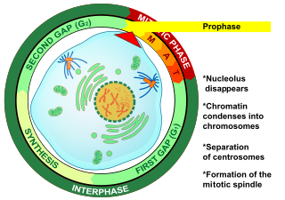
Prophase is the first stage of cell division in both mitosis and meiosis. Beginning after interphase, DNA has already been replicated when the cell enters prophase. The main occurrences in prophase are the condensation of the chromatin reticulum and the disappearance of the nucleolus.

<span class="mw-page-title-main">Genetic recombination</span> Production of offspring with combinations of traits that differ from those found in either parent

Genetic recombination is the exchange of genetic material between different organisms which leads to production of offspring with combinations of traits that differ from those found in either parent. In eukaryotes, genetic recombination during meiosis can lead to a novel set of genetic information that can be further passed on from parents to offspring. Most recombination occurs naturally and can be classified into two types: (1) interchromosomal recombination, occurring through independent assortment of alleles whose loci are on different but homologous chromosomes ; & (2) intrachromosomal recombination, occurring through crossing over.

<span class="mw-page-title-main">Homologous chromosome</span> Chromosomes that pair in fertilization

A couple of homologous chromosomes, or homologs, are a set of one maternal and one paternal chromosome that pair up with each other inside a cell during fertilization. Homologs have the same genes in the same loci, where they provide points along each chromosome that enable a pair of chromosomes to align correctly with each other before separating during meiosis. This is the basis for Mendelian inheritance, which characterizes inheritance patterns of genetic material from an organism to its offspring parent developmental cell at the given time and area.

<span class="mw-page-title-main">Synapsis</span> Biological phenomenon in meiosis

Synapsis is the pairing of two chromosomes that occurs during meiosis. It allows matching-up of homologous pairs prior to their segregation, and possible chromosomal crossover between them. Synapsis takes place during prophase I of meiosis. When homologous chromosomes synapse, their ends are first attached to the nuclear envelope. These end-membrane complexes then migrate, assisted by the extranuclear cytoskeleton, until matching ends have been paired. Then the intervening regions of the chromosome are brought together, and may be connected by a protein-RNA complex called the synaptonemal complex. During synapsis, autosomes are held together by the synaptonemal complex along their whole length, whereas for sex chromosomes, this only takes place at one end of each chromosome.

<span class="mw-page-title-main">Chromomere</span>

A chromomere, also known as an idiomere, is one of the serially aligned beads or granules of a eukaryotic chromosome, resulting from local coiling of a continuous DNA thread. Chromomeres are regions of chromatin that have been compacted through localized contraction. In areas of chromatin with the absence of transcription, condensing of DNA and protein complexes will result in the formation of chromomeres. It is visible on a chromosome during the prophase of meiosis and mitosis. Giant banded (Polytene) chromosomes resulting from the replication of the chromosomes and the synapsis of homologs without cell division is a process called endomitosis. These chromosomes consist of more than 1000 copies of the same chromatid that are aligned and produce alternating dark and light bands when stained. The dark bands are the chromomere.

<span class="mw-page-title-main">Bivalent (genetics)</span>

A bivalent is one pair of chromosomes in a tetrad. A tetrad is the association of a pair of homologous chromosomes physically held together by at least one DNA crossover. This physical attachment allows for alignment and segregation of the homologous chromosomes in the first meiotic division. In most organisms, each replicated chromosome elicits formation of DNA double-strand breaks during the leptotene phase. These breaks are repaired by homologous recombination, that uses the homologous chromosome as a template for repair. The search for the homologous target, helped by numerous proteins collectively referred as the synaptonemal complex, cause the two homologs to pair, between the leptotene and the pachytene phases of meiosis I.

Chromosome segregation is the process in eukaryotes by which two sister chromatids formed as a consequence of DNA replication, or paired homologous chromosomes, separate from each other and migrate to opposite poles of the nucleus. This segregation process occurs during both mitosis and meiosis. Chromosome segregation also occurs in prokaryotes. However, in contrast to eukaryotic chromosome segregation, replication and segregation are not temporally separated. Instead segregation occurs progressively following replication.

<span class="mw-page-title-main">TRIP13</span> Protein-coding gene in the species Homo sapiens

TRIP13 is a mammalian gene that encodes the thyroid receptor-interacting protein 13. In budding yeast, the analog for TRIP13 is PCH2. TRIP13 is a member of the AAA+ ATPase family, a family known for mechanical forces derived from ATP hydrolase reactions. The TRIP13 gene has been shown to interact with a variety of proteins and implicated in a few diseases, notably interacting with the ligand binding domain of thyroid hormone receptors, and may play a role in early-stage non-small cell lung cancer. However, recent evidence implicates TRIP13 in various cell cycle phases, including meiosis G2/Prophase and during the Spindle Assembly checkpoint (SAC). Evidence shows regulation to occur through the HORMA domains, including Hop1, Rev7, and Mad2. Of note, Mad2's involvement in the SAC is shown to be affected by TRIP13 Due to TRIP13's role in cell cycle arrest and progression, it may present opportunity as a therapeutic candidate for cancers.

<span class="mw-page-title-main">MSH4</span> Protein-coding gene in the species Homo sapiens

MutS protein homolog 4 is a protein that in humans is encoded by the MSH4 gene.

<span class="mw-page-title-main">SYCP3</span> Protein-coding gene in the species Homo sapiens

Synaptonemal complex protein 3 is a protein that in humans is encoded by the SYCP3 gene. It is a component of the synaptonemal complex formed between homologous chromosomes during the prophase of meiosis.

<span class="mw-page-title-main">HORMAD1</span> Protein-coding gene in the species Homo sapiens

HORMA domain-containing protein 1 (HORMAD1) also known as cancer/testis antigen 46 (CT46) is a protein that in humans is encoded by the HORMAD1 gene.

<span class="mw-page-title-main">Chiasma (genetics)</span>

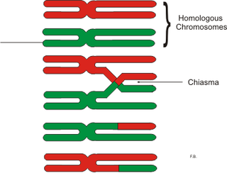
In genetics, a chiasma is the point of contact, the physical link, between two (non-sister) chromatids belonging to homologous chromosomes. At a given chiasma, an exchange of genetic material can occur between both chromatids, what is called a chromosomal crossover, but this is much more frequent during meiosis than mitosis. In meiosis, absence of a chiasma generally results in improper chromosomal segregation and aneuploidy.

<span class="mw-page-title-main">Meiotic recombination checkpoint</span>

The meiotic recombination checkpoint monitors meiotic recombination during meiosis, and blocks the entry into metaphase I if recombination is not efficiently processed.

The leptotene stage, also known as the leptonema, is the first of five substages of prophase I in meiosis. The term leptonema derives from Greek words meaning "thin threads". A cell destined to become a gamete enters the leptotene stage after its chromosomes are duplicated during interphase. During the leptotene stage those duplicated chromosomes—each consisting of two sister chromatids—condense from diffuse chromatin into long, thin strands that are more visible within the nucleoplasm. The next stage of prophase I in meiosis is the zygotene stage.

<span class="mw-page-title-main">STAG3 (gene)</span> Protein-coding gene in the species Homo sapiens

Stromal antigen 3 is a protein that in humans is encoded by the STAG3 gene. STAG3 protein is a component of a cohesin complex that regulates the separation of sister chromatids specifically during meiosis. STAG3 appears to be paramount in sister-chromatid cohesion throughout the meiotic process in human oocytes and spermatocytes.

Achiasmate Meiosis refers to meiosis without chiasmata, which are structures that are necessary for recombination to occur and that usually aid in the segregation of non-sister homologs. The pachytene stage of prophase I typically results in the formation of chiasmata between homologous non-sister chromatids in the tetrad chromosomes that form. The formation of a chiasma is also referred to as crossing over. When two homologous chromatids cross over, they form a chiasma at the point of their intersection. However, it has been found that there are cases where one or more pairs of homologous chromosomes do not form chiasmata during pachynema. Without a chiasma, no recombination between homologs can occur.

Glenna Shirleen Roeder is a geneticist known for identifying and characterizing the yeast genes that regulate the process of meiosis with particular emphasis on synapsis.

Abby F. Dernburg is a professor of Cell and Developmental Biology at the University of California, Berkeley, an Investigator of the Howard Hughes Medical Institute, and a Faculty Senior Scientist at Lawrence Berkeley National Laboratory.

References

  1. Page SL, Hawley RS (2004-10-08). "The genetics and molecular biology of the synaptonemal complex". Annual Review of Cell and Developmental Biology. 20 (1): 525–58. doi:10.1146/annurev.cellbio.19.111301.155141. PMID   15473851.
  2. Yang F, Wang PJ (2009). "The Mammalian synaptonemal complex: a scaffold and beyond". Genome Dynamics. 5: 69–80. doi:10.1159/000166620. ISBN   978-3-8055-8967-3. PMID   18948708.
  3. 1 2 3 Bolcun-Filas E, Hall E, Speed R, Taggart M, Grey C, de Massy B, et al. (February 2009). "Mutation of the mouse Syce1 gene disrupts synapsis and suggests a link between synaptonemal complex structural components and DNA repair". PLOS Genetics. 5 (2): e1000393. doi: 10.1371/journal.pgen.1000393 . PMC   2640461 . PMID   19247432.
  4. Moses, Montrose J. (1968-12-01). "Synaptinemal complex". Annual Review of Genetics. 2 (1): 363–412. doi:10.1146/annurev.ge.02.120168.002051. ISSN   0066-4197.
  5. Zickler D, Kleckner N (1999-12-01). "Meiotic chromosomes: integrating structure and function". Annual Review of Genetics. 33 (1): 603–754. doi:10.1146/annurev.genet.33.1.603. PMID   10690419.
  6. Lukaszewicz A, Howard-Till RA, Loidl J (November 2013). "Mus81 nuclease and Sgs1 helicase are essential for meiotic recombination in a protist lacking a synaptonemal complex". Nucleic Acids Research. 41 (20): 9296–309. doi:10.1093/nar/gkt703. PMC   3814389 . PMID   23935123.
  7. Chi J, Mahé F, Loidl J, Logsdon J, Dunthorn M (March 2014). "Meiosis gene inventory of four ciliates reveals the prevalence of a synaptonemal complex-independent crossover pathway". Molecular Biology and Evolution. 31 (3): 660–72. doi: 10.1093/molbev/mst258 . PMID   24336924.