Related Research Articles
A ligament is the fibrous connective tissue that connects bones to other bones. It is also known as articular ligament, articular larua, fibrous ligament, or true ligament. Other ligaments in the body include the:
Sheath, pronounced, may refer to:

The synovial membrane is a specialized connective tissue that lines the inner surface of capsules of synovial joints and tendon sheath. It makes direct contact with the fibrous membrane on the outside surface and with the synovial fluid lubricant on the inside surface. In contact with the synovial fluid at the tissue surface are many rounded macrophage-like synovial cells and also type B cells, which are also known as fibroblast-like synoviocytes (FLS). Type A cells maintain the synovial fluid by removing wear-and-tear debris. As for the FLS, they produce hyaluronan, as well as other extracellular components in the synovial fluid.

The human shoulder is made up of three bones: the clavicle (collarbone), the scapula, and the humerus as well as associated muscles, ligaments and tendons.
In anatomy, a vinculum is a band of connective tissue, similar to a ligament, that connects a flexor tendon to a phalanx bone. They contain tiny vessels which supply blood to the tendon. In vertebrate anatomy, they are referred to as mesotendons.
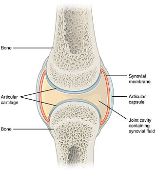
A synovial joint, also known as diarthrosis, joins bones or cartilage with a fibrous joint capsule that is continuous with the periosteum of the joined bones, constitutes the outer boundary of a synovial cavity, and surrounds the bones' articulating surfaces. This joint unites long bones and permits free bone movement and greater mobility. The synovial cavity/joint is filled with synovial fluid. The joint capsule is made up of an outer layer of fibrous membrane, which keeps the bones together structurally, and an inner layer, the synovial membrane, which seals in the synovial fluid.
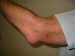
Bursitis is the inflammation of one or more bursae of synovial fluid in the body. They are lined with a synovial membrane that secretes a lubricating synovial fluid. There are more than 150 bursae in the human body. The bursae rest at the points where internal functionaries, such as muscles and tendons, slide across bone. Healthy bursae create a smooth, almost frictionless functional gliding surface making normal movement painless. When bursitis occurs, however, movement relying on the inflamed bursa becomes difficult and painful. Moreover, movement of tendons and muscles over the inflamed bursa aggravates its inflammation, perpetuating the problem. Muscle can also be stiffened.

The digastric muscle is a bilaterally paired suprahyoid muscle located under the jaw. Its posterior belly is attached to the mastoid notch of temporal bone, and its anterior belly is attached to the digastric fossa of mandible; the two bellies are united by an intermediate tendon which is held in a loop that attaches to the hyoid bone. The anterior belly is innervated via the mandibular nerve, and the posterior belly is innervated via the facial nerve. It may act to depress the mandible or elevate the hyoid bone.

The tibialis anterior muscle is a muscle of the anterior compartment of the lower leg. It originates from the upper portion of the tibia; it inserts into the medial cuneiform and first metatarsal bones of the foot. It acts to dorsiflex and invert the foot. This muscle is mostly located near the shin.

Synovial osteochondromatosis (SOC) (synonyms include synovial chondromatosis, primary synovial chondromatosis, synovial chondrometaplasia) is a rare disease that creates a benign change or proliferation in the synovium or joint-lining tissue, which changes to form bone-forming cartilage. In most occurrences, there is only one joint affected, either the knee, the hip, or the elbow. Rarely involves the TMJ.
In human anatomy, the extensor pollicis longus muscle (EPL) is a skeletal muscle located dorsally on the forearm. It is much larger than the extensor pollicis brevis, the origin of which it partly covers and acts to stretch the thumb together with this muscle.

The flexor hallucis longus muscle (FHL) attaches to the plantar surface of phalanx of the great toe and is responsible for flexing that toe. The FHL is one of the three deep muscles of the posterior compartment of the leg, the others being the flexor digitorum longus and the tibialis posterior. The tibialis posterior is the most powerful of these deep muscles. All three muscles are innervated by the tibial nerve which comprises half of the sciatic nerve.

The cuneonavicular joint is a joint (articulation) in the human foot. It is formed between the navicular bone and the three cuneiform bones. The navicular and cuneiform bones are connected by dorsal and plantar ligaments.

The patellar tendon is the distal portion of the common tendon of the quadriceps femoris, which is continued from the patella to the tibial tuberosity. It is also sometimes called the patellar ligament as it forms a bone to bone connection when the patella is fully ossified.

The articular capsule of the knee joint is the wide and lax joint capsule of the knee. It is thin in front and at the side, and contains the patella, ligaments, menisci, and bursae of the knee. The capsule consists of an inner synovial membrane, and an outer fibrous membrane separated by fatty deposits anteriorly and posteriorly.
The posterior compartment of the forearm contains twelve muscles which primarily extend the wrist and digits. It is separated from the anterior compartment by the interosseous membrane between the radius and ulna.

In the human body, the carpal tunnel or carpal canal is a flattened body cavity on the flexor (palmar/volar) side of the wrist, bounded by the carpal bones and flexor retinaculum. It forms the passageway that transmits the median nerve and the tendons of the extrinsic flexor muscles of the hand from the forearm to the hand. There are described cases of the anatomical variant median artery occurrence.

The common synovial sheath for the flexor tendons or the ulnar bursa is a synovial sheath in the carpal tunnel of the human hand.
A tendon sheath is a layer of synovial membrane around a tendon. It permits the tendon to stretch and not adhere to the surrounding fascia. It contains a lubricating fluid that allows for smooth motions of the tendon during muscle contraction and joint movements.
Villonodular synovitis is a type of synovial swelling.
References
| | This human musculoskeletal system article is a stub. You can help Wikipedia by expanding it. |