Rasmussen syndrome is a condition characterized by multiple trichoepitheliomas.

The Leser-Trélat sign is the explosive onset of multiple seborrheic keratoses, often with an inflammatory base. This can be an ominous sign of internal malignancy as part of a paraneoplastic syndrome. In addition to the development of new lesions, preexisting ones frequently increase in size and become symptomatic.

Hidrocystoma is an adenoma of the sweat glands.
Subacute cutaneous lupus erythematosus (SCLE) is a clinically distinct subset of cases of lupus erythematosus that is most often present in white women aged 15 to 40, consisting of skin lesions that are scaly and evolve as polycyclic annular lesions or plaques similar to those of plaque psoriasis.
Fibrofolliculomas are 2 to 4 mm in diameter, dome-shaped, yellowish or skin-colored papules usually located on the head, neck, and upper trunk. They are characteristically seen in Birt–Hogg–Dubé syndrome.
Microvenular hemangioma is an acquired benign vascular neoplasm that presents as an asymptomatic, slowly growing, 0.5- to 2.0 cm reddish lesion on the forearms or other sites of young to middle-aged adults.
Glomeruloid hemangioma is a distinctive vascular neoplasm first described in 1990 when found to be associated with Crow-Fukase syndrome and Castleman's disease.

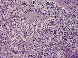
Trichoepithelioma is a neoplasm of the adnexa of the skin. Its appearance is similar to basal cell carcinoma.
Schöpf–Schulz–Passarge syndrome is an autosomal recessive condition with punctate symmetric palmoplantar keratoderma, with the keratoderma and fragility of the nails beginning around age 12. In addition to palmoplantar keratoderma, other symptoms include hypodontia, hypotrichosis, nail dystrophies, and eyelid cysts. Patients may also develop syringofibroadenoma and squamous cell carcinomas.
Clear cell acanthoma is a benign clinical and histological lesion initially described as neoplastic, which some authors now regard as a reactive dermatosis. It usually presents as a moist solitary firm, brown-red, well-circumscribed, 5 mm to 2 cm nodule or plaque on the lower extremities of middle-aged to elderly individuals The lesion has a crusted, scaly peripheral collarette and vascular puncta on the surface. It is characterized by slow growth, and may persist for years. The clinical differential diagnosis includes: dermatofibroma, inflamed seborrheic keratosis, pyogenic granuloma, basal cell carcinoma, squamous cell carcinoma, verruca vulgaris, psoriatic plaque, and melanoma.
Secondary cutaneous amyloidosis is a skin condition that occurs following PUVA therapy and in benign and malignant cutaneous neoplasms in which deopsits of amyloid may be found.

A sebaceous adenoma, a type of adenoma, a cutaneous condition characterized by a slow-growing tumor usually presenting as a pink, flesh-coloured, or yellow papule or nodule.

Spiradenoma, also spiroma or eccrine spiradenoma, is a cutaneous condition that is typically characterized, clinically, as a solitary, deep-seated dermal nodule of approximately one centimeter, occurring on the ventral surface of the body. Spiradenoma lesions are benign sudoriferous tumors, and have also been described as cystic epitheliomas of the sweat glands.

Trichoblastomas are a cutaneous condition characterized by benign neoplasms of follicular germinative cells. Trichoblastic fibroma is a designation used to characterize small nodular trichoblastomas with conspicuous fibrocytic stroma, sometimes constituting over 50% of the lesion.
A trichodiscoma is a cutaneous condition, a benign tumor usually skin colored, most often affecting the face and upper trunk.
Brooke–Fordyce syndrome is a condition characterized by multiple trichoepitheliomas.